Patent application title: Compositions and Methods for Treating and Diagnosing Cancer
Inventors:
Austin Gurney (San Francisco, CA, US)
IPC8 Class: AA61K39395FI
USPC Class:
4241381
Class name: Drug, bio-affecting and body treating compositions immunoglobulin, antiserum, antibody, or antibody fragment, except conjugate or complex of the same with nonimmunoglobulin material binds expression product or fragment thereof of cancer-related gene (e.g., oncogene, proto-oncogene, etc.)
Publication date: 2009-07-30
Patent application number: 20090191205
Inventors list |
Agents list |
Assignees list |
List by place |
Classification tree browser |
Top 100 Inventors |
Top 100 Agents |
Top 100 Assignees |
Usenet FAQ Index |
Documents |
Other FAQs |
Patent application title: Compositions and Methods for Treating and Diagnosing Cancer
Inventors:
Austin GURNEY
Agents:
STERNE, KESSLER, GOLDSTEIN & FOX P.L.L.C.
Assignees:
Origin: WASHINGTON, DC US
IPC8 Class: AA61K39395FI
USPC Class:
4241381
Abstract:
The present invention relates to compositions and methods for
characterizing, diagnosing and treating cancer. In particular, the
present invention identifies LGR5 as a protein over-expressed in solid
tumor stem cell. The present invention further identifies an interaction
between RSPO1 and LGR5 as an alternative pathway for the activation of
beta-catenin signaling. In certain embodiments, the present invention
provides biomolecules that disrupt functional signaling via a LGR
protein, including, in certain embodiments, molecules that inhibit the
interaction between one or more RSPO proteins and one or more LGR
proteins, such as LGR5. In certain embodiments, the present invention
provides methods of treating cancer comprising disrupting functional LGR
signaling and inhibiting growth of a solid tumor comprising solid tumor
stem cells.Claims:
1. An isolated antibody that specifically binds to an extracellular domain
of a human leucine-rich repeat-containing G protein-coupled receptor
(LGR) protein, wherein the antibody:(a) disrupts binding of a human
R-spondin (RSPO) protein to the LGR protein; and/or(b) disrupts RSPO
activation of LGR signaling.
2. The antibody of claim 1, wherein the human RSPO protein is selected from the group consisting of RSPO1, RSPO2, RSPO3, and RSPO4.
3. The antibody of claim 1, wherein the LGR protein is LGR5.
4. The antibody of claim 3, wherein the extracellular domain comprises amino acids 22-564 of human LGR5 (SEQ ID NO: 1).
5. The antibody of claim 1, wherein the LGR protein is LGR4 or LGR6.
6-8. (canceled)
9. A cell line producing the antibody of claim 1.
10. A hybridoma cell line having ATCC deposit number ______.
11. A monoclonal antibody 88M1 produced by the hybridoma cell line of claim 10.
12. An isolated antibody comprising the heavy chain CDRs and/or the light chain CDRs of the antibody of claim 11.
13. An isolated antibody that competes with the antibody of claim 11 in a competitive binding assay.
14. A pharmaceutical composition comprising the antibody of claim 1 and a pharmaceutically acceptable carrier.
15. (canceled)
16. A method of treating cancer or inhibiting growth of a tumor in a human, comprising administering to the human a therapeutically effective amount of the antibody of claim 1.
17-19. (canceled)
20. A method of inhibiting beta-catenin signaling in a tumor cell, comprising contacting said tumor cell with an effective amount of the antibody of claim 1.
21-24. (canceled)
25. An isolated antibody that binds LGR5, comprising:(a) a heavy chain CDR1 comprising SSYAIS (SEQ ID NO:35), a heavy chain CDR2 comprising GIIPIFGMANYAQKFQG (SEQ ID NO:36), and a heavy chain CDR3 comprising YDFITHFDF (SEQ ID NO:37); and/or(b) a light chain CDR1 comprising RASQSVSSNYLA (SEQ ID NO:32), a light chain CDR2 comprising DASNRAT (SEQ ID NO:33), and a light chain CDR3 comprising QQMDDFPM (SEQ ID NO:34).
26-32. (canceled)
33. A method of treating cancer or inhibiting tumor growth in a human, wherein the cancer or tumor comprises cancer stem cells, comprising administering to the human a therapeutically effective amount of the antibody of claim 25.
34. An isolated antibody that binds LGR5, comprising:(a) a heavy chain CDR1 comprising SNYAIG (SEQ ID NO:41), a heavy chain CDR2 comprising GIRPNFGWAKYAQKFQG (SEQ ID NO:42), and a heavy chain CDR3 comprising YGQGHFPSAFDY (SEQ ID NO:43); and/or(b) a light chain CDR1 comprising TGTNSDVGTYNYVH (SEQ ID NO:38), a light chain CDR2 comprising DGSNRPSG (SEQ ID NO:39), and a light chain CDR3 comprising QAYDSHSFNI (SEQ ID NO:40).
35-41. (canceled)
42. A method of treating cancer or inhibiting tumor growth in a human, wherein the cancer or tumor comprises cancer stem cells, comprising administering to the human a therapeutically effective amount of the antibody of claim 34.
43. An isolated antibody that binds LGR5, comprising:(a) a heavy chain variable region having at least about 80% sequence identity to SEQ ID NO:45; and/or(b) a light chain variable region having at least about 80% sequence identity to SEQ ID NO: 44.
44. A polynucleotide comprising a polynucleotide encoding the heavy chain and/or light chain variable region of the antibody of claim 43.
45. A method of treating cancer or inhibiting tumor growth in a human, wherein the cancer or tumor comprises cancer stem cells, comprising administering to the human a therapeutically effective amount of the antibody of claim 43.
46. An isolated antibody that binds LGR5, comprising:(a) a heavy chain variable region having at least about 80% sequence identity to SEQ ID NO:47; and/or(b) a light chain variable region having at least about 80% sequence identity to SEQ ID NO:46.
47. A polynucleotide comprising a polynucleotide encoding the heavy chain and/or light chain variable region of the antibody of claim 46.
48. A method of treating cancer or inhibiting tumor growth in a human, wherein the cancer or tumor comprises cancer stem cells, comprising administering to the human a therapeutically effective amount of the antibody of claim 46.
49. The antibody of claim 1, which inhibits growth of a solid tumor comprising solid tumor stem cells.
Description:
CROSS-REFERENCE TO RELATED APPLICATIONS
[0001]This application claims the priority benefit of U.S. provisional application No. 60/947,611, filed Jul. 2, 2007, which is hereby incorporated by reference herein in its entirety.
BACKGROUND OF THE INVENTION
[0002]1. Field of the Invention
[0003]The present invention relates to the field of oncology and provides novel compositions and methods for diagnosing and treating cancer. In particular, the invention provides the means and methods for characterizing, studying, diagnosing, providing a prognosis, and treating cancers comprising solid tumor cancer stem cells.
[0004]2. Background Art
[0005]Cancer is one of the leading causes of death in the developed world, resulting in over 500,000 deaths per year in the United States alone. Over one million people are diagnosed with cancer in the U.S. each year, and overall it is estimated that more than 1 in 3 people will develop some form of cancer during their lifetime. Though there are more than 200 different types of cancer, four of them--breast, lung, colorectal, and prostate--account for over half of all new cases (Jemal et al., 2003, Cancer J. Clin. 53:5-26).
[0006]Breast cancer is the most common cancer in woman, with an estimate 12% of women at risk of developing the disease during their lifetime. Although mortality rates have decreased due to earlier detection and improved treatments, breast cancer remains a leading cause of death in middle-aged women. Furthermore, metastatic breast cancer is still an incurable disease. On presentation, most patients with metastatic breast cancer have only one or two organ systems affected, but as the disease progresses, multiple sites usually become involved. The most common sites of metastatic involvement are locoregional recurrences in the skin and soft tissues of the chest wall, as well as in axilla and supraclavicular areas. The most common site for distant metastasis is the bone (30-40% of distant metastasis), followed by the lungs and liver. And although only approximately 1-5% of women with newly diagnosed breast cancer have distant metastasis at the time of diagnosis, approximately 50% of patients with local disease eventually relapse with metastasis within five years. At present the median survival from the manifestation of distant metastases is about three years.
[0007]Current methods of diagnosing and staging breast cancer include the tumor-node-metastasis (TNM) system that relies on tumor size, tumor presence in lymph nodes, and the presence of distant metastases as described in the American Joint Committee on Cancer: AJCC Cancer Staging Manual. Philadelphia, Pa.: Lippincott-Raven Publishers, 5th ed., 1997, pp 171-180, and in Harris, J R: "Staging of breast carcinoma" in Harris, J. R., Hellman, S., Henderson, I. C., Kinne D. W. (eds.): Breast Diseases. Philadelphia, Lippincott, 1991. These parameters are used to provide a prognosis and select an appropriate therapy. The morphologic appearance of the tumor may also be assessed but because tumors with similar histopathologic appearance can exhibit significant clinical variability, this approach has serious limitations. Finally, assays for cell surface markers can be used to divide certain tumors types into subclasses. For example, one factor considered in the prognosis and treatment of breast cancer is the presence of the estrogen receptor (ER) as ER-positive breast cancers typically respond more readily to hormonal therapies such as tamoxifen or aromatase inhibitors than ER-negative tumors. Yet these analyses, though useful, are only partially predictive of the clinical behavior of breast tumors, and there is much phenotypic diversity present in breast cancers that current diagnostic tools fail to detect and current therapies fail to treat.
[0008]Prostate cancer is the most common cancer in men in the developed world, representing an estimated 33% of all new cancer cases in the U.S., and is the second most frequent cause of death (Jemal et al., 2003, CA Cancer J. Clin. 53:5-26). Since the introduction of the prostate specific antigen (PSA) blood test, early detection of prostate cancer has dramatically improved survival rates, and the five year survival rate for patients with local and regional stage prostate cancers at the time of diagnosis is nearing 100%. Yet more than 50% of patients will eventually develop locally advanced or metastatic disease (Muthuramalingam et al., 2004, Clin. Oncol. 16:505-16).
[0009]Currently radical prostatectomy and radiation therapy provide curative treatment for the majority of localized prostate tumors. However, therapeutic options are very limited for advanced cases. For metastatic disease, androgen ablation with luteinising hormone-releasing hormone (LHRH) agonist alone or in combination with anti-androgens is the standard treatment. Yet despite maximal androgen blockage, the disease nearly always progresses with the majority developing androgen-independent disease. At present there is no uniformly accepted treatment for hormone refractory prostate cancer, and chemotherapeutic regimes are commonly used (Muthuramalingam et al., 2004, Clin. Oncol. 16:505-16; Trojan et al., 2005, Anticancer Res. 25:551-61).
[0010]Lung cancer is the most common cancer worldwide, the third most commonly diagnosed cancer in the United States, and by far the most frequent cause of cancer deaths (Spiro et al., 2002, Am. J. Respir. Crit. Care Med. 166:1166-96; Jemal et al., 2003, CA Cancer J. Clin. 53:5-26). Cigarette smoking is believed responsible for an estimated 87% of all lung cancers making it the most deadly preventable disease. Lung cancer is divided into two major types that account for over 90% of all lung cancers: small cell lung cancer (SCLC) and non-small cell lung cancer (NSCLC). SCLC accounts for 15-20% of cases and is characterized by its origin in large central airways and histological composition of sheets of small cells with little cytoplasm. SCLC is more aggressive than NSCLC, growing rapidly and metastasizing early and often. NSCLC accounts for 80-85% of all cases and is further divided into three major subtypes based on histology: adenocarcinoma, squamous cell carcinoma (epidermoid carcinoma), and large cell undifferentiated carcinoma.
[0011]Lung cancer typically presents late in its course, and thus has a median survival of only 6-12 months after diagnosis and an overall 5 year survival rate of only 5-10%. Although surgery offers the best chance of a cure, only a small fraction of lung cancer patients are eligible with the majority relying on chemotherapy and radiotherapy. Despite attempts to manipulate the timing and dose intensity of these therapies, survival rates have increased little over the last 15 years (Spiro et al., 2002, Am. J. Respir. Crit. Care Med. 166:1166-96).
[0012]Colorectal cancer is the third most common cancer and the fourth most frequent cause of cancer deaths worldwide (Weitz et al., 2005, Lancet 365:153-65). Approximately 5-10% of all colorectal cancers are hereditary with one of the main forms being familial adenomatous polyposis (FAP), an autosomal dominant disease in which about 80% of affected individuals contain a germline mutation in the adenomatous polyposis coli (APC) gene. Colorectal carcinoma has a tendency to invade locally by circumferential growth and elsewhere by lymphatic, hematogenous, transperitoneal, and perineural spread. The most common site of extralymphatic involvement is the liver, with the lungs the most frequently affected extra-abdominal organ. Other sites of hematogenous spread include the bones, kidneys, adrenal glands, and brain.
[0013]The current staging system for colorectal cancer is based on the degree of tumor penetration through the bowel wall and the presence or absence of nodal involvement. This staging system is defined by three major Duke's classifications: Duke's A disease is confined to submucosa layers of colon or rectum; Duke's B disease has tumors that invade through muscularis propria and can penetrate the wall of the colon or rectum; and Duke's C disease includes any degree of bowel wall invasion with regional lymph node metastasis. While surgical resection is highly effective for early stage colorectal cancers, providing cure rates of 95% in Duke's A patients, the rate is reduced to 75% in Duke's B patients and the presence of positive lymph node in Duke's C disease predicts a 60% likelihood of recurrence within five years. Treatment of Duke's C patients with a post surgical course of chemotherapy reduces the recurrence rate to 40%-50%, and is now the standard of care for these patients.
[0014]Epithelial carcinomas of the head and neck arise from the mucosal surfaces in the head and neck area and are typically squamous cell in origin. This category includes tumors of the paranasal sinuses, the oral cavity, and the nasopharynx, oropharynx, hypopharynx, and larynx.
[0015]The annual number of new cases of head and neck cancers in the United States is approximately 40,000 per year, accounting for about 5 percent of adult malignancies. Head and neck cancers are more common in some other countries, and the worldwide incidence probably exceeds half a million cases annually. In North American and Europe, the tumors usually arise from the oral cavity, oropharynx, or larynx, whereas nasopharyngeal cancer is more common in the Mediterranean countries and in the Far East.
[0016]Traditional modes of therapy (radiation therapy, chemotherapy, and hormonal therapy), while useful, have been limited by the emergence of treatment-resistant cancer cells. Clearly, new approaches are needed to identify targets for treating head and neck cancer and cancer generally.
[0017]Cancer arises from dysregulation of the mechanisms that control normal tissue development and maintenance, and increasingly stem cells are thought to play a central role (Beachy et al., 2004, Nature 432:324). During normal animal development, cells of most or all tissues are derived from normal precursors, called stem cells (Morrison et al., 1997, Cell 88:287-98; Morrison et al., 1997, Curr. Opin. Immunol. 9:216-21; Morrison et al., 1995, Annu. Rev. Cell. Dev. Biol. 11:35-71). Stem cells are cells that: (1) have extensive proliferative capacity; 2) are capable of asymmetric cell division to generate one or more kinds of progeny with reduced proliferative and/or developmental potential; and (3) are capable of symmetric cell divisions for self-renewal or self-maintenance. The best-known example of adult cell renewal by the differentiation of stem cells is the hematopoietic system where developmentally immature precursors (hematopoietic stem and progenitor cells) respond to molecular signals to form the varied blood and lymphoid cell types. Other cells, including cells of the gut, breast ductal system, and skin are constantly replenished from a small population of stem cells in each tissue, and recent studies suggest that most other adult tissues also harbor stem cells, including the brain.
[0018]Solid tumors are composed of heterogeneous cell populations. For example, breast cancers are a mixture of cancer cells and normal cells, including mesenchymal (stromal) cells, inflammatory cells, and endothelial cells. Classic models of cancer hold that phenotypically distinct cancer cell populations all have the capacity to proliferate and give rise to a new tumor. In the classical model, tumor cell heterogeneity results from environmental factors as well as ongoing mutations within cancer cells resulting in a diverse population of tumorigenic cells. This model rests on the idea that all populations of tumor cells would have some degree of tumorigenic potential. (Pandis et al., 1998, Genes, Chromosomes & Cancer 12:122-129; Kuukasjrvi et al., 1997, Cancer Res. 57:1597-1604; Bonsing et al., 1993, Cancer 71:382-391; Bonsing et al., 2000, Genes Chromosomes & Cancer 82: 173-183; Beerman H et al., 1991, Cytometry. 12:147-54; Aubele M & Werner M, 1999, Analyt. Cell. Path. 19:53; Shen L et al., 2000, Cancer Res. 60:3884).
[0019]An alternative model for the observed solid tumor cell heterogeneity is that solid tumors result from a "solid tumor stem cell" (or "cancer stem cell" from a solid tumor) that subsequently undergoes chaotic development through both symmetric and asymmetric rounds of cell division. In this stem cell model, solid tumors contain a distinct and limited (possibly even rare) subset of cells that share properties with normal "stem cells" in that they extensively proliferate and efficiently give rise both to additional solid tumor stem cells (self-renewal) and to the majority of within a solid tumor that lack tumorigenic potential. Indeed, mutations within a long-lived stem cell population can initiate the formation of cancer stem cells that underlie the growth and maintenance of tumors and whose presence contributes to the failure of current therapeutic approaches.
[0020]The stem cell nature of cancer was first revealed in the blood cancer, acute myeloid leukemia (AML) (Lapidot et al., 1994, Nature 17:645-8). More recently it has been demonstrated that malignant human breast tumors similarly harbor a small, distinct population of cancer stem cells enriched for the ability to form tumors in immunodeficient mice. An ESA+, CD44+, CD24-/low, Lin- cell population was found to be 50-fold enriched for tumorigenic cells compared to unfractionated tumor cells (Al-Hajj et al., 2003, PNAS 100:3983-8). Furthermore, a similar population is also present in colon cancers. The ability to prospectively isolate the tumorigenic cancer cells has permitted precise investigation of critical biological pathways that underlie tumorigenicity in these cells, and thus promises the development of better diagnostic assays and therapeutics for cancer patients. It is toward this purpose and the other purposes described herein that this invention is directed.
[0021]All publications, patents, patent applications, internet sites, and accession numbers/database sequences including both polynucleotide and polypeptide sequences cited herein are hereby incorporated by reference herein in their entirety for all purposes to the same extent as if each individual publication, patent, patent application, internet site, or accession number/database sequence were specifically and individually indicated to be so incorporated by reference.
BRIEF SUMMARY OF THE INVENTION
[0022]The present invention relates to compositions and methods in the field of oncology. In particular, the present invention is based, in part, on the discovery that an LGR (leucine-rich repeat-containing, G protein-coupled receptor) protein, such as LGR5 (leucine-rich repeat-containing, G protein-coupled receptor 5) is a protein over-expressed in solid tumor cancer stem cells, and thus is a cancer stem cell marker useful in the characterization, study, diagnosis, and treatment of cancer. The present invention further identifies an interaction between the R-spondin RSPO1 and LGR5 as an alternative pathway for the activation of beta-catenin signaling, suggesting functional blocking of LGR5 can inhibit tumor growth. Interactions between LGR5 and each of the additional RSPO proteins RSPO2, RSPO3, and RSPO4 have now likewise been identified.
[0023]As such, in certain embodiments, the present invention provides biomolecules that disrupt functional signaling via a LGR protein, including, in certain embodiments, molecules that inhibit the interaction between R-spondin (RSPO) proteins and an LGR protein such as LGR5. In certain embodiments, the biomolecules are antibodies. For instance, in certain embodiments, the biomolecules may be antibodies that specifically bind to the extracellular domain of at least one human LGR protein. In certain embodiments, the present invention provides antibodies that specifically bind to an extracellular domain of a human LGR protein and inhibit growth of tumor cells. In certain embodiments, the LGR protein is LGR5. The present invention further provides methods of treating cancer comprising cancer stem cells. In certain embodiments, the method of treating cancer comprises administering a therapeutically effective amount of an antibody that specifically binds an extracellular domain of a LGR protein. In certain embodiments, the LGR protein is LGR5. The present invention further provides for a method of treating cancer in a human and/or inhibiting growth of a tumor in a human comprising administering to the human a therapeutically effective amount of an agent that (a) disrupts the binding of a human RSPO protein to a human LGR protein and/or (b) disrupts RSPO activation of LGR signaling. In some embodiments, the agent is an antibody. In certain embodiments, the agent binds a human LGR protein. In certain alternative embodiments, the agent is an antibody that specifically binds to the extracellular domain of at least one human LGR protein. In certain alternative embodiments, the agent is an antibody that specifically binds to the extracellular domain of two or more human LGR proteins In certain embodiments, the LGR protein is LGR5. In certain embodiments, the cancer or tumor comprises cancer stem cells.
[0024]In addition, the present invention provides a method of inhibiting beta-catenin signaling in a tumor cell, comprising contacting the tumor cell with an agent that (a) disrupts the binding of a human RSPO protein to a human LGR protein and/or (b) disrupts RSPO activation of LGR signaling. In certain embodiments, the agent binds a human LGR protein. In certain alternative embodiments, the agent is an antibody that specifically binds to the extracellular domain of at least one human LGR protein. In certain alternative embodiments, the agent is an antibody that specifically binds to the extracellular domain of two or more human LGR proteins. In certain embodiments, the LGR protein is LGR5. In certain embodiments, the method is an in vitro method. In certain embodiments, the method is an in vivo method.
[0025]The present invention further provides antibodies that bind to an extracellular domain of a human LGR protein and are capable of inhibiting growth of a solid tumor (e.g., a solid tumor comprising solid tumor stem cells) by (a) disrupting binding of a human RSPO protein to a human LGR protein; (b) disrupt RSPO activation of LGR signaling; and/or (c) inhibiting beta-catenin signaling. The present invention also provides antibodies that (a) bind to an extracellular domain of a human LGR protein; (b) disrupt binding of a human RSPO protein to a human LGR protein; (c) disrupt RSPO activation of LGR signaling; (d) inhibit beta-catenin signaling; and/or (e) are capable of inhibiting growth of a solid tumor (e.g., a solid tumor comprising solid tumor stem cells). In certain embodiments, the antibodies specifically bind to the extracellular domain of a human LGR protein. In certain embodiments, the human LGR protein is LGR5. In certain embodiments, the human RSPO protein is RSPO1. In some alternative embodiments, the human RSPO protein is RSPO2, RSPO3, or RSPO4. Cell lines producing the antibodies and compositions comprising the antibodies are further provided. Methods of using therapeutically effective amounts of compositions comprising the antibodies for treating cancer, including, but not limited to, by inhibiting growth of a tumor, are further provided. Methods using the antibodies, in vivo or in vitro, to inhibit beta-catenin signaling are also provided.
[0026]The invention further provides a monoclonal anti-LGR5 antibody 88M1 produced by a hybridoma cell line having ATCC deposit number ______. Antibodies are also provided that specifically bind LGR5 and (a) comprise a heavy chain variable region and/or a light chain variable region having at least about 95% sequence identity to the heavy chain variable region and/or the light chain variable region (respectively) of 88M1; (b) comprise the heavy chain and/or light chain CDRs of 88M1; (c) bind to an epitope capable of binding 88M1; and/or (d) compete with 88M1 in a competitive binding assay. Cells lines producing the antibodies (including, but not limited to, the hybridoma cell line having ATCC deposit number ______) and compositions comprising the antibodies are further provided. Methods of using therapeutically effective amounts of compositions comprising the antibodies for treating cancer, including, but not limited to, by inhibiting growth of a tumor, are further provided. Methods using the antibodies, in vivo or in vitro, to inhibit beta-catenin signaling are also provided.
[0027]The invention further provides an antibody that binds LGR5, comprising: (a) a heavy chain CDR1 comprising SSYAIS (SEQ ID NO:35), a heavy chain CDR2 comprising GIIPIFGMANYAQKFQG (SEQ ID NO:36), and/or a heavy chain CDR3 comprising YDFITHFDF (SEQ ID NO:37); and/or (b) a light chain CDR1 comprising RASQSVSSNYLA (SEQ ID NO:32), a light chain CDR2 comprising DASNRAT (SEQ ID NO:33), and/or a light chain CDR3 comprising QQMDDFPM (SEQ ID NO:34). Polynucleotides comprising a polynucleotide encoding a heavy chain or light chain variable region of the antibody are further provided. Vectors and cells comprising the polynucleotides or antibodies are also provided, as are methods of treating cancer and/or inhibiting tumor growth in a human comprising administering a therapeutically effective amount of the antibody to the human. In some embodiments, the cancer or tumor comprises cancer stem cells.
[0028]The invention further provides an antibody that binds LGR5, comprising: (a) a heavy chain CDR1 comprising SNYAIG (SEQ ID NO:41), a heavy chain CDR2 comprising GIRPNFGWAKYAQKFQG (SEQ ID NO:42), and/or a heavy chain CDR3 comprising YGQGHFPSAFDY (SEQ ID NO:43); and/or (b) a light chain CDR1 comprising TGTNSDVGTYNYVH (SEQ ID NO:38), a light chain CDR2 comprising DGSNRPSG (SEQ ID NO:39), and/or a light chain CDR3 comprising QAYDSHSFNI (SEQ ID NO:40). Polynucleotides comprising a polynucleotide encoding a heavy chain or light chain variable region of the antibody are further provided. Vectors and cells comprising the polynucleotides or antibodies are also provided, as are methods of treating cancer and/or inhibiting tumor growth in a human comprising administering a therapeutically effective amount of the antibody to the human. In some embodiments, the cancer or tumor comprises cancer stem cells.
[0029]The invention further provides an antibody that binds LGR5, comprising: (a) a heavy chain variable region having at least about 80% sequence identity to SEQ ID NO:45; and/or (b) a light chain variable region having at least about 80% sequence identity to SEQ ID NO:44. Polynucleotides comprising a polynucleotide encoding such heavy chain or light chain variable regions are also provided. Vectors and cells comprising the polynucleotides or antibodies are also provided, as are methods of treating cancer and/or inhibiting tumor growth in a human comprising administering a therapeutically effective amount of the antibody to the human. In some embodiments, the cancer or tumor comprises cancer stem cells.
[0030]The invention further provides an antibody that binds LGR5, comprising: (a) a heavy chain variable region having at least about 80% sequence identity to SEQ ID NO:47; and/or (b) a light chain variable region having at least about 80% sequence identity to SEQ ID NO:46. Polynucleotides comprising a polynucleotide encoding such heavy chain or light chain variable regions are also provided. Vectors and cells comprising the polynucleotides or antibodies are also provided, as are methods of treating cancer and/or inhibiting tumor growth in a human comprising administering a therapeutically effective amount of the antibody to the human. In some embodiments, the cancer or tumor comprises cancer stem cells.
[0031]The present invention further provides methods of identifying and/or isolating cancer stem cells (e.g., based on expression of LGR5), screening for anti-cancer agents, and screening patients for suitability for treatment with the agents described herein.
[0032]Where aspects or embodiments of the invention are described in terms of a Markush group or other grouping of alternatives, the present invention encompasses not only the entire group listed as a whole, but also each member of the group individually and all possible subgroups of the main group, and also the main group absent one or more of the group members. The present invention also envisages the explicit exclusion of one or more of any of the group members in the claimed invention.
BRIEF DESCRIPTION OF THE FIGURES
[0033]FIG. 1. LGR5 Is Overexpressed In Solid Tumor Cancer Stem Cells. Cells from human colon tumors were sorted by FACS into a tumorigenic "TG" fraction (right bars) containing cancer stem cells and a non-tumorigenic "NTG" fraction (left bars). mRNA was isolated from these fraction and microarray data was generated. LGR5 demonstrated higher mRNA expression in the TG cancer stem cell fraction from three independent human colon tumors (right bar of each set).
[0034]FIG. 2. LGR5 Is Overexpressed In Human Epithelial Tumors. Shown is microarray data for LGR5 mRNA expression from a large number of human tumors compared to tissue samples from normal human tissues. Expression level of LGR5 in individual patient samples is indicated by vertical dash lines within the horizontal axis for each indicated tissue type. LGR5 is overexpressed in most tumor samples relative to the expression in the corresponding normal tissue.
[0035]FIG. 3. LGR6 Shows Altered Expression In Human Epithelial Tumors. Shown is microarray data for LGR6 mRNA expression from a large number of human tumors compared to tissue samples from normal human tissues. Expression level of LGR6 in individual patient samples is indicated by vertical dash lines within the horizontal axis for each indicated tissue type. LGR6 expression shows altered expression in many tumor samples relative to the expression in the corresponding normal tissue.
[0036]FIG. 4. RSPO1 Activates Beta-Catenin Signaling. Luciferase activity (y-axis) from an 8×TCF luciferase reporter was measured following exposure to RSPO1-Fc in the indicated concentration x-axis). RSPO1-Fc induced luciferase activity from the beta-catenin responsive promoter in a dose dependent manner.
[0037]FIG. 5. Soluble LGR5 (LGR5-Fc) Inhibits the Induction of Beta-Catenin Signaling By RSPO1. Luciferase activity (y-axis) from cells transfected with an 8×TCF luciferase reporter was measured in response to exposure to control medium (squares, no RSPO) or RSPO1-Fc in combination with increasing concentrations of soluble LGR5 (diamonds, RSPO 2.5 ug).
[0038]FIG. 6. Soluble LGR5, but not Soluble FZD10, Inhibits the Synergistic Induction of Beta-Catenin Signaling by RSPO1 and Wnt3A. A) Soluble LGR5 inhibits the synergistic induction of beta-catenin signaling by RSPO1 and Wnt3A. Luciferase activity (y-axis) from cells transfected with an 8×TCF luciferase reporter was measured in response to exposure to control medium (diamonds, LCM); RSPO1 and LCM (squares, RSPO+LCM); Wnt3A (triangles); and RSPO1 plus Wnt3A (crosses). Increasing concentrations of soluble LGR5 (x-axis) reduced the synergistic induction of luciferase activity by RSPO1 and Wnt3A. B) Soluble FZD10 does not inhibit the synergistic induction of beta-catenin signaling by RSPO1 and Wnt3A. Luciferase activity (y-axis) from cells transfected with an 8×TCF luciferase reporter was measured in response to exposure to control medium (diamonds, LCM); RSPO1 and LCM (squares, RSPO+LCM); Wnt3A (triangles); and RSPO1 plus Wnt3A (crosses). Increasing concentrations of soluble LGR5 (x-axis) reduced the synergistic induction of luciferase activity by RSPO1 and Wnt3A.
[0039]FIG. 7. RSPO1 Activates Beta-Catenin Signaling via Binding to LGR5. A) HEK 293 cells transiently transfected with RSPO1-CD4TM and GFP were incubated with LGR5-Fc, LRP6FL-Fc, LRP6E1-2-Fc, or FZD1-10-Fc as indicated. FACS based on GFP (x-axis) and Fc fusion protein binding (y-axis) demonstrated binding between RSPO1 and LGR5 (top left). RSPO1 only weakly bound LRP6 and failed to interact with any FZD. B) HEK 293 cells transiently transfected with FLAG-LGR5-CD4TM and GFP were incubated in the presence of heparin with (in duplicate) RSPO1-Fc (top), FZD8-Fc (middle), or a FLAG antibody as a positive control (bottom). FACS based on GFP (x-axis) and Fc fusion protein binding (y-axis) demonstrated binding between RSPO1 and LGR5 but not FZD8.C) All RSPO family members are able to bind to LGR5. HEK 293 cells transiently transfected with FLAG-LGR5-CD4TM and GFP were incubated in the presence of heparin with RSPO1-Fc, RSPO2-Fc, RSPO3-Fc, RSPO4-Fc, FZD8-Fc, or a FLAG antibody as a positive control as indicated. FACS based on GFP (x-axis) and Fc fusion protein binding (y-axis) demonstrated binding between each RSPO family member and LGR5 as indicated by FACS signal within the upper right hand boxed quadrant of each FACS plot.
[0040]FIG. 8. Identification of mAbs to LGR5. HEK 293 cells transiently transfected with FLAG-LGR5-CD4TM and GFP were incubated with an irrelevant antibody as a negative (IgG1 control), or with anti-FLAG antibody as positive control for LGR5 expression, or a mAbs to LGR5 (88M1, 88M5), followed by incubation with PE-conjugated fluorescent anti-mAb secondary reagent. Samples were then analyzed by flow cytometry. 88M1 and 88M5 were found to display specific LGR5 binding.
[0041]FIG. 9. Identification of mAb that inhibits RSPO binding to LGR5. HEK 293 cells transiently transfected with FLAG-LGR5-CD4TM and GFP. Binding of fusion protein RSPO1-fc to transfected cells was detected by incubation PE-conjugated anti-human-fc. The impact of anti-LGR5 antibodies 88M1 and 88R20 on RPSO binding was assessed by incubation of the cells with 88M1 as indicated and analysis with flow cytometry. The experiment shows that 88M1 and 88R20 reduced the RPSO1 binding to LGR5.
DETAILED DESCRIPTION OF THE INVENTION
[0042]The present invention provides compositions and methods for characterizing, studying, diagnosing, and treating cancer. In particular, the present invention provides LGR5 as a marker of solid tumor cancer stem cells and identifies a novel interaction between LGR5 and an RSPO protein, RSPO1, (as well as RSPO2, RSPO3, and RSPO4) as an alternative pathway for the activation of beta-catenin signaling. Manipulation of this LGR5 signaling pathway, including disruption of functional LGR5 signaling, provides novel compositions and methods for the treatment of cancer.
[0043]This invention is based in part on the discovery of solid tumor stem cells (also referred to as cancer stem cells or solid tumor cancer stem cells) as a distinct and limited subset of cells within the heterogenous cell population of established solid tumors. These cancer stem cells share the properties of normal stem cells in that they extensively proliferate and efficiently give rise both to additional solid tumor stem cells (self-renewal) and to the majority of tumor cells of a solid tumor that lack tumorigenic potential. Identification of cancer stem cells relies both on 1) their expression of a unique pattern of cell-surface receptors used to isolate them from the bulk of non-tumorigenic tumor cells and 2) their properties of self-renewal and proliferation as assessed in xenograft animal models.
[0044]In certain embodiments, the invention thus provides a method for selectively targeting diagnostic or therapeutic agents to cancer stem cells. In certain embodiments, the invention also provides an agent, such as a biomolecule, that is selectively targeted to cancer stem cells (e.g. directed to one of the colon cancer stem cell cancer markers disclosed herein). In certain embodiments, the stem cell cancer marker targeted is part of a self-renewal or cell survival pathway. In certain embodiments, the present invention provides methods for screening for anti-cancer agents; for the testing of anti-cancer therapies; for the development of drugs targeting novel pathways; for the identification of new anti-cancer therapeutic targets; the identification and diagnosis of malignant cells in pathology specimens; for the testing and assaying of solid tumor stem cell drug sensitivity; for the measurement of specific factors that predict drug sensitivity; and for the screening of patients (e.g., as an adjunct for mammography).
[0045]Additional guidance regarding cancer stem cells is provided in Published PCT patent application WO 02/12447 by the Regents of the University of Michigan and PCT patent application PCT/US02/39191 by the Regents of the University of Michigan, both of which are incorporated herein by reference.
[0046]The present invention identifies cancer stem cell expression as comprising elevated levels of LGR5 (leucine-rich repeat-containing G protein-coupled receptor 5) compared to non-tumorigenic tumor cells. LGR5 is a member of a small family of orphan seven transmembrane domain proteins with relatively large extracellular domains that includes LGR4, LGR5, and LGR6.
[0047]The present invention further identifies an interaction between RSPO1 and LGR5 that activates an alternative beta-catenin signaling pathway. R-spondins (RSPO) are a family of four small secreted proteins that have recently been recognized to stimulate beta-catenin in a manner similar to Wnt signaling. Interestingly, Wnt and RSPO proteins show profound synergism. Recently RSPO activation of beta-catenin has been suggested to be mediated through members of the Frizzled receptor family and the LRP5,6 co-receptor family (Nam et al., 2006, JBC 281:13247-57). The present invention identifies LGR5 as a receptor for RSPO.
[0048]The Wnt signaling pathway has long been implicated in cancer due to the presence of mutations activating the pathway in certain tumors (e.g. APC mutations in colon cancer) and the ability of certain WNTs to drive cancer when expressed as constitutive transgenes or following retroviral insertion (e.g. the wWnt1 breast tumor model). However, actual proof that the Wnt proteins themselves drive any spontaneous human tumors has proven surprisingly elusive.
[0049]The present invention identifies an alternative pathway via RSPO proteins and LGR proteins that can lead to activated beta-catenin in tumor cells. Without being bound by theory, the model suggests that the members of the LGR receptor family may function as a "rheostat" that gates the level of beta-catenin in response to Wnt due to the observed profound synergism demonstrated by R-spondin and Wnt in inducing beta-catenin. Because tumors exhibit markedly elevated levels of LGR5, they may consequently demonstrate elevated beta-catenin in the face of "normal" levels of Wnt proteins.
[0050]Based in part on these discoveries, the prevent invention provides, in certain embodiments, agents that disrupt the binding of at least one human RSPO protein to at least one LGR protein (e.g., LGR5). In certain embodiments, the agents disrupt RSPO activation of LGR signaling. In further embodiments, the agents inhibit tumor growth, including the growth of solid tumors comprising cancer stem cells. In some embodiments, the agents are antibodies that specifically bind at least one LGR protein. In some embodiments, the agents are antibodies that specifically bind two or more LGR proteins. Compositions comprising these agents and their use in the treatment of cancers (especially, but not limited to, those involving cancer stem cells) are further provided.
[0051]Other features, objects, and advantages of the invention will be apparent from the detailed description below.
DEFINITIONS
[0052]To facilitate an understanding of the present invention, a number of terms and phrases are defined below:
[0053]An "antibody" is an immunoglobulin molecule that recognizes and specifically binds to a target, such as a protein, polypeptide, peptide, carbohydrate, polynucleotide, lipid, etc., through at least one antigen recognition site within the variable region of the immunoglobulin molecule. As used herein, the term is used in the broadest sense and encompasses intact polyclonal antibodies, intact monoclonal antibodies, antibody fragments (such as Fab, Fab', F(ab')2, and Fv fragments), single chain Fv (scFv) mutants, multispecific antibodies such as bispecific antibodies generated from at least two intact antibodies, fusion proteins comprising an antibody portion, and any other modified immunoglobulin molecule comprising an antigen recognition site so long as the antibodies exhibit the desired biological activity. An antibody can be of any the five major classes of immunoglobulins: IgA, IgD, IgE, IgG, and IgM, or subclasses (isotypes) thereof (e.g. IgG1, IgG2, IgG3, IgG4, IgA1 and IgA2), based on the identity of their heavy-chain constant domains referred to as alpha, delta, epsilon, gamma, and mu, respectively. The different classes of immunoglobulins have different and well known subunit structures and three-dimensional configurations. Antibodies can be naked or conjugated to other molecules such as toxins, radioisotopes, etc.
[0054]As used herein, the term "antibody fragments" refers to a portion of an intact antibody. Examples of antibody fragments include, but are not limited to, linear antibodies; single-chain antibody molecules; Fc or Fc' peptides, Fab and Fab fragments, and multispecific antibodies formed from antibody fragments.
[0055]As used herein, "humanized" forms of non-human (e.g., murine) antibodies are chimeric antibodies that contain minimal sequence, or no sequence, derived from non-human immunoglobulin. For the most part, humanized antibodies are human immunoglobulins (recipient antibody) in which residues from a hypervariable region of the recipient are replaced by residues from a hypervariable region of a non-human species (donor antibody) such as mouse, rat, rabbit or nonhuman primate having the desired specificity, affinity, and capacity. In some instances, Fv framework region (FR) residues of the human immunoglobulin are replaced by corresponding non-human residues. Furthermore, humanized antibodies can comprise residues that are not found in the recipient antibody or in the donor antibody. These modifications are generally made to further refine antibody performance. In general, the humanized antibody will comprise substantially all of at least one, and typically two, variable domains, in which all or substantially all of the hypervariable loops correspond to those of a nonhuman immunoglobulin and all or substantially all of the FR residues are those of a human immunoglobulin sequence. The humanized antibody can also comprise at least a portion of an immunoglobulin constant region (Fc), typically that of a human immunoglobulin. Examples of methods used to generate humanized antibodies are described in U.S. Pat. No. 5,225,539 to Winter et al. (herein incorporated by reference).
[0056]The term "human antibody" as used herein means an antibody produced by a human or an antibody having an amino acid sequence corresponding to an antibody produced by a human made using any of the techniques known in the art. This definition of a human antibody includes intact or full-length antibodies, fragments thereof, and/or antibodies comprising at least one human heavy and/or light chain polypeptide such as, for example, an antibody comprising murine light chain and human heavy chain polypeptides.
[0057]"Hybrid antibodies" are immunoglobulin molecules in which pairs of heavy and light chains from antibodies with different antigenic determinant regions are assembled together so that two different epitopes or two different antigens can be recognized and bound by the resulting tetramer.
[0058]The term "chimeric antibodies" refers to antibodies wherein the amino acid sequence of the immunoglobulin molecule is derived from two or more species. Typically, the variable region of both light and heavy chains corresponds to the variable region of antibodies derived from one species of mammals (e.g. mouse, rat, rabbit, etc) with the desired specificity, affinity, and capability while the constant regions are homologous to the sequences in antibodies derived from another (usually human) to avoid eliciting an immune response in that species.
[0059]The term "epitope" or "antigenic determinant" are used interchangeably herein and refer to that portion of an antigen capable of being recognized and specifically bound by a particular antibody. When the antigen is a polypeptide, epitopes can be formed both from contiguous amino acids and noncontiguous amino acids juxtaposed by tertiary folding of a protein. Epitopes formed from contiguous amino acids are typically retained upon protein denaturing, whereas epitopes formed by tertiary folding are typically lost upon protein denaturing. An epitope typically includes at least 3, and more usually, at least 5 or 8-10 amino acids in a unique spatial conformation. An antigenic determinant can compete with the intact antigen (i.e., the "immunogen" used to elicit the immune response) for binding to an antibody.
[0060]That an antibody "specifically binds" to or shows "specific binding" towards an epitope means that the antibody reacts or associates more frequently, more rapidly, with greater duration, and/or with greater affinity with the epitope than with alternative substances. As used herein, "specifically binds" means that an antibody binds to a protein with a KD of at least about 0.1 mM, at least about 1 μM, at least about 0.1 μM or better, or 0.01 μM or better. It is understood that an antibody or binding moiety that specifically binds to a first target may or may not specifically bind to a second target. As such, "specific binding" does not necessarily require (although it can include) exclusive binding, i.e. binding to a single target. Generally, but not necessarily, reference to binding means specific binding.
[0061]As used herein, the terms "non-specific binding" and "background binding" when used in reference to the interaction of an antibody and a protein or peptide refer to an interaction that is not dependent on the presence of a particular structure (i.e., the antibody is binding to proteins in general rather that a particular structure such as an epitope).
[0062]As used herein, the term "receptor binding domain" refers to any native ligand for a receptor, including cell adhesion molecules, or any region or derivative of such native ligand retaining at least a qualitative receptor binding ability of a corresponding native ligand.
[0063]As used herein, the term "antibody-immunoadhesin chimera" comprises a molecule that combines at least one binding domain of an antibody with at least one immunoadhesin. Examples include, but are not limited to, the bispecific CD4-IgG chimeras described in Berg et al., PNAS (USA) (1991) and Charnow et al., J. Immunol., 153:4268 (1994), both of which are hereby incorporated by reference.
[0064]"Enriched", as in an enriched population of cells, can be defined phenotypically based upon the increased number of cells having a particular marker (e.g. as shown in Table 1) in a fractionated set of cells as compared with the number of cells having the marker in the unfractionated set of cells. However, the term "enriched" can be defined functionally by tumorigenic function as the minimum number of cells that form tumors at limit dilution frequency in test mice. For example, if 500 tumor stem cells form tumors in 63% of test animals, but 5000 unfractionated tumor cells are required to form tumors in 63% of test animals, then the solid tumor stem cell population is 10-fold enriched for tumorigenic activity. The stem cell cancer markers of the present invention can be used to generate enriched populations of cancer stem cells. In some embodiments, the stem cell population is enriched at least 1.4 fold relative to unfractionated tumor cells. In other embodiments, the stem cell population is enriched 2 fold to 10 fold relative to unfractionated tumor cells. In further embodiments, the stem cell population is enriched 20 fold relative to unfractionated tumor cells.
[0065]"Isolated" in regard to cells, refers to a cell that is removed from its natural environment (such as in a solid tumor) and that is isolated or separated, and is at least about 30%, 50%, 75% free, or about 90% free, from other cells with which it is naturally present, but which lack the marker based on which the cells were isolated. The stem cell cancer markers of the present invention can be used to generate isolated populations of cancer stem cells.
[0066]As used herein, the terms "cancer" and "cancerous" refer to or describe the physiological condition in mammals in which a population of cells are characterized by unregulated cell growth. Examples of cancer include, but are not limited to, carcinoma, lymphoma, blastoma, sarcoma, and leukemia. More particular examples of such cancers include squamous cell cancer, small-cell lung cancer, non-small cell lung cancer, adenocarcinoma of the lung, squamous carcinoma of the lung, cancer of the peritoneum, hepatocellular cancer, gastrointestinal cancer, pancreatic cancer, glioblastoma, cervical cancer, ovarian cancer, liver cancer, bladder cancer, hepatoma, breast cancer, colon cancer, colorectal cancer, endometrial or uterine carcinoma, salivary gland carcinoma, kidney cancer, liver cancer, prostate cancer, vulval cancer, thyroid cancer, hepatic carcinoma and various types of head and neck cancer.
[0067]"Metastasis" as used herein refers to the process by which a cancer spreads or transfers from the site of origin to other regions of the body with the development of a similar cancerous lesion at the new location. A "metastatic" or "metastasizing" cell is one that loses adhesive contacts with neighboring cells and migrates via the bloodstream or lymph from the primary site of disease to invade neighboring body structures.
[0068]As used herein, the term "subject" refers to any animal (e.g., a mammal), including, but not limited to, humans, non-human primates, rodents, and the like, which is to be the recipient of a particular treatment. Typically, the terms "subject" and "patient" are used interchangeably herein in reference to a human subject.
[0069]As used herein, the term "subject suspected of having cancer" refers to a subject that presents one or more symptoms indicative of a cancer (e.g., a noticeable lump or mass) or is being screened for a cancer (e.g., during a routine physical). A subject suspected of having cancer can also have one or more risk factors. A subject suspected of having cancer has generally not been tested for cancer. However, a "subject suspected of having cancer" encompasses an individual who has received an initial diagnosis but for whom the stage of cancer is not known. The term further includes people who once had cancer (e.g., an individual in remission).
[0070]As used herein, the term "subject at risk for cancer" refers to a subject with one or more risk factors for developing a specific cancer. Risk factors include, but are not limited to, gender, age, genetic predisposition, environmental exposure, previous incidents of cancer, preexisting non-cancer diseases, and lifestyle.
[0071]As used herein, the term "characterizing cancer in a subject" refers to the identification of one or more properties of a cancer sample in a subject, including but not limited to, the presence of benign, pre-cancerous or cancerous tissue, the stage of the cancer, and the subject's prognosis. Cancers can be characterized by the identification of the expression of one or more cancer marker genes, including but not limited to, the cancer markers disclosed herein.
[0072]The terms "cancer stem cell", "tumor stem cell", or "solid tumor stem cell" are used interchangeably herein and refer to a population of cells from a solid tumor that: (1) have extensive proliferative capacity; (2) are capable of asymmetric cell division to generate one or more kinds of differentiated progeny with reduced proliferative or developmental potential; (3) are capable of symmetric cell divisions for self-renewal or self-maintenance; and, (4) are capable of forming palpable tumors upon serial transplantation in a xenograft model. The properties of enhanced proliferative capacity and asymmetric and symmetric cell division of "cancer stem cells", "tumor stem cells" or "solid tumor stem cells" confer on those cancer stem cells the ability to form palpable tumors upon serial transplantation into an immunocompromised mouse compared to the majority of tumor cells that fail to generate tumors. Cancer stem cells undergo self-renewal versus differentiation in a chaotic manner to form tumors with abnormal cell types that can change over time as mutations occur. The solid tumor stem cells of the present invention differ from the "cancer stem line" provided by U.S. Pat. No. 6,004,528. In that patent, the "cancer stem line" is defined as a slow growing progenitor cell type that itself has few mutations but which undergoes symmetric rather than asymmetric cell divisions as a result of tumorigenic changes that occur in the cell's environment. This "cancer stem line" hypothesis thus proposes that highly mutated, rapidly proliferating tumor cells arise largely as a result of an abnormal environment, which causes relatively normal stem cells to accumulate and then undergo mutations that cause them to become tumor cells. U.S. Pat. No. 6,004,528 proposes that such a model can be used to enhance the diagnosis of cancer. The solid tumor stem cell model is fundamentally different than the "cancer stem line" model and as a result exhibits utilities not offered by the "cancer stem line" model. First, solid tumor stem cells are not "mutationally spared". The "mutationally spared cancer stem line" described by U.S. Pat. No. 6,004,528 can be considered a pre-cancerous lesion, while the solid tumor stem cells described by this invention are cancer cells that themselves contain the mutations that are responsible for tumorigenesis. That is, the solid tumor stem cells ("cancer stem cells") of the invention would be included among the highly mutated cells that are distinguished from the "cancer stem line" in U.S. Pat. No. 6,004,528. Second, the genetic mutations that lead to cancer can be largely intrinsic within the solid tumor stem cells as well as being environmental. The solid tumor stem cell model predicts that isolated solid tumor stem cells can give rise to additional tumors upon transplantation (thus explaining metastasis) while the "cancer stem line" model would predict that transplanted "cancer stem line" cells would not be able to give rise to a new tumor, since it was their abnormal environment that was tumorigenic. Indeed, the ability to transplant dissociated, and phenotypically isolated human solid tumor stem cells to mice (into an environment that is very different from the normal tumor environment), where they still form new tumors, distinguishes the present invention from the "cancer stem line" model. Third, solid tumor stem cells likely divide both symmetrically and asymmetrically, such that symmetric cell division is not an obligate property. Fourth, solid tumor stem cells can divide rapidly or slowly, depending on many variables, such that a slow proliferation rate is not a defining characteristic.
[0073]As used herein "tumorigenic" refers to the functional features of a solid tumor stem cell including the properties of self-renewal (giving rise to additional tumorigenic cancer stem cells) and proliferation to generate all other tumor cells (giving rise to differentiated and thus non-tumorigenic tumor cells) that allow solid tumor stem cells to form a tumor. These properties of self-renewal and proliferation to generate all other tumor cells confer on the cancer stem cells of this invention the ability to form palpable tumors upon serial transplantation into an immunocompromised mouse compared to the majority of tumor cells that are unable to form tumors upon serial transplantation. Tumor cells, i.e. non-tumorigenic tumor cells, may form a tumor upon transplantation into an immunocompromised mouse a limited number of times (for example one or two times) after obtaining the tumor cells from a solid tumor.
[0074]As used herein, the terms "stem cell cancer marker(s)", "cancer stem cell marker(s)", "tumor stem cell marker(s)", or "solid tumor stem cell marker(s)" refer to a gene or genes or a protein, polypeptide, or peptide expressed by the gene or genes whose expression level, alone or in combination with other genes, is correlated with the presence of tumorigenic cancer cells compared to non-tumorigenic cells. The correlation can relate to either an increased or decreased expression of the gene (e.g. increased or decreased levels of mRNA or the peptide encoded by the gene).
[0075]As used herein, the terms "unfractionated tumor cells", "presorted tumor cells", "bulk tumor cells", and their grammatical equivalents are used interchangeably to refer to a tumor cell population isolated from a patient sample (e.g. a tumor biopsy or pleural effusion) that has not been segregated, or fractionated, based on cell surface marker expression.
[0076]As used herein, the terms "non-ESA+CD44+ tumor cells", "non-ESA+44+". "sorted non-tumorigenic tumor cells", "non-stem cells" and their grammatical equivalents are used interchangeably to refer to a tumor population from which ESA+CD44+ cancer stem cells have been segregated, or removed, based on cell surface marker expression.
[0077]As used herein, the term "gene expression" refers to the process of converting genetic information encoded in a gene into RNA (e.g., mRNA, rRNA, tRNA, or snRNA) through "transcription" of the gene (e.g., via the enzymatic action of an RNA polymerase), and for protein encoding genes, into protein through "translation" of mRNA. Gene expression can be regulated at many stages in the process. "Up-regulation" or "activation" refers to regulation that increases the production of gene expression products (e.g., RNA or protein), while "down-regulation" or "repression" refers to regulation that decrease production. Molecules (e.g., transcription factors) that are involved in up-regulation or down-regulation are often called "activators" and "repressors," respectively.
[0078]The terms "high levels", "increased levels", "high expression", "increased expression", "elevated levels" or "upregulated expression" in regards to gene expression are used herein interchangeably to refer to expression of a gene in a cell or population of cells, particularly a cancer stem cell or population of cancer stem cells, at levels higher than the expression of that gene in a second cell or population of cells, for example, unfractionated colon tumor cells or non-ESA+44+ colon tumor cells. "Elevated levels" of gene expression refers to expression of a gene in a cancer stem cell or population of cancer stem cells at levels twice that or more of expression levels of the same gene in unfractionated colon tumor cells or non-ESA+44+ colon tumor cells. "Elevated levels" of gene expression also refers to expression of a gene in a cancer stem cell or population of cancer stem cells at levels six times that or more of expression levels of the same gene in unfractionated colon tumor cells or non-ESA+44+ colon tumor cells. "Elevated levels" of gene expression can be determined by detecting increased amounts of a polynucleotide (mRNA, cDNA, etc.) in cancer stem cells compared to unfractionated colon tumor cells or non-ESA+44+ colon tumor cells by, for example, quantitative RT-PCR or microarray analysis. Alternatively "elevated levels" of gene expression can be determined by detecting increased amounts of a protein in cancer stem cells compared to unfractionated colon tumor cells or non-ESA+44+ colon tumor cells by, for example, ELISA, Western blot, quantitative immunofluorescence.
[0079]The term "undetectable levels" or "loss of expression" in regards to gene expression as used herein refers to expression of a gene in a cell or population of cells, particularly a cancer stem cell or population of cancer stem cells, at levels that cannot be distinguished from background using conventional techniques such that no expression is identified. "Undetectable levels" of gene expression can be determined by the inability to detect levels of a polynucleotide (mRNA, cDNA, etc.) in cancer stem cells above background by, for example, quantitative RT-PCR or microarray analysis. Alternatively "undetectable levels" of gene expression can be determined by the inability to detect levels of a protein in cancer stem cells above background by, for example, ELISA, Western blot, or immunofluorescence.
[0080]As used herein, the terms "low levels", "decreased levels", "low expression", "reduced expression" or "decreased expression" in regards to gene expression are used herein interchangeably to refer to expression of a gene in a cell or population of cells, particularly a cancer stem cell or population of cancer stem cells, at levels less than the expression of that gene in a second cell or population of cells, for example unfractionated colon tumor cells or non-ESA+44+ colon tumor cells. "Low levels" of gene expression refers to expression of a gene in a cancer stem cell or population of cancer stem cells at levels: 1) half that or below expression levels of the same gene in unfractionated colon tumor cells or non-ESA+44+ colon tumor cells and 2) at the lower limit of detection using conventional techniques. "Low levels" of gene expression can be determined by detecting decreased to nearly undetectable amounts of a polynucleotide (mRNA, cDNA, etc.) in cancer stem cells compared to unfractionated colon tumor cells or non-ESA+44+ colon tumor cells by, for example, quantitative RT-PCR or microarray analysis. Alternatively "low levels" of gene expression can be determined by detecting decreased to nearly undetectable amounts of a protein in cancer stem cells compared to unfractionated colon tumor cells or non-ESA+44+ colon tumor cells by, for example, ELISA, Western blot, or quantitative immunofluorescence.
[0081]As used herein, the term "a reagent that specifically detects expression levels" refers to reagents used to detect the expression of one or more genes (e.g., including but not limited to, the cancer markers of the present invention). Examples of suitable reagents include but are not limited to, nucleic acid probes capable of specifically hybridizing to the gene of interest, aptamers, PCR primers capable of specifically amplifying the gene of interest, and antibodies capable of specifically binding to proteins expressed by the gene of interest. Other non-limiting examples can be found in the description and examples below.
[0082]As used herein, the term "detecting a decreased or increased expression relative to non-cancerous control" refers to measuring the level of expression of a gene (e.g., the level of mRNA or protein) relative to the level in a non-cancerous control sample. Gene expression can be measured using any suitable method, including but not limited to, those described herein.
[0083]As used herein, "providing a diagnosis" or "diagnostic information" refers to any information that is useful in determining whether a patient has a disease or condition and/or in classifying the disease or condition into a phenotypic category or any category having significance with regards to the prognosis of or likely response to treatment (either treatment in general or any particular treatment) of the disease or condition. Similarly, diagnosis refers to providing any type of diagnostic information, including, but not limited to, whether a subject is likely to have a condition (such as a tumor), information related to the nature or classification of a tumor as for example a high risk tumor or a low risk tumor, information related to prognosis and/or information useful in selecting an appropriate treatment. Selection of treatment can include the choice of a particular chemotherapeutic agent or other treatment modality such as surgery or radiation or a choice about whether to withhold or deliver therapy.
[0084]As used herein, the terms "providing a prognosis", "prognostic information", or "predictive information" refer to providing information regarding the impact of the presence of cancer (e.g., as determined by the diagnostic methods of the present invention) on a subject's future health (e.g., expected morbidity or mortality, the likelihood of getting cancer, and the risk of metastasis).
[0085]As used herein, the term "post surgical tumor tissue" refers to cancerous tissue (e.g., biopsy tissue) that has been removed from a subject (e.g., during surgery).
[0086]As used herein, the term "subject diagnosed with a cancer" refers to a subject who has been tested and found to have cancerous cells. The cancer can be diagnosed using any suitable method, including but not limited to, biopsy, x-ray, blood test, and the diagnostic methods of the present invention.
[0087]As used herein, the terms "biopsy tissue", "patient sample", "tumor sample", and "cancer sample" refer to a sample of cells, tissue or fluid that is removed from a subject for the purpose of determining if the sample contains cancerous tissue, including cancer stem cells or for determining gene expression profile of that cancerous tissue. In some embodiment, biopsy tissue or fluid is obtained because a subject is suspected of having cancer. The biopsy tissue or fluid is then examined for the presence or absence of cancer, cancer stem cells, and/or cancer stem cell gene signature expression.
[0088]As used herein, the term "gene transfer system" refers to any means of delivering a composition comprising a nucleic acid sequence to a cell or tissue. For example, gene transfer systems include, but are not limited to, vectors (e.g., retroviral, adenoviral, adeno-associated viral, and other nucleic acid-based delivery systems), microinjection of naked nucleic acid, polymer-based delivery systems (e.g., liposome-based and metallic particle-based systems), biolistic injection, and the like. As used herein, the term "viral gene transfer system" refers to gene transfer systems comprising viral elements (e.g., intact viruses, modified viruses and viral components such as nucleic acids or proteins) to facilitate delivery of the sample to a desired cell or tissue. As used herein, the term "adenovirus gene transfer system" refers to gene transfer systems comprising intact or altered viruses belonging to the family Adenoviridae.
[0089]As used herein, the term "site-specific recombination target sequences" refers to nucleic acid sequences that provide recognition sequences for recombination factors and the location where recombination takes place.
[0090]As used herein, the term "nucleic acid molecule" refers to any nucleic acid containing molecule, including but not limited to, DNA or RNA. The term encompasses sequences that include any of the known base analogs of DNA and RNA including, but not limited to, 4-acetylcytosine, 8-hydroxy-N6-methyladenosine, aziridinylcytosine, pseudoisocytosine, 5-(carboxyhydroxylmethyl) uracil, 5-fluorouracil, 5-bromouracil, 5-carboxymethylaminomethyl-2-thiouracil, 5-carboxymethyl-aminomethyluracil, dihydrouracil, inosine, N6-isopentenyladenine, 1-methyladenine, 1-methylpseudo-uracil, 1-methylguanine, 1-methylinosine, 2,2-dimethylguanine, 2-methyladenine, 2-methylguanine, 3-methylcytosine, 5-methylcytosine, N6-methyladenine, 7-methylguanine, 5-methylaminomethyluracil, 5-methoxyaminomethyl-2-thiouracil, beta-D-mannosylqueosine, 5'-methoxycarbonylmethyluracil, 5-methoxyuracil, 2-methylthio-N6-isopentenyladenine, uracil-5-oxyacetic acid methylester, uracil-5-oxyacetic acid, oxybutoxosine, pseudouracil, queosine, 2-thiocytosine, 5-methyl-2-thiouracil, 2-thiouracil, 4-thiouracil, 5-methyluracil, N-uracil-5-oxyacetic acid methylester, uracil-5-oxyacetic acid, pseudouracil, queosine, 2-thiocytosine, and 2,6-diaminopurine.
[0091]The term "gene" refers to a nucleic acid (e.g., DNA) sequence that comprises coding sequences necessary for the production of a polypeptide, precursor, or RNA (e.g., rRNA, tRNA). The polypeptide can be encoded by a full length coding sequence or by any portion of the coding sequence so long as the desired activity or functional properties (e.g., enzymatic activity, ligand binding, signal transduction, immunogenicity, etc.) of the full-length or fragment are retained. The term also encompasses the coding region of a structural gene and the sequences located adjacent to the coding region on both the 5' and 3' ends for a distance of about 1 kb or more on either end such that the gene corresponds to the length of the full-length mRNA. Sequences located 5' of the coding region and present on the mRNA are referred to as 5' non-translated sequences. Sequences located 3' or downstream of the coding region and present on the mRNA are referred to as 3' non-translated sequences. The term "gene" encompasses both cDNA and genomic forms of a gene. A genomic form or clone of a gene contains the coding region interrupted with non-coding sequences termed "introns" or "intervening regions" or "intervening sequences." Introns are segments of a gene that are transcribed into nuclear RNA (hnRNA); introns can contain regulatory elements such as enhancers. Introns are removed or "spliced out" from the nuclear or primary transcript; introns therefore are absent in the messenger RNA (mRNA) transcript. The mRNA functions during translation to specify the sequence or order of amino acids in a nascent polypeptide.
[0092]As used herein, the term "heterologous gene" refers to a gene that is not in its natural environment. For example, a heterologous gene includes a gene from one species introduced into another species. A heterologous gene also includes a gene native to an organism that has been altered in some way (e.g., mutated, added in multiple copies, linked to non-native regulatory sequences, etc). Heterologous genes are distinguished from endogenous genes in that the heterologous gene sequences are typically joined to DNA sequences that are not found naturally associated with the gene sequences in the chromosome or are associated with portions of the chromosome not found in nature (e.g., genes expressed in loci where the gene is not normally expressed).
[0093]As used herein, the term "gene expression" refers to the process of converting genetic information encoded in a gene into RNA (e.g., mRNA, rRNA, tRNA, or snRNA) through "transcription" of the gene (e.g., via the enzymatic action of an RNA polymerase), and for protein encoding genes, into protein through "translation" of mRNA. Gene expression can be regulated at many stages in the process. "Up-regulation" or "activation" refers to regulation that increases the production of gene expression products (e.g., RNA or protein), while "down-regulation" or "repression" refers to regulation that decrease production. Molecules (e.g., transcription factors) that are involved in up-regulation or down-regulation are often called "activators" and "repressors," respectively.
[0094]In addition to containing introns, genomic forms of a gene can also include sequences located on both the 5' and 3' end of the sequences that are present on the RNA transcript. These sequences are referred to as "flanking" sequences or regions (these flanking sequences are located 5' or 3' to the non-translated sequences present on the mRNA transcript). The 5' flanking region can contain regulatory sequences such as promoters and enhancers that control or influence the transcription of the gene. The 3' flanking region can contain sequences that direct the termination of transcription, post-transcriptional cleavage and polyadenylation.
[0095]The term "siRNAs" refers to short interfering RNAs. In some embodiments, siRNAs comprise a duplex, or double-stranded region, of about 18-25 nucleotides long; often siRNAs contain from about two to four unpaired nucleotides at the 3' end of each strand. At least one strand of the duplex or double-stranded region of a siRNA is substantially homologous to or substantially complementary to a target RNA molecule. The strand complementary to a target RNA molecule is the "antisense strand;" the strand homologous to the target RNA molecule is the "sense strand," and is also complementary to the siRNA antisense strand. siRNAs can also contain additional sequences; non-limiting examples of such sequences include linking sequences, or loops, as well as stem and other folded structures. siRNAs appear to function as key intermediaries in triggering RNA interference in invertebrates and in vertebrates, and in triggering sequence-specific RNA degradation during posttranscriptional gene silencing in plants.
[0096]The term "RNA interference" or "RNAi" refers to the silencing or decreasing of gene expression by siRNAs. It is the process of sequence-specific, post-transcriptional gene silencing in animals and plants, initiated by siRNA that is homologous in its duplex region to the sequence of the silenced gene. The gene can be endogenous or exogenous to the organism, present integrated into a chromosome or present in a transfection vector that is not integrated into the genome. The expression of the gene is either completely or partially inhibited. RNAi can also be considered to inhibit the function of a target RNA; the function of the target RNA can be complete or partial.
[0097]As used herein, the terms "nucleic acid molecule encoding," "DNA sequence encoding," and "DNA encoding" refer to the order or sequence of deoxyribonucleotides along a strand of deoxyribonucleic acid. The order of these deoxyribonucleotides determines the order of amino acids along the polypeptide (protein) chain. The DNA sequence thus codes for the amino acid sequence.
[0098]As used herein, the terms "an oligonucleotide having a nucleotide sequence encoding a gene" and "polynucleotide having a nucleotide sequence encoding a gene," means a nucleic acid sequence comprising the coding region of a gene or in other words the nucleic acid sequence that encodes a gene product. The coding region can be present in a cDNA, genomic DNA or RNA form. When present in a DNA form, the oligonucleotide or polynucleotide can be single-stranded (i.e., the sense strand) or double-stranded. Suitable control elements such as enhancers/promoters, splice junctions, polyadenylation signals, etc. can be placed in close proximity to the coding region of the gene if needed to permit proper initiation of transcription and/or correct processing of the primary RNA transcript. Alternatively, the coding region utilized in the expression vectors of the present invention can contain endogenous enhancers/promoters, splice junctions, intervening sequences, polyadenylation signals, etc. or a combination of both endogenous and exogenous control elements.
[0099]As used herein the term "portion" when in reference to a nucleotide sequence (as in "a portion of a given nucleotide sequence") refers to fragments of that sequence. The fragments can range in size from four nucleotides to the entire nucleotide sequence minus one nucleotide (10 nucleotides, 20, 30, 40, 50, 100, 200, etc.).
[0100]The phrases "hybridizes", "selectively hybridizes", or "specifically hybridizes" refer to the binding or duplexing of a molecule only to a particular nucleotide sequence under stringent hybridization conditions when that sequence is present in a complex mixture (e.g., a library of DNAs or RNAs). See, e.g., Andersen (1998) Nucleic Acid Hybridization Springer-Verlag; Ross (ed. 1997) Nucleic Acid Hybridization Wiley.
[0101]The phrase "stringent hybridization conditions" refers to conditions under which a probe will hybridize to its target subsequence, typically in a complex mixture of nucleic acid, but to no other sequences. Stringent conditions are sequence-dependent and will be different in different circumstances. Longer sequences hybridize specifically at higher temperatures. An extensive guide to the hybridization of nucleic acids is found in Tijssen, Techniques in Biochemistry and Molecular Biology--Hybridization with Nucleic Probes, "Overview of principles of hybridization and the strategy of nucleic acid assays" (1993). Generally, stringent conditions are selected to be about 5-10° C. lower than the thermal melting point (Tm) for the specific sequence at a defined ionic strength. The Tm is the temperature (under defined ionic strength, pH, and nucleic concentration) at which 50% of the probes complementary to the target hybridize to the target sequence at equilibrium (as the target sequences are present in excess, at Tm, 50% of the probes are occupied at equilibrium). Stringent conditions will be those in which the salt concentration is less than about 1.0 M sodium ion, typically about 0.01 to 1.0 M sodium ion concentration (or other salts) at pH 7.0 to 8.3 and the temperature is at least about 30° C. for short probes (e.g., 10 to 50 nucleotides) and at least about 60° C. for long probes (e.g., greater than 50 nucleotides). Stringent conditions can also be achieved with the addition of destabilizing agents such as formamide. For high stringency hybridization, a positive signal is at least two times background, or 10 times background hybridization. Exemplary high stringency or stringent hybridization conditions include: 50% formamide, 5×SSC, and 1% SDS incubated at 42° C. or 5×SSC and 1% SDS incubated at 65° C., with a wash in 0.2×SSC and 0.1% SDS at 65° C. For PCR, a temperature of about 36° C. is typical for low stringency amplification, although annealing temperatures can vary from about 32° C. to about 48° C. depending on primer length. For high stringency PCR amplification, a temperature of about 62° C. is typical, although high stringency annealing temperatures can range from about 50° C. to about 65° C., depending on the primer length and specificity. Typical cycle conditions for both high and low stringency amplifications include a denaturation phase of 90° C. to 95° C. for 30-120 sec, an annealing phase lasting 30-120 sec, and an extension phase of about 72° C. for 1-2 min.
[0102]The terms "in operable combination," "in operable order," and "operably linked" as used herein refer to the linkage of nucleic acid sequences in such a manner that a nucleic acid molecule capable of directing the transcription of a given gene and/or the synthesis of a desired protein molecule is produced. The term also refers to the linkage of amino acid sequences in such a manner so that a functional protein is produced.
[0103]The term "isolated" when used in relation to a nucleic acid, as in "an isolated oligonucleotide" or "isolated polynucleotide" refers to a nucleic acid sequence that is identified and separated from at least one component or contaminant with which it is ordinarily associated in its natural source. Isolated nucleic acid is such present in a form or setting that is different from that in which it is found in nature. In contrast, non-isolated nucleic acids as nucleic acids such as DNA and RNA found in the state they exist in nature. For example, a given DNA sequence (e.g., a gene) is found on the host cell chromosome in proximity to neighboring genes; RNA sequences, such as a specific mRNA sequence encoding a specific protein, are found in the cell as a mixture with numerous other mRNAs that encode a multitude of proteins. However, isolated nucleic acid encoding a given protein includes, by way of example, such nucleic acid in cells ordinarily expressing the given protein where the nucleic acid is in a chromosomal location different from that of natural cells, or is otherwise flanked by a different nucleic acid sequence than that found in nature. The isolated nucleic acid, oligonucleotide, or polynucleotide can be present in single-stranded or double-stranded form. When an isolated nucleic acid, oligonucleotide or polynucleotide is to be utilized to express a protein, the oligonucleotide or polynucleotide will contain at a minimum the sense or coding strand (i.e., the oligonucleotide or polynucleotide can be single-stranded), but can contain both the sense and anti-sense strands (i.e., the oligonucleotide or polynucleotide can be double-stranded).
[0104]Similarly, in certain embodiments, the term "isolated" when used in relation to a polypeptide, as in "an isolated polypeptide" or "an isolated antibody," refers to a polypeptide (or antibody) that is separated from at least one component or contaminant with which it is ordinarily associated in its original source. Isolated antibodies or other isolated polypeptides arethus present in a form or setting that is different from that in which they are found in nature. In certain embodiments, an isolated polypeptide (e.g., antibody) is substantially pure.
[0105]As used herein, "substantially pure" refers to material which is at least 50% pure (i.e., free from contaminants), preferably at least 90% pure, more preferably at least 95% pure, still more preferably at least 98% pure, or most preferably at least 99% pure.
[0106]"Amino acid sequence" and terms such as "polypeptide", "protein", or "peptide" are not meant to limit the amino acid sequence to the complete, native amino acid sequence associated with the recited protein molecule.
[0107]The term "native protein" as used herein to indicate that a protein does not contain amino acid residues encoded by vector sequences; that is, the native protein contains only those amino acids found in the protein as it occurs in nature. A native protein can be produced by recombinantly or can be isolated from a naturally occurring source.
[0108]As used herein the term "portion" when in reference to a protein (as in "a portion of a given protein") refers to fragments of that protein. The fragments can range in size from four amino acid residues to the entire amino acid sequence minus one amino acid.
[0109]The term "Southern blot," refers to the analysis of DNA on agarose or acrylamide gels to fractionate the DNA according to size followed by transfer of the DNA from the gel to a solid support, such as nitrocellulose or a nylon membrane. The immobilized DNA is then probed with a labeled probe to detect DNA species complementary to the probe used. The DNA can be cleaved with restriction enzymes prior to electrophoresis. Following electrophoresis, the DNA can be partially depurinated and denatured prior to or during transfer to the solid support. Southern blots are a standard tool of molecular biologists (J. Sambrook et al., Molecular Cloning: A Laboratory Manual, Cold Spring Harbor Press, NY, pp 9.31-9.58 [1989]).
[0110]The term "Northern blot," as used herein refers to the analysis of RNA by electrophoresis of RNA on agarose gels to fractionate the RNA according to size followed by transfer of the RNA from the gel to a solid support, such as nitrocellulose or a nylon membrane. The immobilized RNA is then probed with a labeled probe to detect RNA species complementary to the probe used. Northern blots are a standard tool of molecular biologists (J. Sambrook, et al., supra, pp 7.39-7.52 [1989]).
[0111]The term "Western blot" refers to the analysis of protein(s) (or polypeptides) immobilized onto a support such as nitrocellulose or a membrane. The proteins are run on acrylamide gels to separate the proteins, followed by transfer of the protein from the gel to a solid support, such as nitrocellulose or a nylon membrane. The immobilized proteins are then exposed to antibodies with reactivity against an antigen of interest. The binding of the antibodies can be detected by various methods, including the use of radiolabeled antibodies.
[0112]The term "transgene" as used herein refers to a foreign gene that is placed into an organism by, for example, introducing the foreign gene into newly fertilized eggs or early embryos. The term "foreign gene" refers to any nucleic acid (e.g., gene sequence) that is introduced into the genome of an animal by experimental manipulations and can include gene sequences found in that animal so long as the introduced gene does not reside in the same location as does the naturally occurring gene.
[0113]As used herein, the term "vector" is used in reference to nucleic acid molecules that transfer DNA segment(s) from one cell to another. The term "vehicle" is sometimes used interchangeably with "vector." Vectors are often derived from plasmids, bacteriophages, or plant or animal viruses.
[0114]The term "expression vector" as used herein refers to a recombinant DNA molecule containing a desired coding sequence and appropriate nucleic acid sequences necessary for the expression of the operably linked coding sequence in a particular host organism. Nucleic acid sequences necessary for expression in prokaryotes usually include a promoter, an operator (optional), and a ribosome binding site, often along with other sequences. Eukaryotic cells are known to utilize promoters, enhancers, and termination and polyadenylation signals.
[0115]As used herein, the term "in vitro" refers to an artificial environment and to processes or reactions that occur within an artificial environment. In vitro environments can consist of, but are not limited to, test tubes and cell culture. The term "in vivo" refers to the natural environment (e.g., an animal or a cell) and to processes or reaction that occur within a natural environment.
[0116]The terms "test compound" and "candidate compound" refer to any chemical entity, pharmaceutical, drug, and the like that is a candidate for use to treat or prevent a disease, illness, sickness, or disorder of bodily function (e.g., cancer). Test compounds comprise both known and potential therapeutic compounds. A test compound can be determined to be therapeutic by screening using the screening methods of the present invention. In some embodiments of the present invention, test compounds include antisense compounds.
[0117]As used herein, the term "sample" is used in its broadest sense. In one sense, it is meant to include a specimen or culture obtained from any source, as well as biological and environmental samples. Biological samples can be obtained from animals (including humans) and encompass fluids, solids, tissues, and gases. Biological samples include blood products, such as plasma, serum and the like. Environmental samples include environmental material such as surface matter, soil, water, crystals and industrial samples. Such examples are not however to be construed as limiting the sample types applicable to the present invention.
[0118]By "specific binding" or "unique binding" is intended when an agent binds only to a particular ligand, receptor, or antigen. By "selective binding" is intended when an agent preferably binds to a ligand, receptor, or antigen over others by a magnitude of about two-fold or great, about five-fold or greater, about eight-fold or greater, or about ten-fold or greater.
[0119]As used herein, "about" refers to plus or minus 10% of the indicated number. For example, "about 10%" indicates a range of 9% to 11%.
[0120]Two polynucleotide or polypeptide sequences are said to be "identical" or have "identity" if the sequence of nucleotides or amino acids in the two sequences is the same when aligned for maximum correspondence as described below. Comparisons between two sequences are typically performed by comparing the sequences over a comparison window to identify and compare local regions of sequence similarity.
[0121]In some embodiments, the "percentage of identity" is determined by comparing two optimally aligned sequences over a window of comparison of at least 20 positions, wherein the portion of the polynucleotide or polypeptide sequence in the comparison window may comprise additions or deletions (i.e. gaps) of 20 percent or less, usually 5 to 15 percent, or 10 to 12 percent, as compared to the reference sequences (which does not comprise additions or deletions) for optimal alignment of the two sequences. The percentage is calculated by determining the number of positions at which the identical nucleic acid bases or amino acid residues occur in both sequences to yield the number of matched positions, dividing the number of matched positions by the total number of positions in the reference sequence (i.e., the window size) and multiplying the results by 100 to yield the percentage of sequence identity. In some embodiments, the comparison window may be smaller (e.g., 7 or 10 amino acids).
[0122]Optimal alignment of sequences for comparison may be conducted using the Megalign program in the Lasergene suite of bioinformatics software (DNASTAR, Inc., Madison, Wis.), using default parameters. Alternatively, the % (amino acid) identity may be obtained using one of the publicly available BLAST or BLAST-2 programs. The WU-BLAST-2 computer program (Altschul et al., Methods in Enzymology 266:460-480 (1996)). Percent (amino acid) sequence identity may also be determined using the sequence comparison program NCBI-BLAST2 (Altschul et al., Nucleic Acids Res. 25:3389-3402 (1997)). The BLAST program is based on the alignment method of Karlin and Altschul. Proc. Natl. Acad. Sci. USA 87:2264-2268 (1990) and as discussed in Altschul, et al., J. Mol. Biol. 215:403-410 (1990); Karlin And Altschul, Proc. Natl. Acad. Sci. USA 90:5873-5877 (1993); and Altschul et al., Nucleic Acids Res. 25:3389-3402 (1997).
[0123]In certain embodiments, terms such as "treating" or "treatment" or "to treat" refer to both 1) therapeutic measures that cure, slow down, lessen symptoms of, and/or halt progression of a diagnosed pathologic condition or disorder and 2) prophylactic or preventative measures that prevent or slow the development of a targeted pathologic condition or disorder. Thus, those in need of treatment include those already with the disorder; those prone to have the disorder; those who may have had the disorder and in whom the disorder may recur; and, those in whom the disorder is to be prevented. In certain embodiments, a subject is successfully "treated" if the patient shows one or more of the following: a reduction in the number of or complete absence of cancer cells; a reduction in the tumor size; inhibition of or an absence of cancer cell infiltration into peripheral organs including the spread of cancer into soft tissue and bone; inhibition of or an absence of tumor metastasis; inhibition or an absence of tumor growth; relief of one or more symptoms associate with the specific cancer; reduced morbidity and/or mortality; improvement in quality of life; a reduction in the number of or complete absence of cancer stem cells; a decrease in the proportion of cancer stem cells in a solid tumor (relative to cells in the tumor that are not cancer stem cells); inhibit the proliferation of cancer stem cells; and a delay in or an absence of relapse.
[0124]In certain embodiments, the term "therapeutically effective amount" refers to an amount of an antibody, polypeptide, polynucleotide, small organic molecule, or other drug effective to "treat" a disease or disorder in a subject. In the case of cancer, the therapeutically effective amount of the drug can, in certain embodiments, reduce the number of cancer cells; reduce the number of cancer stem cells; reduce the proportion of cancer stem cells in a solid tumor (relative to tumor cells that are not cancer stem cells); reduce the tumor size; inhibit or stop cancer cell infiltration into peripheral organs; inhibit and/or stop tumor metastasis; inhibit and stop tumor growth; relieve to some extent one or more of the symptoms associated with the cancer; inhibit the proliferation of cancer stem cells; or result in a combination of such effects on cancer cells.
[0125]The terms "inhibit" and "inhibiting" are used interchangeably herein with "disrupt" and "disrupting."
Solid Tumor Stem Cell Cancer Markers
[0126]The present invention provides markers whose expression is differentially expressed in colon cancer stem cells compared to unfractionated colon tumor cells or non-ESA+44+ colon tumor cells. Such markers find use in the diagnosis and treatment (e.g., therapeutic targeting) of various cancers, including breast and colon cancer. In certain embodiments, the solid tumor stem cell marker is LGR5.
[0127]In some embodiments, the present invention provides methods for detection of expression of stem cell cancer markers (e.g., breast cancer stem cell cancer markers). In some embodiments, expression is measured directly (e.g., at the RNA or protein level). In some embodiments, expression is detected in tissue samples (e.g., biopsy tissue). In other embodiments, expression is detected in bodily fluids (e.g., including but not limited to, plasma, serum, whole blood, mucus, and urine). The present invention further provides panels and kits for the detection of markers. In some embodiments, the presence of a stem cell cancer marker is used to provide a prognosis to a subject. The information provided is also used to direct the course of treatment. For example, if a subject is found to have a marker indicative of a solid tumor stem cell, additional therapies (e.g., hormonal or radiation therapies) can be started at an earlier point when they are more likely to be effective (e.g., before metastasis). In addition, if a subject is found to have a tumor that is not responsive to hormonal therapy, the expense and inconvenience of such therapies can be avoided.
[0128]The present invention is not limited to the markers described above. Any suitable marker that correlates with cancer or the progression of cancer can be utilized. Additional markers are also contemplated to be within the scope of the present invention. Any suitable method can be utilized to identify and characterize cancer markers suitable for use in the methods of the present invention, including but not limited to, those described in illustrative Example 1 below. For example, in some embodiments, markers identified as being up or down-regulated in solid tumor stem cells using the gene expression microarray methods of the present invention are further characterized using tissue microarray, immunohistochemistry, Northern blot analysis, siRNA or antisense RNA inhibition, mutation analysis, investigation of expression with clinical outcome, as well as other methods disclosed herein.
[0129]In some embodiments, the present invention provides a panel for the analysis of a plurality of markers. The panel allows for the simultaneous analysis of multiple markers correlating with carcinogenesis and/or metastasis. Depending on the subject, panels can be analyzed alone or in combination in order to provide the best possible diagnosis and prognosis. Markers for inclusion on a panel are selected by screening for their predictive value using any suitable method, including but not limited to, those described in the illustrative examples below.
[0130]1. Detection of RNA
[0131]In some embodiments, detection of solid tumor stem cell cancer markers are detected by measuring the expression of corresponding mRNA in a tissue sample (e.g., breast cancer tissue). mRNA expression can be measured by any suitable method, including but not limited to, those disclosed below.
[0132]In some embodiments, RNA is detection by Northern blot analysis. Northern blot analysis involves the separation of RNA and hybridization of a complementary labeled probe.
[0133]In still further embodiments, RNA (or corresponding cDNA) is detected by hybridization to an oligonucleotide probe). A variety of hybridization assays using a variety of technologies for hybridization and detection are available. For example, in some embodiments, TaqMan assay (PE Biosystems, Foster City, Calif.; See e.g., U.S. Pat. Nos. 5,962,233 and 5,538,848, each of which is herein incorporated by reference) is utilized. The assay is performed during a PCR reaction. The TaqMan assay exploits the 5'-3' exonuclease activity of the AMPLITAQ GOLD DNA polymerase. A probe consisting of an oligonucleotide with a 5'-reporter dye (e.g., a fluorescent dye) and a 3'-quencher dye is included in the PCR reaction. During PCR, if the probe is bound to its target, the 5'-3' nucleolytic activity of the AMPLITAQ GOLD polymerase cleaves the probe between the reporter and the quencher dye. The separation of the reporter dye from the quencher dye results in an increase of fluorescence. The signal accumulates with each cycle of PCR and can be monitored with a fluorimeter.
[0134]In yet other embodiments, reverse-transcriptase PCR (RT-PCR) is used to detect the expression of RNA. In RT-PCR, RNA is enzymatically converted to complementary DNA or "cDNA" using a reverse transcriptase enzyme. The cDNA is then used as a template for a PCR reaction. PCR products can be detected by any suitable method, including but not limited to, gel electrophoresis and staining with a DNA specific stain or hybridization to a labeled probe. In some embodiments, the quantitative reverse transcriptase PCR with standardized mixtures of competitive templates method described in U.S. Pat. Nos. 5,639,606, 5,643,765, and 5,876,978 (each of which is herein incorporated by reference) is utilized.
[0135]2. Detection of Protein
[0136]In other embodiments, gene expression of stem cell cancer markers such as LGR5 is detected by measuring the expression of the corresponding protein or polypeptide. Protein expression can be detected by any suitable method. In some embodiments, proteins are detected by immunohistochemistry. In other embodiments, proteins are detected by their binding to an antibody raised against the protein. The generation of antibodies is described below.
[0137]Antibody binding is detected by techniques known in the art (e.g., radioimmunoassay, ELISA (enzyme-linked immunosorbant assay), "sandwich" immunoassays, immunoradiometric assays, gel diffusion precipitation reactions, immunodiffusion assays, in situ immunoassays (e.g., using colloidal gold, enzyme or radioisotope labels, for example), Western blots, precipitation reactions, agglutination assays (e.g., gel agglutination assays, hemagglutination assays, etc.), complement fixation assays, immunofluorescence assays, protein A assays, and immunoelectrophoresis assays, etc.
[0138]In one embodiment, antibody binding is detected by detecting a label on the primary. antibody. In another embodiment, the primary antibody is detected by detecting binding of a secondary antibody or reagent to the primary antibody. In a further embodiment, the secondary antibody is labeled. Many methods are known in the art for detecting binding in an immunoassay and are within the scope of the present invention.
[0139]In some embodiments, an automated detection assay is utilized. Methods for the automation of immunoassays include those described in U.S. Pat. Nos. 5,885,530, 4,981,785, 6,159,750, and 5,358,691, each of which is herein incorporated by reference. In some embodiments, the analysis and presentation of results is also automated. For example, in some embodiments, software that generates a prognosis based on the presence or absence of a series of proteins corresponding to cancer markers is utilized.
[0140]In other embodiments, the immunoassay described in U.S. Pat. Nos. 5,599,677 and 5,672,480; each of which is herein incorporated by reference.
[0141]3. cDNA Microarray Technology
[0142]cDNA microarrays consist of multiple (usually thousands) of different cDNAs spotted (usually using a robotic spotting device) onto known locations on a solid support, such as a glass microscope slide. The cDNAs are typically obtained by PCR amplification of plasmid library inserts using primers complementary to the vector backbone portion of the plasmid or to the gene itself for genes where sequence is known. PCR products suitable for production of microarrays are typically between 0.5 and 2.5 kB in length. Full length cDNAs, expressed sequence tags (ESTs), or randomly chosen cDNAs from any library of interest can be chosen. ESTs are partially sequenced cDNAs as described, for example, in Hillier, et al., 1996, 6:807-828. Although some ESTs correspond to known genes, frequently very little or no information regarding any particular EST is available except for a small amount of 3' and/or 5' sequence and, possibly, the tissue of origin of the mRNA from which the EST was derived. As will be appreciated by one of ordinary skill in the art, in general the cDNAs contain sufficient sequence information to uniquely identify a gene within the human genome. Furthermore, in general the cDNAs are of sufficient length to hybridize, selectively, specifically or uniquely, to cDNA obtained from mRNA derived from a single gene under the hybridization conditions of the experiment.
[0143]In a typical microarray experiment, a microarray is hybridized with differentially labeled RNA, DNA, or cDNA populations derived from two different samples. Most commonly RNA (either total RNA or poly A+ RNA) is isolated from cells or tissues of interest and is reverse transcribed to yield cDNA. Labeling is usually performed during reverse transcription by incorporating a labeled nucleotide in the reaction mixture. Although various labels can be used, most commonly the nucleotide is conjugated with the fluorescent dyes Cy3 or Cy5. For example, Cy5-dUTP and Cy3-dUTP can be used. cDNA derived from one sample (representing, for example, a particular cell type, tissue type or growth condition) is labeled with one fluorophore while cDNA derived from a second sample (representing, for example, a different cell type, tissue type, or growth condition) is labeled with the second fluorophore. Similar amounts of labeled material from the two samples are cohybridized to the microarray. In the case of a microarray experiment in which the samples are labeled with Cy5 (which fluoresces red) and Cy3 (which fluoresces green), the primary data (obtained by scanning the microarray using a detector capable of quantitatively detecting fluorescence intensity) are ratios of fluorescence intensity (red/green, R/G). These ratios represent the relative concentrations of cDNA molecules that hybridized to the cDNAs represented on the microarray and thus reflect the relative expression levels of the mRNA corresponding to each cDNA/gene represented on the microarray.
[0144]Each microarray experiment can provide tens of thousands of data points, each representing the relative expression of a particular gene in the two samples. Appropriate organization and analysis of the data is of key importance, and various computer programs that incorporate standard statistical tools have been developed to facilitate data analysis. One basis for organizing gene expression data is to group genes with similar expression patterns together into clusters. A method for performing hierarchical cluster analysis and display of data derived from microarray experiments is described in Eisen et al., 1998, PNAS 95:14863-14868. As described therein, clustering can be combined with a graphical representation of the primary data in which each data point is represented with a color that quantitatively and qualitatively represents that data point. By converting the data from a large table of numbers into a visual format, this process facilitates an intuitive analysis of the data. Additional information and details regarding the mathematical tools and/or the clustering approach itself can be found, for example, in Sokal & Sneath, Principles of numerical taxonomy, xvi, 359, W. H. Freeman, San Francisco, 1963; Hartigan, Clustering algorithms, xiii, 351, Wiley, New York, 1975; Paull et al., 1989, J. Natl. Cancer Inst. 81:1088-92; Weinstein et al. 1992, Science 258:447-51; van Osdol et al., 1994, J. Natl. Cancer Inst. 86:1853-9; and Weinstein et al., 1997, Science, 275:343-9.
[0145]Further details of the experimental methods used in the present invention are found in the Examples. Additional information describing methods for fabricating and using microarrays is found in U.S. Pat. No. 5,807,522, which is herein incorporated by reference. Instructions for constructing microarray hardware (e.g., arrayers and scanners) using commercially available parts can be found at http://cmgm.stanford.edu/pbr-own/ and in Cheung et al., 1999, Nat. Genet. Supplement 21:15-19, which are herein incorporated by reference. Additional discussions of microarray technology and protocols for preparing samples and performing microarray experiments are found in, for example, DNA arrays for analysis of gene expression, Methods Enzymol, 303:179-205, 1999; Fluorescence-based expression monitoring using microarrays, Methods Enzymol, 306: 3-18, 1999; and M. Schena (ed.), DNA Microarrays: A Practical Approach, Oxford University Press, Oxford, UK, 1999. Descriptions of how to use an arrayer and the associated software are found at http://cmgm.stanford.edu/pbrown/mguide/a-rrayerHTML/ArrayerDocs.html, which is herein incorporated by reference.
[0146]4. Data Analysis
[0147]In some embodiments, a computer-based analysis program is used to translate the raw data generated by the detection assay (e.g., the presence, absence, or amount of a given marker or markers) into data of predictive value for a clinician. The clinician can access the predictive data using any suitable means. Thus, in some embodiments, the present invention provides the further benefit that the clinician, who is not likely to be trained in genetics or molecular biology, need not understand the raw data. The data is presented directly to the clinician in its most useful form. The clinician is then able to immediately utilize the information in order to optimize the care of the subject.
[0148]The present invention contemplates any method capable of receiving, processing, and transmitting the information to and from laboratories conducting the assays, information provides, medical personal, and subjects. For example, in some embodiments of the present invention, a sample (e.g., a biopsy or a serum or urine sample) is obtained from a subject and submitted to a profiling service (e.g., clinical lab at a medical facility, genomic profiling business, etc.), located in any part of the world (e.g., in a country different than the country where the subject resides or where the information is ultimately used) to generate raw data. Where the sample comprises a tissue or other biological sample, the subject can visit a medical center to have the sample obtained and sent to the profiling center, or subjects can collect the sample themselves and directly send it to a profiling center. Where the sample comprises previously determined biological information, the information can be directly sent to the profiling service by the subject (e.g., an information card containing the information can be scanned by a computer and the data transmitted to a computer of the profiling center using an electronic communication system). Once received by the profiling service, the sample is processed and a profile is produced (e.g., expression data), specific for the diagnostic or prognostic information desired for the subject.
[0149]The profile data is then prepared in a format suitable for interpretation by a treating clinician. For example, rather than providing raw expression data, the prepared format can represent a diagnosis or risk assessment for the subject, along with recommendations for particular treatment options. The data can be displayed to the clinician by any suitable method. For example, in some embodiments, the profiling service generates a report that can be printed for the clinician (e.g., at the point of care) or displayed to the clinician on a computer monitor.
[0150]In some embodiments, the information is first analyzed at the point of care or at a regional facility. The raw data is then sent to a central processing facility for further analysis and/or to convert the raw data to information useful for a clinician or patient. The central processing facility provides the advantage of privacy (all data is stored in a central facility with uniform security protocols), speed, and uniformity of data analysis. The central processing facility can then control the fate of the data following treatment of the subject. For example, using an electronic communication system, the central facility can provide data to the clinician, the subject, or researchers.
[0151]In some embodiments, the subject is able to directly access the data using the electronic communication system. The subject can chose further intervention or counseling based on the results. In some embodiments, the data is used for research use. For example, the data can be used to further optimize the inclusion or elimination of markers as useful indicators of a particular condition or stage of disease.
[0152]5. Kits
[0153]In yet other embodiments, the present invention provides kits for the detection and characterization of cancer, or for modulating the activity of a peptide expressed by one or more cancer stem cell markers such as LGR5. In some embodiments, the kits contain antibodies specific for a cancer marker, in addition to detection reagents and buffers. In other embodiments, the kits contain reagents specific for the detection of mRNA or cDNA (e.g., oligonucleotide probes or primers). In some embodiments, the kits contain all of the components necessary and/or sufficient to perform a detection assay, including all controls, directions for performing assays, and any necessary software for analysis and presentation of results.
[0154]Another embodiment of the present invention comprises a kit to test for the presence of the polynucleotides or proteins, e.g. in a tissue sample or in a body fluid, of a solid tumor stem cell gene signature, such as the alpha-catenin signature. The kit can comprise, for example, an antibody for detection of a polypeptide or a probe for detection of a polynucleotide. In addition, the kit can comprise a reference or control sample; instructions for processing samples, performing the test and interpreting the results; and buffers and other reagents necessary for performing the test. In certain embodiments the kit comprises a panel of antibodies for detecting expression of one or more of the proteins encoded by the genes of the alpha-catenin signature. In other embodiments the kit comprises pairs of primers for detecting expression of one or more of the genes of the solid tumor stem cell gene signature. In other embodiments the kit comprises a cDNA or oligonucleotide array for detecting expression of one or more of the genes of the solid tumor stem cell gene signature.
[0155]6. In Vivo Imaging
[0156]In some embodiments, in vivo imaging techniques are used to visualize the expression of cancer markers in an animal (e.g., a human or non-human mammal). For example, in some embodiments, cancer marker mRNA or protein is labeled using a labeled antibody specific for the cancer marker. A specifically bound and labeled antibody can be detected in an individual using an in vivo imaging method, including, but not limited to, radionuclide imaging, positron emission tomography, computerized axial tomography, X-ray or magnetic resonance imaging method, fluorescence detection, and chemiluminescent detection. Methods for generating antibodies to the cancer markers of the present invention are described below.
[0157]The in vivo imaging methods of the present invention are useful in the diagnosis of cancers that express the solid tumor stem cell cancer markers of the present invention (e.g., in breast cancer). In vivo imaging is used to visualize the presence of a marker indicative of the cancer. Such techniques allow for diagnosis without the use of an unpleasant biopsy. The in vivo imaging methods of the present invention are also useful for providing prognoses to cancer patients. For example, the presence of a marker indicative of cancer stem cells can be detected. The in vivo imaging methods of the present invention can further be used to detect metastatic cancers in other parts of the body.
[0158]In some embodiments, reagents (e.g., antibodies) specific for the cancer markers of the present invention are fluorescently labeled. The labeled antibodies are introduced into a subject (e.g., orally or parenterally). Fluorescently labeled antibodies are detected using any suitable method (e.g., using the apparatus described in U.S. Pat. No. 6,198,107, herein incorporated by reference).
[0159]In other embodiments, antibodies are radioactively labeled. The use of antibodies for in vivo diagnosis is well known in the art. Sumerdon et al., (Nucl. Med. Biol 17:247-254 [1990] have described an optimized antibody-chelator for the radioimmunoscintographic imaging of tumors using Indium-111 as the label. Griffin et al., (J Clin One 9:631-640 [1991]) have described the use of this agent in detecting tumors in patients suspected of having recurrent colorectal cancer. The use of similar agents with paramagnetic ions as labels for magnetic resonance imaging is known in the art (Lauffer, Magnetic Resonance in Medicine 22:339-342 [1991]). The label used will depend on the imaging modality chosen. Radioactive labels such as Indium-111, Technetium-99m, or Iodine-131 can be used for planar scans or single photon emission computed tomography (SPECT). Positron emitting labels such as Fluorine-19 can also be used for positron emission tomography (PET). For MRI, paramagnetic ions such as Gadolinium (III) or Manganese (II) can be used.
[0160]Radioactive metals with half-lives ranging from 1 hour to 3.5 days are available for conjugation to antibodies, such as scandium-47 (3.5 days) gallium-67 (2.8 days), gallium-68 (68 minutes), technetium-99m (6 hours), and indium-111 (3.2 days), of which gallium-67, technetium-99m, and indium-111 are preferable for gamma camera imaging, gallium-68 is preferable for positron emission tomography.
[0161]A useful method of labeling antibodies with such radiometals is by means of a bifunctional chelating agent, such as diethylenetriaminepentaacetic acid (DTPA), as described, for example, by Khaw et al. (Science 209:295 [1980]) for In-111 and Tc-99m, and by Scheinberg et al. (Science 215:1511 [1982]). Other chelating agents can also be used, but the 1-(p-carboxymethoxybenzyl)EDTA and the carboxycarbonic anhydride of DTPA are advantageous because their use permits conjugation without affecting the antibody's immunoreactivity substantially.
[0162]Another method for coupling DPTA to proteins is by use of the cyclic anhydride of DTPA, as described by Hnatowich et al. (Int. J. Appl. Radiat. Isot. 33:327 [1982]) for labeling of albumin with In-111, but which can be adapted for labeling of antibodies. A suitable method of labeling antibodies with Tc-99m which does not use chelation with DPTA is the pretinning method of Crockford et al., (U.S. Pat. No. 4,323,546, herein incorporated by reference).
[0163]A method of labeling immunoglobulins with Tc-99m is that described by Wong et al. (Int. J. Appl. Radiat. Isot., 29:251 [1978]) for plasma protein, and recently applied successfully by Wong et al. (J. Nucl. Med., 23:229 [1981]) for labeling antibodies.
[0164]In the case of the radiometals conjugated to the specific antibody, it is likewise desirable to introduce as high a proportion of the radiolabel as possible into the antibody molecule without destroying its immunospecificity. A further improvement can be achieved by effecting radiolabeling in the presence of the specific stem cell cancer marker of the present invention, to insure that the antigen binding site on the antibody will be protected.
[0165]In still further embodiments, in vivo biophotonic imaging (Xenogen, Almeda, Calif.) is utilized for in vivo imaging. This real-time in vivo imaging utilizes luciferase. The luciferase gene is incorporated into cells, microorganisms, and animals (e.g., as a fusion protein with a cancer marker of the present invention). When active, it leads to a reaction that emits light. A CCD camera and software is used to capture the image and analyze it.
Therapeutic Agents
[0166]The present invention provides a variety of therapeutic agents. In certain embodiments, the agents bind at least one human LGR protein. In certain alternative embodiments, the agents bind two or more human LGR proteins. In some embodiments, the agents disrupt (partially or wholly) the binding of at least one RSPO protein (e.g., RSPO1, RSPO2, RSPO3, and/or RSPO4) to at least one LGR protein (e.g., LGR4, LGR5, and/or LGR6). In certain embodiments, the agents disrupt RSPO-activated LGR signaling, such as LGR5 signaling. In certain embodiments, the agents disrupt beta-catenin signaling.
[0167]In certain embodiments, the therapeutic agent is a biomolecule. In certain embodiments, the therapeutic agent or biomolecule is an antibody, such as an antibody that binds at least one LGR protein. Thus, the therapeutic agent or biomolecule may be an antibody that specifically binds LGR5. In certain alternative embodiments, the therapeutic agent or biomolecule is an antibody that specifically binds LGR4 or LGR6.
[0168]In certain alternative embodiments, the therapeutic agent is an antisense oligonucleotide, an siRNA molecule, or a ribozyme.
[0169]In some embodiments, the present invention provides therapies for cancer (e.g., breast cancer). In some embodiments, the therapies target cancer markers.
[0170]The present invention provides an antibody that specifically binds at least one human LGR protein. In certain embodiments, the antibody specifically binds at least one human LGR protein selected from the group consisting of LGR4, LGR5, and LGR6. In certain embodiments, the antibody specifically binds LGR5. In certain embodiments, the antibody specifically binds two or more human LGR proteins selected from the group consisting of LGR4, LGR5, and LGR6. In certain embodiments, the antibody that specifically binds at least one human LGR protein, also disrupts binding of at least one RSPO protein (e.g., RSPO1, RSPO2, RSPO3, and/or RSPO4) to the at least one human LGR protein (e.g., LGR5). In certain embodiments, the antibody that specifically binds at least one human LGR protein is characterized by an ability to disrupt RSPO activation of LGR signaling and/or an ability to disrupt beta-catenin signaling. In certain embodiments, the antibody that specifically binds at least one human LGR protein is characterized by the ability to inhibit tumor growth, such as the growth of a solid tumor comprising solid tumor stem cells. For example, in some embodiments, the antibody that specifically binds at least one human LGR protein, disrupts or inhibits RSPO binding to LGR, and inhibits tumor growth. In certain alternative embodiments, the antibody that specifically binds at least one LGR protein, also disrupts RSPO activation of LGR signaling and inhibits tumor growth. In certain alternative embodiments, the antibody that specifically binds at least one LGR protein, also disrupts RSPO activation of LGR signaling and/or beta-catenin signaling and inhibits tumor growth. (In certain embodiments, the inhibition of tumor growth provided by an antibody may, but need not necessarily be, a result of RSPO activation of LGR signaling. In certain embodiments, the inhibition of tumor growth provided by an antibody may, but need not necessarily be, a result of inhibition of the binding of an RSPO protein to an LGR protein.)
[0171]In certain embodiments, an anti-LGR antibody (or other agent) that disrupts binding of an RSPO protein to an LGR protein, disrupts at least about 25%, at least about 50%, at least about 60%, at least about 70%, at least about 80%, or at least about 90% of the binding of the RSPO protein to an LGR protein in an in vitro or in vivo assay.
[0172]Likewise, in certain embodiments, an anti-LGR antibody (or other agent) that disrupts (a) RSPO activation of LGR signaling and/or (b) beta-catenin signaling, disrupts at least about 25%, at least about 50%, at least about 60%, at least about 70%, at least about 80%, or at least about 90% of the signaling in an in vitro or in vivo assay.
[0173]In certain embodiments, the present invention provides an isolated antibody that specifically binds to an extracellular domain of a human LGR protein and inhibits growth of a solid tumor comprising solid tumor stem cells. In certain embodiments, the extracellular domain comprises amino acids 22-564 of human LGR5 (SEQ ID NO: 1). In certain embodiments, the antibody is a monoclonal antibody. In certain embodiments, the antibody is a human antibody.
[0174]In certain embodiments, the present invention provides an isolated antibody that specifically binds to an extracellular domain of a human LGR protein and disrupts RSPO activation of LGR signaling. In certain embodiments, the extracellular domain comprises amino acids 22-564 of human LGR5 (SEQ ID NO: 1). In certain embodiments, the antibody is a monoclonal antibody. In certain embodiments, the antibody is a human antibody.
[0175]The invention further provides a monoclonal anti-LGR5 antibody 88M1. The 88M1 monoclonal antibody is produced by a hybridoma cell line deposited with the American Type Culture collection (ATCC), 10801 University Blvd, Manassas, Va., 20110, USA, on Jul. ______, 2008, in accordance with the Budapest Treaty, under ATCC deposit number ______. Antibodies that specifically bind human LGR5 and (a) comprise a heavy chain variable region that has at least about 95% sequence identity (e.g., at least about 98% or about 100% sequence identity) to the heavy chain variable region of 88M1; (b) comprise a light chain variable region that has at least about 95% (e.g., at least about 98% or about 100% sequence identity) sequence identity to the light chain variable region of 88M1; (c) comprise the heavy chain CDRs of 88M1; (d) comprise the light chain CDRs of 88M1; (e) bind to an epitope capable of binding 88M1; and/or (f) compete with 88M1 in a competitive binding assay are also provided. Cells lines producing the antibodies (including, but not limited to, the hybridoma cell line having ATCC deposit number ______) and compositions comprising the antibodies are further provided. Polynucleotides encoding the light chain variable region and/or the heavy chain variable region of the monoclonal antibodies, and vectors and cells comprising the polynucleotides are also provided.
[0176]The invention further provides an antibody that binds LGR5, comprising: (a) a heavy chain CDR1 comprising SSYAIS (SEQ ID NO:35), a heavy chain CDR2 comprising GIIPIFGMANYAQKFQG (SEQ ID NO:36), and/or a heavy chain CDR3 comprising YDFITHFDF (SEQ ID NO:37); and/or (b) a light chain CDR1 comprising RASQSVSSNYLA (SEQ ID NO:32), a light chain CDR2 comprising DASNRAT (SEQ ID NO:33), and/or a light chain CDR3 comprising QQMDDFPM (SEQ ID NO:34). In some embodiments, the antibody (a) disrupts binding of a human R-spondin (RSPO) protein to LGR5; and/or (b) disrupts RSPO activation of LGR5 signaling.
[0177]The invention further provides an antibody that binds LGR5, comprising: (a) a heavy chain CDR1 comprising SNYAIG (SEQ ID NO:41), a heavy chain CDR2 comprising GIRPNFGWAKYAQKFQG (SEQ ID NO:42), and/or a heavy chain CDR3 comprising YGQGHFPSAFDY (SEQ ID NO:43); and/or (b) a light chain CDR1 comprising TGTNSDVGTYNYVH (SEQ ID NO:38), a light chain CDR2 comprising DGSNRPSG (SEQ ID NO:39), and/or a light chain CDR3 comprising QAYDSHSFNI (SEQ ID NO:40). In some embodiments, the antibody (a) disrupts binding of a human R-spondin (RSPO) protein to LGR5; and/or (b) disrupts RSPO activation of LGR5 signaling.
[0178]The invention further provides an antibody that binds LGR5, comprising: (a) a heavy chain variable region having at least about 80% sequence identity to SEQ ID NO:45; and/or (b) a light chain variable region having at least about 80% sequence identity to SEQ ID NO:44. In certain embodiments, the antibody comprises a heavy chain variable region having at least about 85%, at least about 90%, at least about 95%, at least about 98%, or 100% sequence identity to SEQ ID NO:45. In certain embodiments, the antibody comprises a light chain variable region having at least about 85%, at least about 90%, at least about 95%, at least about 98%, or 100% sequence identity to SEQ ID NO:44. In certain embodiments, the antibody (a) disrupts binding of a human R-spondin (RSPO) protein to LGR5; and/or (b) disrupts RSPO activation of LGR5 signaling. Polynucleotides comprising a polynucleotide encoding such heavy chain or light chain variable regions are also provided.
[0179]In certain embodiments, the antibody comprises the heavy chain variable region of SEQ ID NO: 45 and the light chain variable region of SEQ ID NO: 44. The invention also provides antibodies that compete for binding to LGR5 with such an antibody in a competitive binding assay.
[0180]The invention further provides an antibody that binds LGR5, comprising: (a) a heavy chain variable region having at least about 80% sequence identity to SEQ ID NO:47; and/or (b) a light chain variable region having at least about 80% sequence identity to SEQ ID NO:46. In certain embodiments, the antibody comprises a heavy chain variable region having at least about 85%, at least about 90%, at least about 95%, at least about 98%, or 100% sequence identity to SEQ ID NO:47. In certain embodiments, the antibody comprises a light chain variable region having at least about 85%, at least about 90%, at least about 95%, at least about 98%, or 100% sequence identity to SEQ ID NO:46. In certain embodiments, the antibody (a) disrupts binding of a human R-spondin (RSPO) protein to LGR5; and/or (b) disrupts RSPO activation of LGR5 signaling. Polynucleotides comprising a polynucleotide encoding such heavy chain or light chain variable regions are also provided.
[0181]In certain embodiments, the antibody comprises the heavy chain variable region of SEQ ID NO: 47 and the light chain variable region of SEQ ID NO: 46. The invention also provides antibodies that compete for binding to LGR5 with such an antibody in a competitive binding assay.
[0182]Competition assays can be used to determine whether two antibodies bind the same epitope by recognizing identical or sterically overlapping epitopes. Any method known in the art for determining competitive binding (such as e.g., the immunoassays described elsewhere herein) may be used.
[0183]In vitro and in vivo assays for screening candidate therapeutic agents that have the ability to specifically bind a particular LGR protein are well known in the art. Immunoassays which can be used for assessing binding by antibodies include, for example, competitive and noncompetitive assays systems using techniques such as Western blots, radioimmunoassay, ELISA (enzyme-linked immunosorbent assay), "sandwich" immunoassays, immunoprecipitation assays, precipitin reactions, gel diffusion precipitin reactions, immunodiffusion assays, agglutination assays, complement-fixation assays, immunoradiometric assays, fluorescent immunoassays, and protein A immunoassays. The use of FACS analysis to determine specific binding to a target LGR protein is outlined in the specific example, Example 3 below.
[0184]In addition, the binding affinity of an antibody to an LGR protein and the off-rate of the antibody to LGR interaction can be determined by competitive binding assays. One example of a competitive binding assay is a radioimmunoassay comprising the incubation of labeled LGR or RSPO protein with the antibody of interest in the presence of increasing amounts of unlabeled LGR or RSPO protein, and the detection of the antibody bound to the labeled LGR protein. The affinity of the antibody for the LGR protein and the binding off-rates can then be determined from the data by Scatchard plot analysis. Competition with a second antibody (e.g., 88M1) can also be determined using radioimmunoassays. For example, the LGR or RSPO protein is incubated with the antibody of interest conjugated to a labeled compound in the presence of increasing amounts of an unlabeled second antibody. Alternatively, the binding affinity of an antibody to an LGR or RSPO protein and the on- and off-rates of an antibody-LGR interaction can be determined by surface plasmon resonance, such as BIAcore. In certain embodiments, the anti-LGR antibodies can be targeted to and accumulate on the membrane of an LGR-expressing cell.
[0185]Additional assays known in the art for assessing the binding or other interaction of a candidate therapeutic agent (including those which are not antibodies) with a protein such as an LGR are described below in the section entitled "Drug screening."
[0186]Assays suitable for determining whether a candidate therapeutic agent (such as an anti-LGR antibody) is capable of blocking binding of an RSPO protein to an LGR protein are likewise well known in the art. Examples of such competitive binding assays are described elsewhere herein. An example of the use of a FACS-based competitive binding assay to determine the ability of an antibody to LGR5 to at least partially block binding of RSPO1 to LGR5 is provided in the specific example, Example 3, below.
[0187]In addition, assays for determining whether a particular candidate therapeutic agent is capable of disrupting RSPO activation of LGR signaling (e.g., LGR5 signaling) and/or is capable of disrupting beta-catenin signaling are also known in the art. For examples assays employing the use of reporter genes operably linked to a beta-catenin responsive promoter may be used to measure the level of beta-catenin signaling in the presence of an anti-LGR antibody. See, e.g., the luciferase assays described in the specific example Example 2 below.
[0188]In vitro and in vivo assays for screening candidate therapeutic agents that target an LGR protein for anti-tumor and/or anti-cancer stem cell efficacy will be apparent to one skilled in the art. Exemplary assays known in the art are provided below in the section entitled "Drug Screening" and in the specific example, Example 4, below. In addition further guidance regarding assessing anti-tumor and anti-cancer stem cell efficacy are provided in International Patent Publication No. WO 08/042,236, U.S. Patent Publication No. US 2007/0117751, and U.S. Patent Publication Nos. US 2008/0131434, each of which is hereby incorporated by reference herein in its entirety.
Antibodies (Including Antibody Fragments)
[0189]As described above, in certain embodiments, the therapeutic agents are antibodies, such as antibodies to a human LGR protein. In addition, the present invention provides antibodies useful for other purposes, such as for diagnostic or screening purposes. In certain embodiments, the antibodies described herein (including, but not limited to, therapeutic antibodies) are isolated. In certain embodiments, the antibodies described herein are substantially pure.
[0190]In some embodiments the antibodies (whether for use in therapy or other purposes) are monoclonal antibodies. In certain embodiments, the antibodies are chimeric, humanized, or human antibodies. The invention further provides bispecific antibodies. In certain embodiments, the antibodies are antibody fragments, such as Fab fragments.
[0191]In certain embodiments, the present invention provides isolated antibodies against a cancer stem cell marker (e.g., LGR5). The antibody, or antibody fragment, can be any monoclonal or polyclonal antibody that specifically recognizes the described colon cancer stem cell marker. In some embodiments, the present invention provides monoclonal antibodies, or fragments thereof, that specifically bind to a colon cancer stem cell marker polypeptide described herein. In some embodiments, the monoclonal antibodies, or fragments thereof, are chimeric or humanized antibodies that specifically bind to the extracellular domain of a colon cancer stem cell marker polypeptide described herein. In other embodiments, the monoclonal antibodies, or fragments thereof, are human antibodies that specifically bind to the extracellular domain of a colon cancer stem cell marker polypeptide described herein.
[0192]The antibodies against a cancer stem cell marker find use in the experimental, diagnostic and therapeutic methods described herein. In certain embodiments, the antibodies of the present invention are used to detect the expression of a colon cancer stem cell marker protein in biological samples such as, for example, a patient tissue biopsy, pleural effusion, or blood sample. Tissue biopsies can be sectioned and protein detected using, for example, immunofluorescence or immunohistochemistry. Alternatively, individual cells from a sample are isolated, and protein expression detected on fixed or live cells by FACS analysis. Furthermore, the antibodies can be used on protein arrays to detect expression of a colon cancer stem cell marker, for example, on tumor cells, in cell lysates, or in other protein samples. In other embodiments, the antibodies of the present invention are used to inhibit the growth of tumor cells by contacting the antibodies with tumor cells either in vitro cell based assays or in vivo animal models. In still other embodiments, the antibodies are used to treat cancer in a human patient by administering a therapeutically effective amount of an antibody against a colon cancer stem cell marker.
[0193]Polyclonal antibodies can be prepared by any known method. Polyclonal antibodies can be raised by immunizing an animal (e.g. a rabbit, rat, mouse, donkey, etc) by multiple subcutaneous or intraperitoneal injections of the relevant antigen (a purified peptide fragment, full-length recombinant protein, fusion protein, etc) optionally conjugated to keyhole limpet hemocyanin (KLH), serum albumin, etc. diluted in sterile saline and combined with an adjuvant (e.g. Complete or Incomplete Freund's Adjuvant) to form a stable emulsion. The polyclonal antibody is then recovered from blood, ascites and the like, of an animal so immunized. Collected blood is clotted, and the serum decanted, clarified by centrifugation, and assayed for antibody titer. The polyclonal antibodies can be purified from serum or ascites according to standard methods in the art including affinity chromatography, ion-exchange chromatography, gel electrophoresis, dialysis, etc.
[0194]Monoclonal antibodies can be prepared using hybridoma methods, such as those described by Kohler and Milstein (1975) Nature 256:495. Using the hybridoma method, a mouse, hamster, or other appropriate host animal, is immunized as described above to elicit the production by lymphocytes of antibodies that will specifically bind to an immunizing antigen. Alternatively, lymphocytes can be immunized in vitro. Following immunization, the lymphocytes are isolated and fused with a suitable myeloma cell line using, for example, polyethylene glycol, to form hybridoma cells that can then be selected away from unfused lymphocytes and myeloma cells. Hybridomas that produce monoclonal antibodies directed specifically against a chosen antigen as determined by immunoprecipitation, immunoblotting, or by an in vitro binding assay such as radioimmunoassay (RIA) or enzyme-linked immunosorbent assay (ELISA) can then be propagated either in vitro culture using standard methods (Goding, Monoclonal Antibodies: Principles and Practice, Academic Press, 1986) or in vivo as ascites tumors in an animal. The monoclonal antibodies can then be purified from the culture medium or ascites fluid as described for polyclonal antibodies above.
[0195]Alternatively monoclonal antibodies can also be made using recombinant DNA methods as described in U.S. Pat. No. 4,816,567. The polynucleotides encoding a monoclonal antibody are isolated, such as from mature B-cells or hybridoma cell, such as by RT-PCR using oligonucleotide primers that specifically amplify the genes encoding the heavy and light chains of the antibody, and their sequence is determined using conventional procedures. The isolated polynucleotides encoding the heavy and light chains are then cloned into suitable expression vectors, which when transfected into host cells such as E. coli cells, simian COS cells, Chinese hamster ovary (CHO) cells, or myeloma cells that do not otherwise produce immunoglobulin protein, monoclonal antibodies are generated by the host cells. Also, recombinant monoclonal antibodies or fragments thereof of the desired species can be isolated from phage display libraries as described (McCafferty et al., 1990, Nature, 348:552-554; Clackson et al., 1991, Nature, 352:624-628; and Marks et al., 1991, J. Mol. Biol., 222:581-597).
[0196]The polynucleotide(s) encoding a monoclonal antibody can further be modified in a number of different manners using recombinant DNA technology to generate alternative antibodies. In one embodiment, the constant domains of the light and heavy chains of, for example, a mouse monoclonal antibody can be substituted 1) for those regions of, for example, a human antibody to generate a chimeric antibody or 2) for a non-immunoglobulin polypeptide to generate a fusion antibody. In other embodiments, the constant regions are truncated or removed to generate the desired antibody fragment of a monoclonal antibody. Furthermore, site-directed or high-density mutagenesis of the variable region can be used to optimize specificity, affinity, etc. of a monoclonal antibody.
[0197]In some embodiments, of the present invention the monoclonal antibody against a colon cancer stem cell marker is a humanized antibody. Humanized antibodies are antibodies that contain minimal sequences from non-human (e.g. murine) antibodies within the variable regions. Such antibodies are used therapeutically to reduce antigenicity and HAMA (human anti-mouse antibody) responses when administered to a human subject. In practice, humanized antibodies are typically human antibodies with minimum to no non-human sequences. A human antibody is an antibody produced by a human or an antibody having an amino acid sequence corresponding to an antibody produced by a human.
[0198]Humanized antibodies can be produced using various techniques known in the art. An antibody can be humanized by substituting the CDR of a human antibody with that of a non-human antibody (e.g. mouse, rat, rabbit, hamster, etc.) having the desired specificity, affinity, and capability (Jones et al., 1986, Nature, 321:522-525; Riechmann et al., 1988, Nature, 332:323-327; Verhoeyen et al., 1988, Science, 239:1534-1536). The humanized antibody can be further modified by the substitution of additional residue either in the Fv framework region and/or within the replaced non-human residues to refine and optimize antibody specificity, affinity, and/or capability.
[0199]Human antibodies can be directly prepared using various techniques known in the art. Immortalized human B lymphocytes immunized in vitro or isolated from an immunized individual that produce an antibody directed against a target antigen can be generated (See, for example, Cole et al., Monoclonal Antibodies and Cancer Therapy, Alan R. Liss, p. 77 (1985); Boerner et al., 1991, J. Immunol., 147 (1):86-95; and U.S. Pat. No. 5,750,373). Also, the human antibody can be selected from a phage library, where that phage library expresses human antibodies (Vaughan et al., 1996, Nature Biotechnology, 14:309-314; Sheets et al., 1998, PNAS, 95:6157-6162; Hoogenboom and Winter, 1991, J. Mol. Biol., 227:381; Marks et al., 1991, J. Mol. Biol., 222:581). Humanized antibodies can also be made in transgenic mice containing human immunoglobulin loci that are capable upon immunization of producing the full repertoire of human antibodies in the absence of endogenous immunoglobulin production. This approach is described in U.S. Pat. Nos. 5,545,807; 5,545,806; 5,569,825; 5,625,126; 5,633,425; and 5,661,016.
[0200]This invention also encompasses bispecific antibodies that specifically recognize a colon cancer stem cell marker. Bispecific antibodies are antibodies that are capable of specifically recognizing and binding at least two different epitopes. The different epitopes can either be within the same molecule (e.g. the same colon cancer stem cell marker polypeptide) or on different molecules such that both, for example, the antibodies can specifically recognize and bind a colon cancer stem cell marker as well as, for example, 1) an effector molecule on a leukocyte such as a T-cell receptor (e.g. CD3) or Fc receptor (e.g. CD64, CD32, or CD16) or 2) a cytotoxic agent as described in detail below. Bispecific antibodies can be intact antibodies or antibody fragments. Techniques for making bispecific antibodies are common in the art (Millstein et al., 1983, Nature 305:537-539; Brennan et al., 1985, Science 229:81; Suresh et al, 1986, Methods in Enzymol. 121:120; Traunecker et al., 1991, EMBO J. 10:3655-3659; Shalaby et al., 1992, J. Exp. Med. 175:217-225; Kostelny et al., 1992, J. Immunol. 148:1547-1553; Gruber et al., 1994, J. Immunol. 152:5368; and U.S. Pat. No. 5,731,168).
[0201]In certain embodiments of the invention, it may be desirable to use an antibody fragment, rather than an intact antibody, to increase tumor penetration, for example. Various techniques are known for the production of antibody fragments. Traditionally, these fragments are derived via proteolytic digestion of intact antibodies (for example Morimoto et al., 1993, Journal of Biochemical and Biophysical Methods 24:107-117 and Brennan et al., 1985, Science, 229:81). However, these fragments are now typically produced directly by recombinant host cells as described above. Thus Fab, Fv, and scFv antibody fragments can all be expressed in and secreted from E. coli or other host cells, thus allowing the production of large amounts of these fragments. Alternatively, such antibody fragments can be isolated from the antibody phage libraries discussed above. The antibody fragment can also be linear antibodies as described in U.S. Pat. No. 5,641,870, for example, and can be monospecific or bispecific. Other techniques for the production of antibody fragments will be apparent.
[0202]It may further be desirable, especially in the case of antibody fragments, to modify an antibody in order to increase its serum half-life. This can be achieved, for example, by incorporation of a salvage receptor binding epitope into the antibody fragment by mutation of the appropriate region in the antibody fragment or by incorporating the epitope into a peptide tag that is then fused to the antibody fragment at either end or in the middle (e.g., by DNA or peptide synthesis).
[0203]The present invention further embraces variants and equivalents which are substantially homologous to the chimeric, humanized and human antibodies, or antibody fragments thereof, set forth herein. These can contain, for example, conservative substitution mutations, i.e. the substitution of one or more amino acids by similar amino acids. For example, conservative substitution refers to the substitution of an amino acid with another within the same general class such as, for example, one acidic amino acid with another acidic amino acid, one basic amino acid with another basic amino acid or one neutral amino acid by another neutral amino acid. What is intended by a conservative amino acid substitution is well known in the art.
[0204]The invention also pertains to immunoconjugates comprising an antibody conjugated to a cytotoxic agent. Cytotoxic agents include chemotherapeutic agents, growth inhibitory agents, toxins (e.g., an enzymatically active toxin of bacterial, fungal, plant, or animal origin, or fragments thereof), radioactive isotopes (i.e., a radioconjugate), etc. Chemotherapeutic agents useful in the generation of such immunoconjugates include, for example, methotrexate, adriamicin, doxorubicin, melphalan, mitomycin C, chlorambucil, daunorubicin or other intercalating agents. Enzymatically active toxins and fragments thereof that can be used include diphtheria A chain, nonbinding active fragments of diphtheria toxin, exotoxin A chain, ricin A chain, abrin A chain, modeccin A chain, alpha-sarcin, Aleurites fordii proteins, dianthin proteins, Phytolaca americana proteins (PAPI, PAPII, and PAP-S), momordica charantia inhibitor, curcin, crotin, sapaonaria officinalis inhibitor, gelonin, mitogellin, restrictocin, phenomycin, enomycin, and the tricothecenes. A variety of radionuclides are available for the production of radioconjugated antibodies including 212Bi, 131I, 131In, 90Y, and 186Re. Conjugates of the antibody and cytotoxic agent are made using a variety of bifunctional protein-coupling agents such as N-succinimidyl-3-(2-pyridyldithiol) propionate (SPDP), iminothiolane (IT), bifunctional derivatives of imidoesters (such as dimethyl adipimidate HCL), active esters (such as disuccinimidyl suberate), aldehydes (such as glutareldehyde), bis-azido compounds (such as bis(p-azidobenzoyl) hexanediamine), bis-diazonium derivatives (such as bis-(p-diazoniumbenzoyl)-ethylenediamine), diisocyanates (such as tolyene 2,6-diisocyanate), and bis-active fluorine compounds (such as 1,5-difluoro-2,4-dinitrobenzene). Conjugates of an antibody and one or more small molecule toxins, such as a calicheamicin, maytansinoids, a trichothene, and CC1065, and the derivatives of these toxins that have toxin activity, can also be used.
[0205]In some embodiments the antibody of the invention contains human Fc regions that are modified to enhance effector function, for example, antigen-dependent cell-mediated cytotoxicity (ADCC) and/or complement dependent cytotoxicity (CDC). This can be achieved by introducing one or more amino acid substitutions in an Fc region of the antibody. For example, cysteine residue(s) can be introduced in the Fc region to allow interchain disulfide bond formation in this region to improve complement-mediated cell killing and antibody-dependent cellular cytotoxicity (ADCC) (Caron et al., 1992, J. Exp Med. 176:1191-1195; Shopes, 1992, Immunol. 148:2918-2922). Homodimeric antibodies with enhanced anti-tumor activity can also be prepared using heterobifunctional cross-linkers as described in Wolff et al., 1993, Cancer Research 53:2560-2565. Alternatively, an antibody can be engineered which has dual Fc regions (Stevenson et al., 1989, Anti-Cancer Drug Design 3:219-230).
[0206]In some embodiments, the present invention provides antibodies that target tumors that express a stem cell cancer marker of the present invention. Any suitable antibody (e.g., monoclonal, polyclonal, or synthetic) can be utilized in the therapeutic methods disclosed herein. In some embodiments, the antibodies used for cancer therapy are humanized antibodies. Methods for humanizing antibodies are well known in the art (See e.g., U.S. Pat. Nos. 6,180,370, 5,585,089, 6,054,297, and 5,565,332; each of which is herein incorporated by reference).
[0207]In some embodiments, the therapeutic antibodies comprise an antibody generated against a stem cell cancer marker of the present invention, wherein the antibody is conjugated to a cytotoxic agent. In such embodiments, a tumor specific therapeutic agent is generated that does not target normal cells, thus reducing many of the detrimental side effects of traditional chemotherapy. For certain applications, it is envisioned that the therapeutic agents will be pharmacologic agents that will serve as useful agents for attachment to antibodies, particularly cytotoxic or otherwise anticellular agents having the ability to kill or suppress the growth or cell division of endothelial cells. The present invention contemplates the use of any pharmacologic agent that can be conjugated to an antibody, and delivered in active form. Exemplary anticellular agents include chemotherapeutic agents, radioisotopes, and cytotoxins. The therapeutic antibodies of the present invention can include a variety of cytotoxic moieties, including but not limited to, radioactive isotopes (e.g., iodine-131, iodine-123, technicium-99m, indium-111, rhenium-188, rhenium-186, gallium-67, copper-67, yttrium-90, iodine-125 or astatine-211), hormones such as a steroid, antimetabolites such as cytosines (e.g., arabinoside, fluorouracil, methotrexate or aminopterin; an anthracycline; mitomycin C), vinca alkaloids (e.g., demecolcine; etoposide; mithramycin), and antitumor alkylating agent such as chlorambucil or melphalan. Other embodiments can include agents such as a coagulant, a cytokine, growth factor, bacterial endotoxin or the lipid A moiety of bacterial endotoxin. For example, in some embodiments, therapeutic agents will include plant-, fungus- or bacteria-derived toxin, such as an A chain toxins, a ribosome inactivating protein, α-sarcin, aspergillin, restrictocin, a ribonuclease, diphtheria toxin or pseudomonas exotoxin, to mention just a few examples. In some embodiments, deglycosylated ricin A chain is utilized.
[0208]In any event, it is proposed that agents such as these can, if desired, be successfully conjugated to an antibody, in a manner that will allow their targeting, internalization, release or presentation to blood components at the site of the targeted tumor cells as required using known conjugation technology (See, e.g., Ghose et al., Methods Enzymol., 93:280 [1983]).
[0209]For example, in some embodiments the present invention provides immunotoxins targeted a stem cell cancer marker of the present invention. Immunotoxins are conjugates of a specific targeting agent typically a tumor-directed antibody or fragment, with a cytotoxic agent, such as a toxin moiety. The targeting agent directs the toxin to, and thereby selectively kills, cells carrying the targeted antigen. In some embodiments, therapeutic antibodies employ crosslinkers that provide high in vivo stability (Thorpe et al., Cancer Res., 48:6396 [1988]).
[0210]In other embodiments, particularly those involving treatment of solid tumors, antibodies are designed to have a cytotoxic or otherwise anticellular effect against the tumor vasculature, by suppressing the growth or cell division of the vascular endothelial cells. This attack is intended to lead to a tumor-localized vascular collapse, depriving the tumor cells, particularly those tumor cells distal of the vasculature, of oxygen and nutrients, ultimately leading to cell death and tumor necrosis.
[0211]In some embodiments, antibody based therapeutics are formulated as pharmaceutical compositions as described below. In some embodiments, administration of an antibody composition of the present invention results in a measurable decrease in cancer (e.g., decrease or elimination of tumor).
[0212]The invention further provides kits and articles of manufacture comprising one or more antibodies. In certain embodiments, the kits comprise at least two antibodies. In certain embodiments, the kits comprise at least two antibodies that specifically bind a human LGR protein.
Drug Screening
[0213]In some embodiments, the present invention provides drug screening assays (e.g., to screen for anticancer drugs). In certain embodiments, the screening methods of the present invention utilize stem cell cancer markers identified using the methods of the present invention. For example, in some embodiments, the present invention provides methods of screening for compound that alter (e.g., increase or decrease) the expression of stem cell cancer marker genes. In some embodiments, candidate compounds are antisense agents or siRNA agents (e.g., oligonucleotides) directed against cancer markers. In other embodiments, candidate compounds are antibodies that specifically bind to a stem cell cancer marker of the present invention. In certain embodiments, libraries of compounds of small molecules are screened using the methods described herein.
[0214]In one screening method, candidate compounds are evaluated for their ability to alter stem cell cancer marker expression by contacting a compound with a cell expressing a stem cell cancer marker and then assaying for the effect of the candidate compounds on expression. In some embodiments, the effect of candidate compounds on expression of a cancer marker gene is assayed by detecting the level of cancer marker mRNA expressed by the cell. mRNA expression can be detected by any suitable method. In other embodiments, the effect of candidate compounds on expression of cancer marker genes is assayed by measuring the level of polypeptide encoded by the cancer markers. The level of polypeptide expressed can be measured using any suitable method, including but not limited to, those disclosed herein. In some embodiments, other changes in cell biology (e.g., apoptosis) are detected.
[0215]Specifically, the present invention provides screening methods for identifying modulators, i.e., candidate or test compounds or agents (e.g., proteins, peptides, peptidomimetics, peptoids, small molecules or other drugs) which bind to, or alter the signaling or function associated with the cancer markers of the present invention, have an inhibitory (or stimulatory) effect on, for example, stem cell cancer marker expression or cancer markers activity, or have a stimulatory or inhibitory effect on, for example, the expression or activity of a cancer marker substrate. Compounds thus identified can be used to modulate the activity of target gene products (e.g., stem cell cancer marker genes) either directly or indirectly in a therapeutic protocol, to elaborate the biological function of the target gene product, or to identify compounds that disrupt normal target gene interactions. Compounds which inhibit the activity or expression of cancer markers are useful in the treatment of proliferative disorders, e.g., cancer, particularly metastatic cancer or eliminating or controlling tumor stem cells to prevent or reduce the risk of cancer.
[0216]In one embodiment, the invention provides assays for screening candidate or test compounds that are substrates of a cancer markers protein or polypeptide or a biologically active portion thereof. In another embodiment, the invention provides assays for screening candidate or test compounds that bind to or modulate the activity of a cancer marker protein or polypeptide or a biologically active portion thereof.
[0217]The test compounds of the present invention can be obtained using any of the numerous approaches in combinatorial library methods known in the art, including biological libraries; peptoid libraries (libraries of molecules having the functionalities of peptides, but with a novel, non-peptide backbone, which are resistant to enzymatic degradation but which nevertheless remain bioactive; see, e.g., Zuckennann et al., J. Med. Chem. 37: 2678-85 [1994]); spatially addressable parallel solid phase or solution phase libraries; synthetic library methods requiring deconvolution; the `one-bead one-compound` library method; and synthetic library methods using affinity chromatography selection. The biological library and peptoid library approaches are preferred for use with peptide libraries, while the other four approaches are applicable to peptide, non-peptide oligomer or small molecule libraries of compounds (Lam (1997) Anticancer Drug Des. 12:145).
[0218]Examples of methods for the synthesis of molecular libraries can be found in the art, for example in: DeWitt et al., Proc. Natl. Acad. Sci. U.S.A. 90:6909 [1993]; Erb et al., Proc. Nad. Acad. Sci. USA 91:11422 [1994]; Zuckermann et al., J. Med. Chem. 37:2678 [1994]; Cho et al., Science 261:1303 [1993]; Carrell et al., Angew. Chem. Int. Ed. Engl. 33.2059 [1994]; Carell et al., Angew. Chem. Int. Ed. Engl. 33:2061 [1994]; and Gallop et al., J. Med. Chem. 37:1233 [1994].
[0219]Libraries of compounds can be presented in solution (e.g., Houghten, Biotechniques 13:412-421 [1992]), or on beads (Lam, Nature 354:82-84 [1991]), chips (Fodor, Nature 364:555-556 [1993]), bacteria or spores (U.S. Pat. No. 5,223,409; herein incorporated by reference), plasmids (Cull et al., Proc. Natl. Acad. Sci. USA 89:18651869 [1992]) or on phage (Scott and Smith, Science 249:386-390 [1990]; Devlin Science 249:404-406 [1990]; Cwirla et al., Proc. Natl. Acad. Sci. 87:6378-6382 [1990]; Felici, J. Mol. Biol. 222:301 [1991]).
[0220]In one embodiment, an assay is a cell-based assay in which a cell that expresses a stem cell cancer marker protein or biologically active portion thereof is contacted with a test compound, and the ability of the test compound to modulate the cancer marker's activity is determined. Determining the ability of the test compound to modulate stem cell cancer marker activity can be accomplished by monitoring, for example, changes in enzymatic activity. The cell, for example, can be of mammalian origin.
[0221]The ability of the test compound to modulate cancer marker binding to a compound, e.g., a stem cell cancer marker substrate, can also be evaluated. This can be accomplished, for example, by coupling the compound, e.g., the substrate, with a radioisotope or enzymatic label such that binding of the compound, e.g., the substrate, to a cancer marker can be determined by detecting the labeled compound, e.g., substrate, in a complex.
[0222]Alternatively, the stem cell cancer marker is coupled with a radioisotope or enzymatic label to monitor the ability of a test compound to modulate cancer marker binding to a cancer markers substrate in a complex. For example, compounds (e.g., substrates) can be labeled with 125I, 35S 14C or 3H, either directly or indirectly, and the radioisotope detected by direct counting of radioemmission or by scintillation counting. Alternatively, compounds can be enzymatically labeled with, for example, horseradish peroxidase, alkaline phosphatase, or luciferase, and the enzymatic label detected by determination of conversion of an appropriate substrate to product.
[0223]The ability of a compound (e.g., a stem cell cancer marker substrate) to interact with a stem cell cancer marker with or without the labeling of any of the interactants can be evaluated. For example, a microphysiometer can be used to detect the interaction of a compound with a cancer marker without the labeling of either the compound or the cancer marker (McConnell et al. Science 257:1906-1912 [1992]). As used herein, a "microphysiometer" (e.g., Cytosensor) is an analytical instrument that measures the rate at which a cell acidifies its environment using a light-addressable potentiometric sensor (LAPS). Changes in this acidification rate can be used as an indicator of the interaction between a compound and cancer markers.
[0224]In yet another embodiment, a cell-free assay is provided in which a cancer marker protein or biologically active portion thereof is contacted with a test compound and the ability of the test compound to bind to the stem cell cancer marker protein or biologically active portion thereof is evaluated. Biologically active portions of the cancer markers proteins to be used in assays of the present invention include fragments that participate in interactions with substrates or other proteins, e.g., fragments with high surface probability scores.
[0225]Cell-free assays involve preparing a reaction mixture of the target gene protein and the test compound under conditions and for a time sufficient to allow the two components to interact and bind, thus forming a complex that can be removed and/or detected.
[0226]The interaction between two molecules can also be detected, e.g., using fluorescence energy transfer (FRET) (see, for example, Lakowicz et al., U.S. Pat. No. 5,631,169; Stavrianopoulos et al., U.S. Pat. No. 4,968,103; each of which is herein incorporated by reference). A fluorophore label is selected such that a first donor molecule's emitted fluorescent energy will be absorbed by a fluorescent label on a second, `acceptor` molecule, which in turn is able to fluoresce due to the absorbed energy.
[0227]Alternately, the `donor` protein molecule can simply utilize the natural fluorescent energy of tryptophan residues. Labels are chosen that emit different wavelengths of light, such that the `acceptor` molecule label can be differentiated from that of the `donor`. Since the efficiency of energy transfer between the labels is related to the distance separating the molecules, the spatial relationship between the molecules can be assessed. In a situation in which binding occurs between the molecules, the fluorescent emission of the `acceptor` molecule label in the assay should be maximal. An FRET binding event can be conveniently measured through standard fluorometric detection means well known in the art (e.g., using a fluorimeter).
[0228]In another embodiment, determining the ability of the stem cell cancer markers protein to bind to a target molecule can be accomplished using real-time Biomolecular Interaction Analysis (BIA) (see, e.g., Sjolander and Urbaniczky, Anal. Chem. 63:2338-2345 [1991] and Szabo et al. Curr. Opin. Struct. Biol. 5:699-705 [1995]). "Surface plasmon resonance" or "BIA" detects biospecific interactions in real time, without labeling any of the interactants (e.g., BIAcore). Changes in the mass at the binding surface (indicative of a binding event) result in alterations of the refractive index of light near the surface (the optical phenomenon of surface plasmon resonance (SPR)), resulting in a detectable signal that can be used as an indication of real-time reactions between biological molecules.
[0229]In one embodiment, the target gene product or the test substance is anchored onto a solid phase. The target gene product/test compound complexes anchored on the solid phase can be detected at the end of the reaction. The target gene product can be anchored onto a solid surface, and the test compound, (which is not anchored), can be labeled, either directly or indirectly, with detectable labels discussed herein.
[0230]It may be desirable to immobilize stem cell cancer markers, an anti-cancer marker antibody or its target molecule to facilitate separation of complexed from non-complexed forms of one or both of the proteins, as well as to accommodate automation of the assay. Binding of a test compound to a stem cell cancer marker protein, or interaction of a cancer marker protein with a target molecule in the presence and absence of a candidate compound, can be accomplished in any vessel suitable for containing the reactants. Examples of such vessels include microtiter plates, test tubes, and micro-centrifuge tubes. In one embodiment, a fusion protein can be provided which adds a domain that allows one or both of the proteins to be bound to a matrix. For example, glutathione-S-transferase-cancer marker fusion proteins or glutathione-S-transferase/target fusion proteins can be adsorbed onto glutathione Sepharose beads (Sigma Chemical, St. Louis, Mo.) or glutathione-derivatized microtiter plates, which are then combined with the test compound or the test compound and either the non-adsorbed target protein or cancer marker protein, and the mixture incubated under conditions conducive for complex formation (e.g., at physiological conditions for salt and pH). Following incubation, the beads or microtiter plate wells are washed to remove any unbound components, the matrix immobilized in the case of beads, complex determined either directly or indirectly, for example, as described above.
[0231]Alternatively, the complexes can be dissociated from the matrix, and the level of cancer markers binding or activity determined using standard techniques. Other techniques for immobilizing either cancer markers protein or a target molecule on matrices include using conjugation of biotin and streptavidin. Biotinylated cancer marker protein or target molecules can be prepared from biotin-NHS (N-hydroxy-succinimide) using techniques known in the art (e.g., biotinylation kit, Pierce Chemicals, Rockford, Ill.), and immobilized in the wells of streptavidin-coated 96 well plates (Pierce Chemical).
[0232]In order to conduct the assay, the non-immobilized component is added to the coated surface containing the anchored component. After the reaction is complete, unreacted components are removed (e.g., by washing) under conditions such that any complexes formed will remain immobilized on the solid surface. The detection of complexes anchored on the solid surface can be accomplished in a number of ways. Where the previously non-immobilized component is pre-labeled, the detection of label immobilized on the surface indicates that complexes were formed. Where the previously non-immobilized component is not pre-labeled, an indirect label can be used to detect complexes anchored on the surface; e.g., using a labeled antibody specific for the immobilized component (the antibody, in turn, can be directly labeled or indirectly labeled with, e.g., a labeled anti-IgG antibody).
[0233]This assay is performed utilizing antibodies reactive with stem cell cancer marker protein or target molecules but which do not interfere with binding of the stem cell cancer markers protein to its target molecule. Such antibodies can be derivatized to the wells of the plate, and unbound target or cancer markers protein trapped in the wells by antibody conjugation. Methods for detecting such complexes, in addition to those described above for the GST-immobilized complexes, include immunodetection of complexes using antibodies reactive with the cancer marker protein or target molecule, as well as enzyme-linked assays which rely on detecting an enzymatic activity associated with the cancer marker protein or target molecule.
[0234]Alternatively, cell free assays can be conducted in a liquid phase. In such an assay, the reaction products are separated from unreacted components, by any of a number of standard techniques, including, but not limited to: differential centrifugation (see, for example, Rivas and Minton, Trends Biochem Sci 18:284-7 [1993]); chromatography (gel filtration chromatography, ion-exchange chromatography); electrophoresis (see, e.g., Ausubel et al., eds. Current Protocols in Molecular Biology 1999, J. Wiley: New York.); and immunoprecipitation (see, for example, Ausubel et al., eds. Current Protocols in Molecular Biology 1999, J. Wiley: New York). Such resins and chromatographic techniques are known (See e.g., Heegaard J. Mol. Recognit. 11:141-8 [1998]; Hageand Tweed J. Chromatogr. Biomed. Sci. Appl 699:499-525 [1997]). Further, fluorescence energy transfer can also be conveniently utilized, as described herein, to detect binding without further purification of the complex from solution.
[0235]The assay can include contacting the stem cell cancer markers protein or biologically active portion thereof with a known compound that binds the cancer marker to form an assay mixture, contacting the assay mixture with a test compound, and determining the ability of the test compound to interact with a cancer marker protein, wherein determining the ability of the test compound to interact with a cancer marker protein includes determining the ability of the test compound to preferentially bind to cancer markers or biologically active portion thereof, or to modulate the activity of a target molecule, as compared to the known compound.
[0236]To the extent that stem cell cancer markers can, in vivo, interact with one or more cellular or extracellular macromolecules, such as proteins, inhibitors of such an interaction are useful. A homogeneous assay can be used can be used to identify inhibitors.
[0237]For example, a preformed complex of the target gene product and the interactive cellular or extracellular binding partner product is prepared such that either the target gene products or their binding partners are labeled, but the signal generated by the label is quenched due to complex formation (see, e.g., U.S. Pat. No. 4,109,496, herein incorporated by reference, that utilizes this approach for immunoassays). The addition of a test substance that competes with and displaces one of the species from the preformed complex will result in the generation of a signal above background. In this way, test substances that disrupt target gene product-binding partner interaction can be identified. Alternatively, cancer markers protein can be used as a "bait protein" in a two-hybrid assay or three-hybrid assay (see, e.g., U.S. Pat. No. 5,283,317; Zervos et al., Cell 72:223-232 [1993]; Madura et al., J. Biol. Chem. 268.12046-12054 [1993]; Bartel et al., Biotechniques 14:920-924 [1993]; Iwabuchi et al., Oncogene 8:1693-1696 [1993]; and Brent WO 94/10300; each of which is herein incorporated by reference), to identify other proteins, that bind to or interact with cancer markers ("cancer marker-binding proteins" or "cancer marker-bp") and are involved in cancer marker activity. Such cancer marker-bps can be activators or inhibitors of signals by the cancer marker proteins or targets as, for example, downstream elements of a cancer markers-mediated signaling pathway.
[0238]Modulators of cancer markers expression can also be identified. For example, a cell or cell free mixture is contacted with a candidate compound and the expression of cancer marker mRNA or protein evaluated relative to the level of expression of stem cell cancer marker mRNA or protein in the absence of the candidate compound. When expression of cancer marker mRNA or protein is greater in the presence of the candidate compound than in its absence, the candidate compound is identified as a stimulator of cancer marker mRNA or protein expression. Alternatively, when expression of cancer marker mRNA or protein is less (i.e., statistically significantly less) in the presence of the candidate compound than in its absence, the candidate compound is identified as an inhibitor of cancer marker mRNA or protein expression. The level of cancer markers mRNA or protein expression can be determined by methods described herein for detecting cancer markers mRNA or protein.
[0239]A modulating agent can be identified using a cell-based or a cell free assay, and the ability of the agent to modulate the activity of a cancer markers protein can be confirmed in vivo, e.g., in an animal such as an animal model for a disease (e.g., an animal with prostate cancer or metastatic prostate cancer; or an animal harboring a xenograft of a prostate cancer from an animal (e.g., human) or cells from a cancer resulting from metastasis of a prostate cancer (e.g., to a lymph node, bone, or liver), or cells from a prostate cancer cell line.
[0240]This invention further pertains to novel agents identified by the above-described screening assays (See e.g., below description of cancer therapies). Accordingly, it is within the scope of this invention to further use an agent identified as described herein (e.g., a cancer marker modulating agent, an antisense cancer marker nucleic acid molecule, a siRNA molecule, a cancer marker specific antibody, or a cancer marker-binding partner) in an appropriate animal model (such as those described herein) to determine the efficacy, toxicity, side effects, or mechanism of action, of treatment with such an agent. Furthermore, novel agents identified by the above-described screening assays can be, e.g., used for treatments as described herein (e.g. to treat a human patient who has cancer).
[0241]In certain embodiments, the present invention provides methods for screening candidate drugs, including, but not limited to, antibodies, for their ability to (a) specifically bind a human LGR protein; (b) disrupt binding between a human RSPO protein and a human LGR protein and/or (c) disrupt RSPO activation of LGR signaling.
Pharmaceutical Compositions and Methods
[0242]The present invention further provides pharmaceutical compositions (e.g., comprising a small molecule, antisense, antibody, or siRNA that, for example, targets the stem cell cancer markers of the present invention). Thus, pharmaceutical compositions comprising one or more of the therapeutic agents described herein, such as the antibodies targeting LGR proteins, are provided. In certain embodiments, the pharmaceutical compositions comprising the one or more therapeutic agents described herein further comprise a pharmaceutically acceptable carrier.
[0243]The pharmaceutical compositions of the present invention can be administered in a number of ways depending upon whether local or systemic treatment is desired and upon the area to be treated. Administration can be topical (including ophthalmic and to mucous membranes including vaginal and rectal delivery), pulmonary (e.g., by inhalation or insufflation of powders or aerosols, including by nebulizer; intratracheal, intranasal, epidermal and transdermal), oral or parenteral. Parenteral administration includes intravenous, intraarterial, subcutaneous, intraperitoneal or intramuscular injection or infusion; or intracranial, e.g., intrathecal or intraventricular, administration.
[0244]Pharmaceutical compositions and formulations for topical administration can include transdermal patches, ointments, lotions, creams, gels, drops, suppositories, sprays, liquids and powders. Conventional pharmaceutical carriers, aqueous, powder or oily bases, thickeners and the like can be necessary or desirable.
[0245]Compositions and formulations for oral administration include powders or granules, suspensions or solutions in water or non-aqueous media, capsules, sachets or tablets. Thickeners, flavoring agents, diluents, emulsifiers, dispersing aids or binders can be desirable.
[0246]Compositions and formulations for parenteral, intrathecal or intraventricular administration can include sterile aqueous solutions that can also contain buffers, diluents and other suitable additives such as, but not limited to, penetration enhancers, carrier compounds and other pharmaceutically acceptable carriers or excipients.
[0247]Pharmaceutical compositions of the present invention include, but are not limited to, solutions, emulsions, and liposome-containing formulations. These compositions can be generated from a variety of components that include, but are not limited to, preformed liquids, self-emulsifying solids and self-emulsifying semisolids.
[0248]The pharmaceutical formulations of the present invention, which can conveniently be presented in unit dosage form, can be prepared according to conventional techniques well known in the pharmaceutical industry. Such techniques include the step of bringing into association the active ingredients with the pharmaceutical carrier(s) or excipient(s). In general the formulations are prepared by uniformly and intimately bringing into association the active ingredients with liquid carriers or finely divided solid carriers or both, and then, if necessary, shaping the product.
[0249]The compositions of the present invention can be formulated into any of many possible dosage forms such as, but not limited to, tablets, capsules, liquid syrups, soft gels, suppositories, and enemas. The compositions of the present invention can also be formulated as suspensions in aqueous, non-aqueous or mixed media. Aqueous suspensions can further contain substances that increase the viscosity of the suspension including, for example, sodium carboxymethylcellulose, sorbitol and/or dextran. The suspension can also contain stabilizers.
[0250]In one embodiment of the present invention the pharmaceutical compositions can be formulated and used as foams. Pharmaceutical foams include formulations such as, but not limited to, emulsions, microemulsions, creams, jellies and liposomes. While basically similar in nature these formulations vary in the components and the consistency of the final product.
[0251]Agents that enhance uptake of oligonucleotides at the cellular level can also be added to the pharmaceutical and other compositions of the present invention. For example, cationic lipids, such as lipofectin (U.S. Pat. No. 5,705,188), cationic glycerol derivatives, and polycationic molecules, such as polylysine (WO 97/30731), also enhance the cellular uptake of oligonucleotides.
[0252]The compositions of the present invention can additionally contain other adjunct components conventionally found in pharmaceutical compositions. Thus, for example, the compositions can contain additional, compatible, pharmaceutically-active materials such as, for example, antipruritics, astringents, local anesthetics or anti-inflammatory agents, or can contain additional materials useful in physically formulating various dosage forms of the compositions of the present invention, such as dyes, flavoring agents, preservatives, antioxidants, opacifiers, thickening agents and stabilizers. However, such materials, when added, should not unduly interfere with the biological activities of the components of the compositions of the present invention. The formulations can be sterilized and, if desired, mixed with auxiliary agents, e.g., lubricants, preservatives, stabilizers, wetting agents, emulsifiers, salts for influencing osmotic pressure, buffers, colorings, flavorings and/or aromatic substances and the like which do not deleteriously interact with the nucleic acid(s) of the formulation.
[0253]The present invention provides pharmaceutical composition comprising (a) one or more of the therapeutic agents described herein and (b) a second anticancer agent. In certain embodiments, the second anticancer agent is a chemotherapeutic agent. Certain embodiments of the invention provide pharmaceutical compositions containing (a) one or more compounds that modulate the activity of a stem cell cancer marker (e.g. antibody, small molecule, siRNA, anti-sense, etc.) and (b) one or more other chemotherapeutic agents.
[0254]Examples of such chemotherapeutic agents include, but are not limited to, anticancer drugs such as daunorubicin, dactinomycin, doxorubicin, bleomycin, mitomycin, nitrogen mustard, chlorambucil, melphalan, cyclophosphamide, 6-mercaptopurine, 6-thioguanine, cytarabine (CA), 5-fluorouracil (5-FU), floxuridine (5-FUdR), methotrexate (MTX), colchicine, vincristine, vinblastine, etoposide, teniposide, cisplatin and diethylstilbestrol (DES). In certain embodiments, the chemotherapeutic agent is irinotecan or paclitaxel. Anti-inflammatory drugs, including but not limited to nonsteroidal anti-inflammatory drugs and corticosteroids, and antiviral drugs, including but not limited to ribivirin, vidarabine, acyclovir and ganciclovir, can also be combined in compositions of the invention. Other chemotherapeutic agents are also within the scope of this invention.
[0255]Two or more combined compounds can be used together or sequentially.
[0256]Dosing is dependent on severity and responsiveness of the disease state to be treated, with the course of treatment lasting from several days to several months, or until a cure is effected or a diminution of the disease state is achieved (e.g. reduction in tumor size). Optimal dosing schedules can be calculated from measurements of drug accumulation in the body of the patient. The administering physician can easily determine optimum dosages, dosing methodologies and repetition rates. Optimum dosages can vary depending on the relative potency of individual oligonucleotides, and can generally be estimated based on EC50s found to be effective in in vitro and in vivo animal models or based on the examples described herein. In general, dosage is from 0.01 μg to 100 g per kg of body weight, and can be given once or more daily, weekly, monthly or yearly. The treating physician can estimate repetition rates for dosing based on measured residence times and concentrations of the drug in bodily fluids or tissues. Following successful treatment, it can be desirable to have the subject undergo maintenance therapy to prevent the recurrence of the disease state, wherein the oligonucleotide is administered in maintenance doses, ranging from 0.01 μg to 100 g per kg of body weight, once or more daily, to once every 20 years.
[0257]The invention provides methods of treating cancer comprising administering one or more of the therapeutic agents described herein to a subject (e.g., human). In certain embodiments, the cancer involves cancer stem cells. In certain embodiments, the cancer treated is breast cancer or colon cancer.
[0258]In some embodiments, the subject treated by the methods described herein has a solid tumor. In some embodiments, the subject treated by the methods described herein has had a solid tumor removed. In certain embodiments, the tumor comprises solid tumor stem cells. In certain embodiments, the tumor is a tumor that overexpresses LGR5 (relative to normal cells of the same tissue type). In certain embodiments, the tumor does not significantly overexpress a Wnt protein relative to normal tissue, and the tumor therefore exhibits normal Wnt expression. In some alternative embodiments, the tumor overexpresses at least one Wnt protein.
[0259]In certain embodiments, a subject having a tumor is screened to identify whether the tumor overexpresses LGR5 or comprises cancer stem cells overexpressing LGR5 prior to administration of the therapeutic agent.
[0260]The invention provides methods of treating cancer and/or inhibiting growth of a tumor in a human, comprising administering to the human a therapeutically effective amount of one or more therapeutic agents or biomolecules described herein. In certain embodiments, the cancer or tumor comprises cancer stem cells.
[0261]The invention provides a method of treating cancer or inhibiting growth of a tumor in a human, comprising administering to the human a therapeutically effective amount of an agent that (a) disrupts the binding of a human R-spondin (RSPO) protein to a human leucine-rich repeat-containing G protein-coupled receptor (LGR); and/or (b) disrupts RSPO activation of LGR signaling.
[0262]The invention further provides a method of treating cancer or inhibiting tumor growth by inhibiting beta-catenin signaling in a tumor cell, comprising contacting said tumor cell with an agent that (a) disrupts the binding of a human R-spondin (RSPO) protein to a human leucine-rich repeat-containing G protein-coupled receptor (LGR); and/or (b) disrupts RSPO activation of LGR signaling. In certain embodiments, the method is an in vivo method. In alternative embodiments, the method is an in vitro method.
[0263]In certain embodiments, the present invention provides a method of treating cancer comprising cancer stem cells, the method comprising administering a therapeutically effective amount of an antibody that specifically binds to an extracellular domain of a human LGR protein. In certain embodiments, the extracellular domain comprises amino acids 22-564 of human LGR5 (SEQ ID NO: 1). In certain embodiments, the antibody is a monoclonal antibody. In certain embodiments, the antibody is a human antibody.
[0264]In certain embodiments, the present invention provides a method of inhibiting tumor growth, the method comprising administering a therapeutically effective amount of an antibody that specifically binds to a human LGR protein. In certain embodiments, the human LGR protein is LGR5. In certain embodiments, the antibody is a monoclonal antibody. In certain embodiments, the antibody is a human antibody. In certain embodiments, the tumor is a tumor comprising solid tumor stem cells. In certain embodiments, the antibody disrupts binding of a human RSPO protein to the human LGR protein. In certain alternative embodiments, the antibody disrupts RSPO activation of LGR signaling and/or disrupts beta-catenin signaling.
[0265]The invention further provides methods of inhibiting the proliferation of cancer stem cells and/or decreasing the number or proportion of cancer stem cells in a subject comprising administering to the subject one or more of the therapeutic agents described herein, including, but not limited to anti-LGR antibodies.
[0266]In certain embodiments, the methods comprising administration of a therapeutic agent to a subject further comprise administration of a second anticancer agent to the subject. The therapeutic agent and second anticancer agent may be administered at the same time (concurrently) or at different times (e.g., sequentially). In certain embodiments, the two agents are administered to the subject as part of the same composition. In certain embodiments, the therapeutic agent is administered to the subject in one composition, whereas the second anticancer agent is administered to the subject in a second composition.
[0267]In certain embodiments, the subjects are mammals. In certain embodiments, the subjects to which the therapeutic agents are administered are humans.
[0268]The present invention further provides kits and articles of manufacture comprising both a therapeutic agent described herein, as well as a second anticancer agent. In certain embodiments the second anticancer agent is a chemotherapeutic agent.
Transgenic Animals Expressing Cancer Marker Genes
[0269]The present invention contemplates the generation of transgenic animals comprising an exogenous cancer marker gene of the present invention or mutants and variants thereof (e.g., truncations or single nucleotide polymorphisms) or knock-outs thereof. In some embodiments, the transgenic animal displays an altered phenotype (e.g., increased or decreased presence of markers) as compared to wild-type animals. Methods for analyzing the presence or absence of such phenotypes include but are not limited to, those disclosed herein. In some embodiments, the transgenic animals further display an increased or decreased growth of tumors or evidence of cancer.
[0270]The transgenic animals of the present invention find use in drug (e.g., cancer therapy) screens. In some embodiments, test compounds (e.g., a drug that is suspected of being useful to treat cancer) and control compounds (e.g., a placebo) are administered to the transgenic animals and the control animals and the effects evaluated.
[0271]The transgenic animals can be generated via a variety of methods. In some embodiments, embryonal cells at various developmental stages are used to introduce transgenes for the production of transgenic animals. Different methods are used depending on the stage of development of the embryonal cell. The zygote is the best target for micro-injection. In the mouse, the male pronucleus reaches the size of approximately 20 micrometers in diameter that allows reproducible injection of 1-2 picoliters (pl) of DNA solution. The use of zygotes as a target for gene transfer has a major advantage in that in most cases the injected DNA will be incorporated into the host genome before the first cleavage (Brinster et al., 1985, PNAS 82:44384442). As a consequence, all cells of the transgenic non-human animal will carry the incorporated transgene. This will in general also be reflected in the efficient transmission of the transgene to offspring of the founder since 50% of the germ cells will harbor the transgene. U.S. Pat. No. 4,873,191 describes a method for the micro-injection of zygotes; the disclosure of this patent is incorporated herein in its entirety.
[0272]In other embodiments, retroviral infection is used to introduce transgenes into a non-human animal. In some embodiments, the retroviral vector is utilized to transfect oocytes by injecting the retroviral vector into the perivitelline space of the oocyte (U.S. Pat. No. 6,080,912, incorporated herein by reference). In other embodiments, the developing non-human embryo can be cultured in vitro to the blastocyst stage. During this time, the blastomeres can be targets for retroviral infection (Janenich, 1976, PNAS 73:1260). Efficient infection of the blastomeres is obtained by enzymatic treatment to remove the zona pellucida (Hogan et al., in Manipulating the Mouse Embryo, Cold Spring Harbor Laboratory Press, Cold Spring Harbor, N.Y. [1986]). The viral vector system used to introduce the transgene is typically a replication-defective retrovirus carrying the transgene (Jahner et al., 1985, PNAS 82:6927). Transfection is easily and efficiently obtained by culturing the blastomeres on a monolayer of virus-producing cells (Stewart, et al., 1987, EMBO J., 6:383).
[0273]Alternatively, infection can be performed at a later stage. Virus or virus-producing cells can be injected into the blastocoele (Jahner et al., 1982, Nature 298:623). Most of the founders will be mosaic for the transgene since incorporation occurs only in a subset of cells that form the transgenic animal. Further, the founder can contain various retroviral insertions of the transgene at different positions in the genome that generally will segregate in the offspring. In addition, it is also possible to introduce transgenes into the germline, albeit with low efficiency, by intrauterine retroviral infection of the midgestation embryo (Jahner et al., supra [1982]). Additional means of using retroviruses or retroviral vectors to create transgenic animals known to the art involve the micro-injection of retroviral particles or mitomycin C-treated cells producing retrovirus into the perivitelline space of fertilized eggs or early embryos (PCT International Application WO 90/08832 [1990], and Haskell and Bowen, 1995, Mol. Reprod. Dev., 40:386).
[0274]In other embodiments, the transgene is introduced into embryonic stem cells and the transfected stem cells are utilized to form an embryo. ES cells are obtained by culturing pre-implantation embryos in vitro under appropriate conditions (Evans et al., 1981, Nature 292:154; Bradley et al., 1984, Nature 309:255; Gossler et al., 1986, PNAS 83:9065; and Robertson et al., 1986, Nature 322:445). Transgenes can be efficiently introduced into the ES cells by DNA transfection by a variety of methods known to the art including calcium phosphate co-precipitation, protoplast or spheroplast fusion, lipofection and DEAE-dextran-mediated transfection. Transgenes can also be introduced into ES cells by retrovirus-mediated transduction or by micro-injection. Such transfected ES cells can thereafter colonize an embryo following their introduction into the blastocoel of a blastocyst-stage embryo and contribute to the germ line of the resulting chimeric animal (for review, See, Jaenisch, Science, 1988, 240:1468). Prior to the introduction of transfected ES cells into the blastocoel, the transfected ES cells can be subjected to various selection protocols to enrich for ES cells which have integrated the transgene assuming that the transgene provides a means for such selection. Alternatively, the polymerase chain reaction can be used to screen for ES cells that have integrated the transgene. This technique obviates the need for growth of the transfected ES cells under appropriate selective conditions prior to transfer into the blastocoel.
[0275]In still other embodiments, homologous recombination is utilized to knock-out gene function or create deletion mutants (e.g., truncation mutants). Methods for homologous recombination are described in U.S. Pat. No. 5,614,396, incorporated herein by reference.
EXPERIMENTAL
[0276]The following examples are provided in order to demonstrate and further illustrate certain embodiments and aspects of the present invention and are not to be construed as limiting the scope thereof.
Example 1
LGR5 is Over-Expressed in Cancer Stem Cells Relative to Non-Tumorigenic Tumor Cells
[0277]Recently it has been demonstrated that malignant human breast tumors harbor a small, distinct population of cancer stem cells that are enriched for the ability to form tumors in immunodeficient mice. An ESA+, CD44+, CD24-/low, Lin- cell population was found to be 50-fold enriched for tumorigenic breast tumor cells compared to unfractionated tumor cells (Al-Hajj et al., 2003, PNAS 100:3983-8). A similar population of ESA+CD44+ cancer stem cells has been identified in colon cancers (U.S. patent application Ser. No. 11/591,019). Microarray analysis of FACS sorted tumorigenic cancer stem cells compared to non-tumorigenic tumor cells has revealed a number of cancer stem cell markers upregulated in cancer stem cells relative to non-tumorigenic tumor cells. (U.S. patent application Ser. Nos. 10/864,207 and 11/050,282).
[0278]These microarray data also revealed that LGR5 is overexpressed in colon cancer stem cells compared to non-tumorigenic tumor cells (FIG. 1). Tumorigenic (TG) colon cancer stem cells were isolated from bulk tumor cells based on cell surface markers using FACS. Specifically, cells were counted, washed twice with HBSS containing 2% heat-inactivated calf serum (HICS) and 25 mM HEPES, and resuspended at 106 cells per 100 ul. Tumor cells were incubated with rat anti-mouse CD3, CD4, CD8, Ter119, Mac1 and Gr1 antibodies conjugated to a magnetic bead and run over a magnetic column to remove mouse hematopoietic cells. Tumors cells were then incubated with a sheep anti-rat antibody conjugated to Cy5.5-PE and the viability dye propidium iodide to detect and remove the remaining mouse hematopoietic and dead cells, respectively. After blocking, the cells were further incubated with fluorescently conjugated antibodies against mouse H-2Kd cells, human ESA (Miltenyi Biotec; Auburn, Calif.) and CD44, (Bioscience, San Diego, Calif.) to remove mouse cells and to positively select human tumor cells expressing ESA and CD44. Flow cytometry was performed on a FACSAria (Becton Dickinson, Franklin Lakes, N.J.) with the use of side scatter and forward scatter profiles to select for single cells. Cy5.5-PE+ and propidium iodide positive cells were first excluded and a fraction of ESA+44+ cells was isolated independently of a fraction of non-ESA+44+ tumor cells.
[0279]Microarray analysis was utilized to identify markers for colon cancer stem cells versus non-tumorigenic tumor cells. Total RNA from FACS sorted tumorigenic cancer stem cells and non-tumorigenic solid tumor cells was isolated using RNasy (Qiagen, Valencia, Calif.) according to the manufacturer's protocol. Probes for microarray analysis were prepared and hybridized to Affymetrix HG-U133 gene chips according to Affymetrix protocols (Affymetrix, Santa Clara, Calif.). Arrays were scanned with an argon-ion laser confocal microscope and the intensity for each probe set on the array was assessed with Affymetrix Microarray Suite 4.0 software according to Affymetrix procedures. Microarray analysis of three different colon cancers (C4, C6, and C9) revealed the over-expression of LGR5 in tumorigenic solid tumor stem cells compared to non-tumorigenic solid tumor cells (FIG. 1).
[0280]Microarray analysis using mRNA isolated from tumors and corresponding normal tissue from a large number of human patients (GeneLogic BioExpress Datasuite) further revealed increased expression of LGR5 as well as LGR6 in human tumors of epithelial origin. Expression of LGR5 in individual patient samples from a wide range of epithelial tumors was compared to expression in normal organ epithelia. LGR5 on chip HG-U133_Plus--2, fragmentID(ChipID) 244395(51), showed increased expression in most tumors but especially in colon, liver, ovary, and lung tumors (FIG. 2). Similarly, LGR6 on chip HG-U133_Plus--2, fragmentID(ChipID) 258288(51), showed altered expression in most epithelial tumors (FIG. 3).
Example 2
RSPO1 Activates Beta-Catenin Signaling Via LGR5
[0281]This example describes the activation of beta-catenin signaling by RSPO1 via LGR5.
[0282]In certain embodiments, an 8×TCF luciferase reporter assay demonstrated RSPO1 activates expression of a beta-catenin responsive promoter. A RSPO1-Fc construct was generated using standard recombinant DNA techniques. Specifically, full-length, human RSPO1 was ligated in-frame to human Fc and the recombinant RSPO1-Fc protein expressed in insect cells using baculovirus. Recombinant RSPO1-Fc was then purified from the conditioned insect medium using protein A chromatography. HEK 293 cells stably transfected with an 8×TCF luciferase reporter were exposed to RSPO1-Fc at increasingly higher concentrations for a total of twelve hours. Reporter cells showed greater luciferase activity in response to increasing concentrations of RSPO1 (FIG. 4).
[0283]The effect of soluble LGR5 on RSPO1 activation of the 8×TCF beta-catenin responsive promoter was assessed (FIG. 5). A soluble LGR5-Fc construct was generated using standard recombinant DNA techniques. Specifically, amino acids 1 to 564 of human LGR5 were ligated in frame to human Fc and the recombinant LGR5-Fc was expressed in insect cells using baculovirus. Cleavage of the LGR5 signal sequence results in a mature LGR5-Fc fusion protein containing amino acids 22-564 of LGR5. HEK 293 cells stably transfected with an 8×TCF luciferase reporter construct were cultured in 96 well plates and exposed to either: control medium; 2.5 ug RSPO1-Fc; or RSPO1-Fc in combination with increasing concentrations of soluble LGR5-Fc for 24 hours. Soluble LGR5 inhibited RSPO1 activation of luciferase activity via the beta-catenin responsive promoter.
[0284]Soluble LGR5 also specifically inhibits the synergistic activation of beta-catenin signaling by RSPO1 and Wnt3B. HEK 293 cells stably transfected with an 8×TCF luciferase reporter construct were cultured in 96 well plates and exposed to either: control medium (LCM, L cell conditioned medium); 2.5 ug RSPO1-Fc in LCM; Wnt3A (Wnt 3A containing L cell conditioned medium); or a combination of RSPO1-Fc and Wnt3A along with increasing concentrations of soluble LGR5-Fc (FIG. 6A) or soluble FZD10-Fc (FIG. 6B) for 24 hours. RSPO1 and Wnt3A act synergistically in activating luciferase activity from the beta-catenin promoter, and soluble LGR5 inhibited this activation (FIG. 6A). In contrast, soluble FZD10 had no effect (FIG. 6B).
[0285]To determine the mechanism by which RSPO1 activates beta-catenin signaling, FACS analysis was used to assess binding between RSPO1 and LGR5. In certain embodiments, binding between soluble LGR5 and cell-surface RSPO1 was determined. A cell-surface RSPO1 protein was generated by ligating full-length human RSPO1 to the transmembrane domain of CD4 using standard recombinant DNA techniques (RSPO1-CD4TM). HEK 293 cells were transiently transfected RSPO1-CD4TM and GFP. After 48 hours, cells were suspended in ice cold PBS containing 2% FCS and then incubated on ice in the presence of LGR5-Fc, LRP6-ECD-Fc (containing the extracellular domain of human LRP6 fused to an Fc domain), LRP6E1-2-Fc (containing amino acids 1-629 of human LRP6 fused to an Fc domain), or various FZD-Fc constructs, including FZD1-10. RSPO1 transfected cells interacted with LGR5 but did not interact with any FZD constructs (FIG. 7A). Only a weak interaction between RSPO1 and the WNT co-receptor LRP6 was detected.
[0286]In certain embodiments, binding between soluble RSPO1 and cell-surface LGR5 was determined. A variant cell-surface LGR5 protein was generated by ligating amino acids 22-564 of LGR5 to an N-terminal FLAG tag and to the transmembrane domain of CD4 using standard recombinant DNA techniques (FLAG-LGR5-CD4TM). HEK 293 cells were transiently transfected with FLAG-LGR5-CD4TM and GFP. After 48 hours, cells were suspended in ice cold PBS containing 2% FCS and heparin and then incubated on ice in the presence of RSPO1-Fc, FZD8-Fc, or a FLAG antibody as a positive control. Soluble RSPO1 interacts with LGR5 transfected cells but soluble FZD8 did not (FIG. 7B).
[0287]To determine whether other RSPO family members also were able to bind to LGR5 additional studies were performed examining the interaction of each RSPO family member with LGR5. HEK 293 cells were transiently transfected with FLAG-LGR5-CD4TM and GFP. After 48 hours, cells were suspended in ice cold PBS containing 2% FCS heparin and then incubated on ice in the presence of RSPO1-Fc, RSPO2-Fc, RSPO3-Fc, RSPO4-Fc, FZD8-Fc, as indicated (FIG. 7C) or a FLAG antibody as a positive control. Each RSPO family member interacted with the LGR5 transfected cells.
Example 3
Generation of Anti-LGR5 Antibodies
[0288]Example 2 identifies an alternative pathway to beta-catenin activation via RSPO1 and LGR5. Blocking the interaction between RSPO and LGR proteins, therefore, could disrupt over-activation of beta-catenin signaling associated with tumorigenicity. In certain embodiments, antibodies against a LGR protein act as a cancer therapeutic by disrupting LGR signaling. In certain embodiments, antibodies against LGR5 disrupt the interaction between RSPO1 and LGR5.
[0289]This example describes the generation of antibodies against LGR5. Similar techniques are used to generate antibodies against LGR4 and LGR6.
[0290]Antigen Production
[0291]In certain embodiments, recombinant full-length or partial protein fragments of human LGR5 or an extracellular domain of human LGR5 are generated as antigens for antibody production. Standard recombinant DNA technology is used to isolate polynucleotides encoding LGR5. These polynucleotides are then ligated in-frame to protein tag sequence, including, for example, a human Fc, a histidine-tag, a FLAG-tag, or other suitable protein tag, and cloned into a transfer plasmid vector for baculovirus mediated expression in insect cells. Standard transfection, infection, and cell culture protocols are then used to produce recombinant insect cells expressing the corresponding LGR5 polypeptides (O'Reilley et al., Baculovirus expression vectors: A Laboratory Manual, Oxford: Oxford University Press (1994)). Antigen protein is purified from insect cell lysates using affinity chromatography. Purified antigen protein is dialyzed against PBS (pH=7), concentrated to approximately 1 mg/ml, and sterile filtered in preparation for immunization.
Immunization
[0292]Mice (n=3) are immunized with purified LGR5 antigen protein (Antibody Solutions; Mountain View, Calif.) using standard techniques. Blood from individual mice is screened approximately 70 days after initial immunization for antigen recognition using ELISA and FACS analysis. The two animals with the highest antibody titers are selected for final antigen boost after which spleen cells are isolated for hybridoma production. Hybridoma cells are plated at 1 cell per well in 96 well plates, and the supernatant from each well screened by ELISA and FACS analysis against antigen protein. Several hybridomas with high antibody titer are selected and scaled up in static flask culture. Antibodies are purified from the hybridoma supernatant using protein A or protein G agarose chromatography and antibodies are tested by FACS sorting of cells expressing LGR5.
[0293]FACS Analysis
[0294]To select monoclonal antibodies produced by hybridoma clones that recognize native cell-surface LGR5 protein, FACs analysis is used. In one example, to facilitate the screening of cells by FACS, an isotype control mouse IgG1κ antibody, and anti-LGR5 monoclonal antibodies are conjugated to Alexa Fluor® 647 (AF647) using Invitrogen kit #A-20186. HEK 293 cells are transiently co-transfected with expression vectors encoding a cell-associated LGR5 construct and GFP. Twenty-four to 48-hours post-transfection cells are collected in suspension and incubated on ice with anti-LGR5 antibodies compared to control IgG1 antibodies to detect background antibody binding. The cells are washed and then sorted by FACS to identify antibody binding to surface expressed LGR5, respectively.
[0295]In one experiment, monoclonal antibodies produced by hybridoma clones that recognize native cell-surface LGR5 protein were selected using FACs analysis. HEK 293 cells were transiently co-transfected with expression vectors encoding a cell-associated LGR5 construct (FLAG-LGR5-CD4TM) and GFP. Twenty-four to 48-hours post-transfection, the cells were collected in suspension and incubated on ice with an irrelevant antibody as a negative (IgG1 control), or with anti-FLAG antibody as positive control for LGR5 expression, or with a mAbs to LGR5 (88M1, 88M5), followed by incubation with PE-conjugated fluorescent anti-mAb secondary reagent. The cells were washed and then sorted by FACS to identify antibody binding to surface expressed LGR5, respectively. In this manner antibodies to LGR5 were identified (FIG. 8). The monoclonal antibodies 88M1 and 88M5 were found to display specific LGR5 binding. Further, one can use FACS analysis to select antibodies that disrupt the interaction between an RSPO protein and an LGR protein (e.g., LGR5). For example, one can measure the binding of an RSPO to LGR5 by flow cytometry in the presence or absence of an antibody to LGR5.
[0296]As shown (FIG. 9), the monoclonal antibody 88M1 and the antibody 88R20 were both identified as anti-LGR5 antibodies that inhibit the binding of RSPO to LGR5. HEK 293 cells were transiently transfected with FLAG-LGR5-CD4TM and GFP. Binding of fusion protein RSPO1-Fc to transfected cells was detected by incubation with PE-conjugated anti-human-Fc. The impact of anti-LGR5 antibody 88M1 and 88R20 on RSPO1-Fc binding was assessed by incubation of the transfected cells with 88M1 or 88R20 as indicated and analysis with flow cytometry. The experiment shows that 88M1 and 88R20 reduced the binding of RSPO1-Fc to LGR5 on the transfected cells
Chimeric Antibodies
[0297]After monoclonal antibodies that specifically recognize LGR5 are identified, these antibodies are modified to overcome the human anti-mouse antibody (HAMA) immune response when rodent antibodies are used as therapeutics agents. The variable regions of the heavy-chain and light-chain of the selected monoclonal antibody are isolated by RT-PCR from hybridoma cells and ligated in-frame to human IgG1 heavy-chain and kappa light chain constant regions, respectively, in mammalian expression vectors. Alternatively a human Ig expression vector such as TCAE 5.3 is used that contains the human IgG1 heavy-chain and kappa light-chain constant region genes on the same plasmid (Preston et al., 1998, Infection & Immunity 66:4137-42). Expression vectors encoding chimeric heavy- and light-chains are then co-transfected into Chinese hamster ovary (CHO) cells for chimeric antibody production. Immunoreactivity and affinity of chimeric antibodies are compared to parental murine antibodies by ELISA and FACS.
Humanized Antibodies
[0298]As chimeric antibody therapeutics are still frequently antigenic, producing a human anti-chimeric antibody (HACA) immune response, chimeric antibodies against LGR5 can require further humanization. To generate humanized antibodies the three short hypervariable sequences, or complementary determining regions (CDRs), of the chimeric antibody heavy- and light-chain variable domains described above are engineered using recombinant DNA technology into the variable domain framework of a human heavy- and light-chain sequences, respectively, and then cloned into a mammalian expression vector for expression in CHO cells. The immunoreactivity and affinity of the humanized antibodies are compared to parental chimeric antibodies by ELISA and FACS. Additionally, site-directed or high-density mutagenesis of the variable region can be used to optimize specificity, affinity, etc. of the humanized antibody.
Human Antibodies
[0299]In alternative embodiments, human antibodies that specifically recognize an extracellular domain of LGR5 are isolated using phage display technology. A synthetic antibody library containing human antibody variable domains (MorphoSys, Munich, Germany) is screened for specific and high affinity recognition of an antigen described above. CDR cassettes in the library are specifically exchanged via unique flanking restriction sites for antibody optimization. Optimized human variable regions are then cloned into an Ig expression vector containing human IgG1 heavy-chain and kappa light-chain for expression of human antibodies in CHO cells.
[0300]A further description of exemplary methods for generating human antibodies to LGR5 is provided in Example 6, below.
Example 4
In Vivo Prevention of Tumor Growth by Targeting LGR5
[0301]This example described the use of biomolecules targeting LGR5 to effect the growth of tumor cells in vivo. In certain embodiments, antibodies against LGR5 are used to inhibit the growth of tumor cells in vivo. Similar techniques are used with biomolecules targeting LGR4 and LGR6.
[0302]Tumor cells from patient samples that have been passaged as a xenograft in mice are prepared for injection into experimental animals. Tumor tissue is removed under sterile conditions, cut up into small pieces, minced completely using sterile blades, and single cell suspensions obtained by enzymatic digestion and mechanical disruption. The resulting tumor pieces are mixed with ultra-pure collagenase III in culture medium (200-250 units of collagenase per mL) and incubated at 37° C. for 3-4 hours with pipetting up and down through a 10-mL pipette every 15-20 min. Digested cells are filtered through a 45 ul nylon mesh, washed with RPMI/20% FBS, and washed twice with HBSS. Dissociated tumor cells are then injected into NOD/SCID mice at 6-8 weeks to elicit tumor growth. In certain embodiments, breast tumor cells are injected at 50,000 cells in 100 ul into the right mammary fat pad (n=10) along with the implantation of an estrogen pellet. In certain embodiments, colon tumor cells are injected at 50,000 cells in 100 ul into the right flank region (n=10).
[0303]In alternative embodiments, dissociated tumor cells are first sorted into tumorigenic and non-tumorigenic cells based on cell surface markers before injection into experimental animals. Specifically, tumor cells dissociated as described above are washed twice with HBSS containing 2% heat-inactivated calf serum (HICS) and resuspended at 106 cells per 100 ul. Antibodies are added and the cells incubated for 20 minutes on ice followed by two washes with HBSS/2% HICS. Antibodies include anti-ESA (Biomeda, Foster City, Calif.), anti-CD44, anti-CD24, and Lineage markers anti-CD2, -CD3, -CD10, -CD16, -CD18, -CD31, -CD64, and -CD140b (collectively referred to as Lin; PharMingen, San Jose, Calif.). Antibodies are directly conjugated to fluorochromes to positively or negatively select cells expressing these markers. Mouse cells are eliminated by selecting against H2Kd+ cells, and dead cells are eliminated by using the viability dye 7AAD. Flow cytometry is performed on a FACSVantage (Becton Dickinson, Franklin Lakes, N.J.). Side scatter and forward scatter profiles are used to eliminate cell clumps. Isolated ESA+, CD44+, CD24-/low, Lin- tumorigenic cells are then injected subcutaneously into NOD/SCID mice to elicit tumor growth.
[0304]In certain embodiments, tumors are allowed to grow to approximately 75 mm2 before treatment begins. In certain embodiments, treatment begins two days following cell injections. In certain embodiments, each injected animal receives 10 mg/kg of an anti-LGR5 antibody (e.g., 88M1) or a control antibody. Animals receive antibody treatment two times per week for a total of 6 to 8 weeks, and tumor size is assessed twice a week. Animals treated with anti-LGR5 antibodies are expected to show significantly reduced tumor cell growth compared to control injected animals.
Example 5
Treatment of Human Cancers by Disruption of LGR5Signaling
[0305]This example describes methods for treating cancer in human patients using biomolecules that disrupt functional LGR5 signaling. In certain embodiments, antibodies against LGR5 are used to inhibit growth of a solid tumor comprising solid tumor stem cells. Similar techniques are used with biomolecules targeting LGR4 and LGR6.
[0306]In certain embodiments, the presence of cancer stem cell marker expression is first determined from a tumor biopsy. Tumor cells from a biopsy from a patient diagnosed with cancer are removed under sterile conditions. In certain embodiments, the tissue biopsy is fresh-frozen in liquid nitrogen, embedded in O.C.T., and cut on a cryostat as 10 um sections onto glass slides. In certain embodiments, the tissue biopsy is formalin-fixed, paraffin-embedded, and cut on a microtome as 10 um section onto glass slides. Sections are incubated with antibodies against a cancer stem cell marker such as LGR5 to detect protein expression. In certain embodiments, the presence of cancer stem cells is determined by FACS. Tissue biopsy samples are cut up into small pieces, minced completely using sterile blades, and cells subject to enzymatic digestion and mechanical disruption to obtain a single cell suspension. Dissociated tumor cells are then incubated with anti-ESA and -CD44 antibodies and the presence of tumor stem cells is determined by flow cytometry.
[0307]Cancer patients whose tumors are diagnosed as expressing a cancer stem cell marker are treated with a molecule that disrupts functional LGR5 signaling. In certain embodiments, the molecule comprises anti-LGR5 antibodies. Humanized or human monoclonal LGR5 antibodies generated as described above are purified and formulated with a suitable pharmaceutical carrier in PBS for injection. Patients are treated once a week for at least 10 weeks, but in certain cases once a week for at least about 14 weeks. Each administration should be a pharmaceutically effective dose about 2 to about 100 mg/ml and in certain cases between about 5 to about 40 mg/ml. Treatment can be administered prior to, concurrently with, or after standard radiotherapy regimens or chemotherapy regimens using one or more chemotherapeutic agent, such as oxaliplatin, fluorouracil, leucovorin, or streptozocin. Patients are monitored to determine whether such treatment has resulted in an anti-tumor response, for example, based on tumor regression, reduction in the incidences of new tumors, lower tumor antigen expression, decreased numbers of cancer stem cells, or other means of evaluating disease prognosis.
Example 6
Human Antibodies Against LGR5
[0308]In certain embodiments, human antibodies that specifically recognize LGR5, were isolated using phage display. Generally, a synthetic antibody library containing human antibody variable domains is panned for specific and high affinity recognition of the extracellular domains of LGR5 receptor. Human variable regions are then cloned into an Ig expression vector containing human IgG2 heavy-chain and light-chain (kappa or lambda) for expression of human antibodies in CHO cells.
[0309]In certain embodiments, phage display identified a specific Fab that binds to the RSPO1 ligand binding domain within LGR5. In certain embodiments, 2×10¹³ Fab displaying phage particles from a human Fab phage library were incubated with cells expressing full length recombinant protein or passively immobilized recombinant protein fragments, non-specific phage were washed off, and then specific phage were eluted with either low pH (cells) or DTT (recombinant protein). The eluted output was used to infect TG1 F+ bacteria, rescued with helper phage, and then Fab display induced with IPTG (0.25 mM). The output of this rescued round one served as the starting point for further selection rounds. In all cases, selections were continued to round 3 and then the output was screened in ELISA for specific Fabs to each antigen.
[0310]The phage display library was panned for binding to the recombinant LGR5 extracellular domain Fc fusion. Fabs were surveyed for their ability to bind to LGR5 and block its interaction with RSPO1. 88R20 and 88R21Fabs specifically bound and blocked LGR5-RSPO1 binding in a FACS based assay. These Fabs were reformatted to full length IgG and their blocking ability was reconfirmed. The sequences of the 88R20 and 88R21 Fabs are provided below.
Sequence CWU
1
471543PRTArtificial SequenceSynthetic LGR5 ECD Antibody 1Gly Ser Ser Pro
Arg Ser Gly Val Leu Leu Arg Gly Cys Pro Thr His1 5
10 15Cys His Cys Glu Pro Asp Gly Arg Met Leu
Leu Arg Val Asp Cys Ser20 25 30Asp Leu
Gly Leu Ser Glu Leu Pro Ser Asn Leu Ser Val Phe Thr Ser35
40 45Tyr Leu Asp Leu Ser Met Asn Asn Ile Ser Gln Leu
Leu Pro Asn Pro50 55 60Leu Pro Ser Leu
Arg Phe Leu Glu Glu Leu Arg Leu Ala Gly Asn Ala65 70
75 80Leu Thr Tyr Ile Pro Lys Gly Ala Phe
Thr Gly Leu Tyr Ser Leu Lys85 90 95Val
Leu Met Leu Gln Asn Asn Gln Leu Arg His Val Pro Thr Glu Ala100
105 110Leu Gln Asn Leu Arg Ser Leu Gln Ser Leu Arg
Leu Asp Ala Asn His115 120 125Ile Ser Tyr
Val Pro Pro Ser Cys Phe Ser Gly Leu His Ser Leu Arg130
135 140His Leu Trp Leu Asp Asp Asn Ala Leu Thr Glu Ile
Pro Val Gln Ala145 150 155
160Phe Arg Ser Leu Ser Ala Leu Gln Ala Met Thr Leu Ala Leu Asn Lys165
170 175Ile His His Ile Pro Asp Tyr Ala Phe
Gly Asn Leu Ser Ser Leu Val180 185 190Val
Leu His Leu His Asn Asn Arg Ile His Ser Leu Gly Lys Lys Cys195
200 205Phe Asp Gly Leu His Ser Leu Glu Thr Leu Asp
Leu Asn Tyr Asn Asn210 215 220Leu Asp Glu
Phe Pro Thr Ala Ile Arg Thr Leu Ser Asn Leu Lys Glu225
230 235 240Leu Gly Phe His Ser Asn Asn
Ile Arg Ser Ile Pro Glu Lys Ala Phe245 250
255Val Gly Asn Pro Ser Leu Ile Thr Ile His Phe Tyr Asp Asn Pro Ile260
265 270Gln Phe Val Gly Arg Ser Ala Phe Gln
His Leu Pro Glu Leu Arg Thr275 280 285Leu
Thr Leu Asn Gly Ala Ser Gln Ile Thr Glu Phe Pro Asp Leu Thr290
295 300Gly Thr Ala Asn Leu Glu Ser Leu Thr Leu Thr
Gly Ala Gln Ile Ser305 310 315
320Ser Leu Pro Gln Thr Val Cys Asn Gln Leu Pro Asn Leu Gln Val
Leu325 330 335Asp Leu Ser Tyr Asn Leu Leu
Glu Asp Leu Pro Ser Phe Ser Val Cys340 345
350Gln Lys Leu Gln Lys Ile Asp Leu Arg His Asn Glu Ile Tyr Glu Ile355
360 365Lys Val Asp Thr Phe Gln Gln Leu Leu
Ser Leu Arg Ser Leu Asn Leu370 375 380Ala
Trp Asn Lys Ile Ala Ile Ile His Pro Asn Ala Phe Ser Thr Leu385
390 395 400Pro Ser Leu Ile Lys Leu
Asp Leu Ser Ser Asn Leu Leu Ser Ser Phe405 410
415Pro Ile Thr Gly Leu His Gly Leu Thr His Leu Lys Leu Thr Gly
Asn420 425 430His Ala Leu Gln Ser Leu Ile
Ser Ser Glu Asn Phe Pro Glu Leu Lys435 440
445Val Ile Glu Met Pro Tyr Ala Tyr Gln Cys Cys Ala Phe Gly Val Cys450
455 460Glu Asn Ala Tyr Lys Ile Ser Asn Gln
Trp Asn Lys Gly Asp Asn Ser465 470 475
480Ser Met Asp Asp Leu His Lys Lys Asp Ala Gly Met Phe Gln
Ala Gln485 490 495Asp Glu Arg Asp Leu Glu
Asp Phe Leu Leu Asp Phe Glu Glu Asp Leu500 505
510Lys Ala Leu His Ser Val Gln Cys Ser Pro Ser Pro Gly Pro Phe
Lys515 520 525Pro Cys Glu His Leu Leu Asp
Gly Trp Leu Ile Arg Ile Gly Val530 535
5402792DNAHomo sapiens 2atgcggcttg ggctgtgtgt ggtggccctg gttctgagct
ggacgcacct caccatcagc 60agccggggga tcaaggggaa aaggcagagg cggatcagtg
ccgaggggag ccaggcctgt 120gccaaaggct gtgagctctg ctctgaagtc aacggctgcc
tcaagtgctc acccaagctg 180ttcatcctgc tggagaggaa cgacatccgc caggtgggcg
tctgcttgcc gtcctgccca 240cctggatact tcgacgcccg caaccccgac atgaacaagt
gcatcaaatg caagatcgag 300cactgtgagg cctgcttcag ccataacttc tgcaccaagt
gtaaggaggg cttgtacctg 360cacaagggcc gctgctatcc agcttgtccc gagggctcct
cagctgccaa tggcaccatg 420gagtgcagta gtcctgcgca atgtgaaatg agcgagtggt
ctccgtgggg gccctgctcc 480aagaagcagc agctctgtgg tttccggagg ggctccgagg
agcggacacg cagggtgcta 540catgcccctg tgggggacca tgctgcctgc tctgacacca
aggagacccg gaggtgcaca 600gtgaggagag tgccgtgtcc tgaggggcag aagaggagga
agggaggcca gggccggcgg 660gagaatgcca acaggaacct ggccaggaag gagagcaagg
aggcgggtgc tggctctcga 720agacgcaagg ggcagcaaca gcagcagcag caagggacag
tggggccact cacatctgca 780gggcctgcct ag
7923263PRTHomo sapiens 3Met Arg Leu Gly Leu Cys
Val Val Ala Leu Val Leu Ser Trp Thr His1 5
10 15Leu Thr Ile Ser Ser Arg Gly Ile Lys Gly Lys Arg
Gln Arg Arg Ile20 25 30Ser Ala Glu Gly
Ser Gln Ala Cys Ala Lys Gly Cys Glu Leu Cys Ser35 40
45Glu Val Asn Gly Cys Leu Lys Cys Ser Pro Lys Leu Phe Ile
Leu Leu50 55 60Glu Arg Asn Asp Ile Arg
Gln Val Gly Val Cys Leu Pro Ser Cys Pro65 70
75 80Pro Gly Tyr Phe Asp Ala Arg Asn Pro Asp Met
Asn Lys Cys Ile Lys85 90 95Cys Lys Ile
Glu His Cys Glu Ala Cys Phe Ser His Asn Phe Cys Thr100
105 110Lys Cys Lys Glu Gly Leu Tyr Leu His Lys Gly Arg
Cys Tyr Pro Ala115 120 125Cys Pro Glu Gly
Ser Ser Ala Ala Asn Gly Thr Met Glu Cys Ser Ser130 135
140Pro Ala Gln Cys Glu Met Ser Glu Trp Ser Pro Trp Gly Pro
Cys Ser145 150 155 160Lys
Lys Gln Gln Leu Cys Gly Phe Arg Arg Gly Ser Glu Glu Arg Thr165
170 175Arg Arg Val Leu His Ala Pro Val Gly Asp His
Ala Ala Cys Ser Asp180 185 190Thr Lys Glu
Thr Arg Arg Cys Thr Val Arg Arg Val Pro Cys Pro Glu195
200 205Gly Gln Lys Arg Arg Lys Gly Gly Gln Gly Arg Arg
Glu Asn Ala Asn210 215 220Arg Asn Leu Ala
Arg Lys Glu Ser Lys Glu Ala Gly Ala Gly Ser Arg225 230
235 240Arg Arg Lys Gly Gln Gln Gln Gln Gln
Gln Gln Gly Thr Val Gly Pro245 250 255Leu
Thr Ser Ala Gly Pro Ala2604732DNAHomo sapiens 4atgcagtttc gccttttctc
ctttgccctc atcattctga actgcatgga ttacagccac 60tgccaaggca accgatggag
acgcagtaag cgagctagtt atgtatcaaa tcccatttgc 120aagggttgtt tgtcttgttc
aaaggacaat gggtgtagcc gatgtcaaca gaagttgttc 180ttcttccttc gaagagaagg
gatgcgccag tatggagagt gcctgcattc ctgcccatcc 240gggtactatg gacaccgagc
cccagatatg aacagatgtg caagatgcag aatagaaaac 300tgtgattctt gctttagcaa
agacttttgt accaagtgca aagtaggctt ttatttgcat 360agaggccgtt gctttgatga
atgtccagat ggttttgcac cattagaaga aaccatggaa 420tgtgtggaag gatgtgaagt
tggtcattgg agcgaatggg gaacttgtag cagaaataat 480cgcacatgtg gatttaaatg
gggtctggaa accagaacac ggcaaattgt taaaaagcca 540gtgaaagaca caataccgtg
tccaaccatt gctgaatcca ggagatgcaa gatgacaatg 600aggcattgtc caggagggaa
gagaacacca aaggcgaagg agaagaggaa caagaaaaag 660aaaaggaagc tgatagaaag
ggcccaggag caacacagcg tcttcctagc tacagacaga 720gctaaccaat aa
7325243PRTHomo sapiens 5Met
Gln Phe Arg Leu Phe Ser Phe Ala Leu Ile Ile Leu Asn Cys Met1
5 10 15Asp Tyr Ser His Cys Gln Gly
Asn Arg Trp Arg Arg Ser Lys Arg Ala20 25
30Ser Tyr Val Ser Asn Pro Ile Cys Lys Gly Cys Leu Ser Cys Ser Lys35
40 45Asp Asn Gly Cys Ser Arg Cys Gln Gln Lys
Leu Phe Phe Phe Leu Arg50 55 60Arg Glu
Gly Met Arg Gln Tyr Gly Glu Cys Leu His Ser Cys Pro Ser65
70 75 80Gly Tyr Tyr Gly His Arg Ala
Pro Asp Met Asn Arg Cys Ala Arg Cys85 90
95Arg Ile Glu Asn Cys Asp Ser Cys Phe Ser Lys Asp Phe Cys Thr Lys100
105 110Cys Lys Val Gly Phe Tyr Leu His Arg
Gly Arg Cys Phe Asp Glu Cys115 120 125Pro
Asp Gly Phe Ala Pro Leu Glu Glu Thr Met Glu Cys Val Glu Gly130
135 140Cys Glu Val Gly His Trp Ser Glu Trp Gly Thr
Cys Ser Arg Asn Asn145 150 155
160Arg Thr Cys Gly Phe Lys Trp Gly Leu Glu Thr Arg Thr Arg Gln
Ile165 170 175Val Lys Lys Pro Val Lys Asp
Thr Ile Pro Cys Pro Thr Ile Ala Glu180 185
190Ser Arg Arg Cys Lys Met Thr Met Arg His Cys Pro Gly Gly Lys Arg195
200 205Thr Pro Lys Ala Lys Glu Lys Arg Asn
Lys Lys Lys Lys Arg Lys Leu210 215 220Ile
Glu Arg Ala Gln Glu Gln His Ser Val Phe Leu Ala Thr Asp Arg225
230 235 240Ala Asn Gln6819DNAHomo
sapiens 6atgcacttgc gactgatttc ttggcttttt atcattttga actttatgga
atacatcggc 60agccaaaacg cctcccgggg aaggcgccag cgaagaatgc atcctaacgt
tagtcaaggc 120tgccaaggag gctgtgcaac atgctcagat tacaatggat gtttgtcatg
taagcccaga 180ctattttttg ctctggaaag aattggcatg aagcagattg gagtatgtct
ctcttcatgt 240ccaagtggat attatggaac tcgatatcca gatataaata agtgtacaaa
atgcaaagct 300gactgtgata cctgtttcaa caaaaatttc tgcacaaaat gtaaaagtgg
attttactta 360caccttggaa agtgccttga caattgccca gaagggttgg aagccaacaa
ccatactatg 420gagtgtgtca gtattgtgca ctgtgaggtc agtgaatgga atccttggag
tccatgcacg 480aagaagggaa aaacatgtgg cttcaaaaga gggactgaaa cacgggtccg
agaaataata 540cagcatcctt cagcaaaggg taacctgtgt cccccaacaa atgagacaag
aaagtgtaca 600gtgcaaagga agaagtgtca gaagggagaa cgaggaaaaa aaggaaggga
gaggaaaaga 660aaaaaaccta ataaaggaga aagtaaagaa gcaatacctg acagcaaaag
tctggaatcc 720agcaaagaaa tcccagagca acgagaaaac aaacagcagc agaagaagcg
aaaagtccaa 780gataaacaga aatcggtatc agtcagcact gtacactag
8197272PRTHomo sapiens 7Met His Leu Arg Leu Ile Ser Trp Leu
Phe Ile Ile Leu Asn Phe Met1 5 10
15Glu Tyr Ile Gly Ser Gln Asn Ala Ser Arg Gly Arg Arg Gln Arg
Arg20 25 30Met His Pro Asn Val Ser Gln
Gly Cys Gln Gly Gly Cys Ala Thr Cys35 40
45Ser Asp Tyr Asn Gly Cys Leu Ser Cys Lys Pro Arg Leu Phe Phe Ala50
55 60Leu Glu Arg Ile Gly Met Lys Gln Ile Gly
Val Cys Leu Ser Ser Cys65 70 75
80Pro Ser Gly Tyr Tyr Gly Thr Arg Tyr Pro Asp Ile Asn Lys Cys
Thr85 90 95Lys Cys Lys Ala Asp Cys Asp
Thr Cys Phe Asn Lys Asn Phe Cys Thr100 105
110Lys Cys Lys Ser Gly Phe Tyr Leu His Leu Gly Lys Cys Leu Asp Asn115
120 125Cys Pro Glu Gly Leu Glu Ala Asn Asn
His Thr Met Glu Cys Val Ser130 135 140Ile
Val His Cys Glu Val Ser Glu Trp Asn Pro Trp Ser Pro Cys Thr145
150 155 160Lys Lys Gly Lys Thr Cys
Gly Phe Lys Arg Gly Thr Glu Thr Arg Val165 170
175Arg Glu Ile Ile Gln His Pro Ser Ala Lys Gly Asn Leu Cys Pro
Pro180 185 190Thr Asn Glu Thr Arg Lys Cys
Thr Val Gln Arg Lys Lys Cys Gln Lys195 200
205Gly Glu Arg Gly Lys Lys Gly Arg Glu Arg Lys Arg Lys Lys Pro Asn210
215 220Lys Gly Glu Ser Lys Glu Ala Ile Pro
Asp Ser Lys Ser Leu Glu Ser225 230 235
240Ser Lys Glu Ile Pro Glu Gln Arg Glu Asn Lys Gln Gln Gln
Lys Lys245 250 255Arg Lys Val Gln Asp Lys
Gln Lys Ser Val Ser Val Ser Thr Val His260 265
2708705DNAHomo sapiens 8atgcgggcgc cactctgcct gctcctgctc gtcgcccacg
ccgtggacat gctcgccctg 60aaccgaagga agaagcaagt gggcactggc ctggggggca
actgcacagg ctgtatcatc 120tgctcagagg agaacggctg ttccacctgc cagcagaggc
tcttcctgtt catccgccgg 180gaaggcatcc gccagtacgg caagtgcctg cacgactgtc
cccctgggta cttcggcatc 240cgcggccagg aggtcaacag gtgcaaaaaa tgtggggcca
cttgtgagag ctgcttcagc 300caggacttct gcatccggtg caagaggcag ttttacttgt
acaaggggaa gtgtctgccc 360acctgcccgc cgggcacttt ggcccaccag aacacacggg
agtgccaggg ggagtgtgaa 420ctgggtccct ggggcggctg gagcccctgc acacacaatg
gaaagacctg cggctcggct 480tggggcctgg agagccgggt acgagaggct ggccgggctg
ggcatgagga ggcagccacc 540tgccaggtgc tttctgagtc aaggaaatgt cccatccaga
ggccctgccc aggagagagg 600agccccggcc agaagaaggg caggaaggac cggcgcccac
gcaaggacag gaagctggac 660cgcaggctgg acgtgaggcc gcgccagccc ggcctgcagc
cctga 7059234PRTHomo sapiens 9Met Arg Ala Pro Leu Cys
Leu Leu Leu Leu Val Ala His Ala Val Asp1 5
10 15Met Leu Ala Leu Asn Arg Arg Lys Lys Gln Val Gly
Thr Gly Leu Gly20 25 30Gly Asn Cys Thr
Gly Cys Ile Ile Cys Ser Glu Glu Asn Gly Cys Ser35 40
45Thr Cys Gln Gln Arg Leu Phe Leu Phe Ile Arg Arg Glu Gly
Ile Arg50 55 60Gln Tyr Gly Lys Cys Leu
His Asp Cys Pro Pro Gly Tyr Phe Gly Ile65 70
75 80Arg Gly Gln Glu Val Asn Arg Cys Lys Lys Cys
Gly Ala Thr Cys Glu85 90 95Ser Cys Phe
Ser Gln Asp Phe Cys Ile Arg Cys Lys Arg Gln Phe Tyr100
105 110Leu Tyr Lys Gly Lys Cys Leu Pro Thr Cys Pro Pro
Gly Thr Leu Ala115 120 125His Gln Asn Thr
Arg Glu Cys Gln Gly Glu Cys Glu Leu Gly Pro Trp130 135
140Gly Gly Trp Ser Pro Cys Thr His Asn Gly Lys Thr Cys Gly
Ser Ala145 150 155 160Trp
Gly Leu Glu Ser Arg Val Arg Glu Ala Gly Arg Ala Gly His Glu165
170 175Glu Ala Ala Thr Cys Gln Val Leu Ser Glu Ser
Arg Lys Cys Pro Ile180 185 190Gln Arg Pro
Cys Pro Gly Glu Arg Ser Pro Gly Gln Lys Lys Gly Arg195
200 205Lys Asp Arg Arg Pro Arg Lys Asp Arg Lys Leu Asp
Arg Arg Leu Asp210 215 220Val Arg Pro Arg
Gln Pro Gly Leu Gln Pro225 23010951PRTHomo sapiens 10Met
Pro Gly Pro Leu Gly Leu Leu Cys Phe Leu Ala Leu Gly Leu Leu1
5 10 15Gly Ser Ala Gly Pro Ser Gly
Ala Ala Pro Pro Leu Cys Ala Ala Pro20 25
30Cys Ser Cys Asp Gly Asp Arg Arg Val Asp Cys Ser Gly Lys Gly Leu35
40 45Thr Ala Val Pro Glu Gly Leu Ser Ala Phe
Thr Gln Ala Leu Asp Ile50 55 60Ser Met
Asn Asn Ile Thr Gln Leu Pro Glu Asp Ala Phe Lys Asn Phe65
70 75 80Pro Phe Leu Glu Glu Leu Gln
Leu Ala Gly Asn Asp Leu Ser Phe Ile85 90
95His Pro Lys Ala Leu Ser Gly Leu Lys Glu Leu Lys Val Leu Thr Leu100
105 110Gln Asn Asn Gln Leu Lys Thr Val Pro
Ser Glu Ala Ile Arg Gly Leu115 120 125Ser
Ala Leu Gln Ser Leu Arg Leu Asp Ala Asn His Ile Thr Ser Val130
135 140Pro Glu Asp Ser Phe Glu Gly Leu Val Gln Leu
Arg His Leu Trp Leu145 150 155
160Asp Asp Asn Ser Leu Thr Glu Val Pro Val His Pro Leu Ser Asn
Leu165 170 175Pro Thr Leu Gln Ala Leu Thr
Leu Ala Leu Asn Lys Ile Ser Ser Ile180 185
190Pro Asp Phe Ala Phe Thr Asn Leu Ser Ser Leu Val Val Leu His Leu195
200 205His Asn Asn Lys Ile Arg Ser Leu Ser
Gln His Cys Phe Asp Gly Leu210 215 220Asp
Asn Leu Glu Thr Leu Asp Leu Asn Tyr Asn Asn Leu Gly Glu Phe225
230 235 240Pro Gln Ala Ile Lys Ala
Leu Pro Ser Leu Lys Glu Leu Gly Phe His245 250
255Ser Asn Ser Ile Ser Val Ile Pro Asp Gly Ala Phe Asp Gly Asn
Pro260 265 270Leu Leu Arg Thr Ile His Leu
Tyr Asp Asn Pro Leu Ser Phe Val Gly275 280
285Asn Ser Ala Phe His Asn Leu Ser Asp Leu His Ser Leu Val Ile Arg290
295 300Gly Ala Ser Met Val Gln Gln Phe Pro
Asn Leu Thr Gly Thr Val His305 310 315
320Leu Glu Ser Leu Thr Leu Thr Gly Thr Lys Ile Ser Ser Ile
Pro Asn325 330 335Asn Leu Cys Gln Glu Gln
Lys Met Leu Arg Thr Leu Asp Leu Ser Tyr340 345
350Asn Asn Ile Arg Asp Leu Pro Ser Phe Asn Gly Cys His Ala Leu
Glu355 360 365Glu Ile Ser Leu Gln Arg Asn
Gln Ile Tyr Gln Ile Lys Glu Gly Thr370 375
380Phe Gln Gly Leu Ile Ser Leu Arg Ile Leu Asp Leu Ser Arg Asn Leu385
390 395 400Ile His Glu Ile
His Ser Arg Ala Phe Ala Thr Leu Gly Pro Ile Thr405 410
415Asn Leu Asp Val Ser Phe Asn Glu Leu Thr Ser Phe Pro Thr
Glu Gly420 425 430Leu Asn Gly Leu Asn Gln
Leu Lys Leu Val Gly Asn Phe Lys Leu Lys435 440
445Glu Ala Leu Ala Ala Lys Asp Phe Val Asn Leu Arg Ser Leu Ser
Val450 455 460Pro Tyr Ala Tyr Gln Cys Cys
Ala Phe Trp Gly Cys Asp Ser Tyr Ala465 470
475 480Asn Leu Asn Thr Glu Asp Asn Ser Leu Gln Asp His
Ser Val Ala Gln485 490 495Glu Lys Gly Thr
Ala Asp Ala Ala Asn Val Thr Ser Thr Leu Glu Asn500 505
510Glu Glu His Ser Gln Ile Ile Ile His Cys Thr Pro Ser Thr
Gly Ala515 520 525Phe Lys Pro Cys Glu Tyr
Leu Leu Gly Ser Trp Met Ile Arg Leu Thr530 535
540Val Trp Phe Ile Phe Leu Val Ala Leu Phe Phe Asn Leu Leu Val
Ile545 550 555 560Leu Thr
Thr Phe Ala Ser Cys Thr Ser Leu Pro Ser Ser Lys Leu Phe565
570 575Ile Gly Leu Ile Ser Val Ser Asn Leu Phe Met Gly
Ile Tyr Thr Gly580 585 590Ile Leu Thr Phe
Leu Asp Ala Val Ser Trp Gly Arg Phe Ala Glu Phe595 600
605Gly Ile Trp Trp Glu Thr Gly Ser Gly Cys Lys Val Ala Gly
Phe Leu610 615 620Ala Val Phe Ser Ser Glu
Ser Ala Ile Phe Leu Leu Met Leu Ala Thr625 630
635 640Val Glu Arg Ser Leu Ser Ala Lys Asp Ile Met
Lys Asn Gly Lys Ser645 650 655Asn His Leu
Lys Gln Phe Arg Val Ala Ala Leu Leu Ala Phe Leu Gly660
665 670Ala Thr Val Ala Gly Cys Phe Pro Leu Phe His Arg
Gly Glu Tyr Ser675 680 685Ala Ser Pro Leu
Cys Leu Pro Phe Pro Thr Gly Glu Thr Pro Ser Leu690 695
700Gly Phe Thr Val Thr Leu Val Leu Leu Asn Ser Leu Ala Phe
Leu Leu705 710 715 720Met
Ala Val Ile Tyr Thr Lys Leu Tyr Cys Asn Leu Glu Lys Glu Asp725
730 735Leu Ser Glu Asn Ser Gln Ser Ser Met Ile Lys
His Val Ala Trp Leu740 745 750Ile Phe Thr
Asn Cys Ile Phe Phe Cys Pro Val Ala Phe Phe Ser Phe755
760 765Ala Pro Leu Ile Thr Ala Ile Ser Ile Ser Pro Glu
Ile Met Lys Ser770 775 780Val Thr Leu Ile
Phe Phe Pro Leu Pro Ala Cys Leu Asn Pro Val Leu785 790
795 800Tyr Val Phe Phe Asn Pro Lys Phe Lys
Glu Asp Trp Lys Leu Leu Lys805 810 815Arg
Arg Val Thr Lys Lys Ser Gly Ser Val Ser Val Ser Ile Ser Ser820
825 830Gln Gly Gly Cys Leu Glu Gln Asp Phe Tyr Tyr
Asp Cys Gly Met Tyr835 840 845Ser His Leu
Gln Gly Asn Leu Thr Val Cys Asp Cys Cys Glu Ser Phe850
855 860Leu Leu Thr Lys Pro Val Ser Cys Lys His Leu Ile
Lys Ser His Ser865 870 875
880Cys Pro Ala Leu Ala Val Ala Ser Cys Gln Arg Pro Glu Gly Tyr Trp885
890 895Ser Asp Cys Gly Thr Gln Ser Ala His
Ser Asp Tyr Ala Asp Glu Glu900 905 910Asp
Ser Phe Val Ser Asp Ser Ser Asp Gln Val Gln Ala Cys Gly Arg915
920 925Ala Cys Phe Tyr Gln Ser Arg Gly Phe Pro Leu
Val Arg Tyr Ala Tyr930 935 940Asn Leu Pro
Arg Val Lys Asp945 95011915PRTHomo sapiens 11Met Gly Arg
Pro Arg Leu Thr Leu Val Cys Gln Val Ser Ile Ile Ile1 5
10 15Ser Ala Arg Asp Leu Ser Met Asn Asn
Leu Thr Glu Leu Gln Pro Gly20 25 30Leu
Phe His His Leu Arg Phe Leu Glu Glu Leu Arg Leu Ser Gly Asn35
40 45His Leu Ser His Ile Pro Gly Gln Ala Phe Ser
Gly Leu Tyr Ser Leu50 55 60Lys Ile Leu
Met Leu Gln Asn Asn Gln Leu Gly Gly Ile Pro Ala Glu65 70
75 80Ala Leu Trp Glu Leu Pro Ser Leu
Gln Ser Leu Arg Leu Asp Ala Asn85 90
95Leu Ile Ser Leu Val Pro Glu Arg Ser Phe Glu Gly Leu Ser Ser Leu100
105 110Arg His Leu Trp Leu Asp Asp Asn Ala Leu
Thr Glu Ile Pro Val Arg115 120 125Ala Leu
Asn Asn Leu Pro Ala Leu Gln Ala Met Thr Leu Ala Leu Asn130
135 140Arg Ile Ser His Ile Pro Asp Tyr Ala Phe Gln Asn
Leu Thr Ser Leu145 150 155
160Val Val Leu His Leu His Asn Asn Arg Ile Gln His Leu Gly Thr His165
170 175Ser Phe Glu Gly Leu His Asn Leu Glu
Thr Leu Asp Leu Asn Tyr Asn180 185 190Lys
Leu Gln Glu Phe Pro Val Ala Ile Arg Thr Leu Gly Arg Leu Gln195
200 205Glu Leu Gly Phe His Asn Asn Asn Ile Lys Ala
Ile Pro Glu Lys Ala210 215 220Phe Met Gly
Asn Pro Leu Leu Gln Thr Ile His Phe Tyr Asp Asn Pro225
230 235 240Ile Gln Phe Val Gly Arg Ser
Ala Phe Gln Tyr Leu Pro Lys Leu His245 250
255Thr Leu Ser Leu Asn Gly Ala Met Asp Ile Gln Glu Phe Pro Asp Leu260
265 270Lys Gly Thr Thr Ser Leu Glu Ile Leu
Thr Leu Thr Arg Ala Gly Ile275 280 285Arg
Leu Leu Pro Ser Gly Met Cys Gln Gln Leu Pro Arg Leu Arg Val290
295 300Leu Glu Leu Ser His Asn Gln Ile Glu Glu Leu
Pro Ser Leu His Arg305 310 315
320Cys Gln Lys Leu Glu Glu Ile Gly Leu Gln His Asn Arg Ile Trp
Glu325 330 335Ile Gly Ala Asp Thr Phe Ser
Gln Leu Ser Ser Leu Gln Ala Leu Asp340 345
350Leu Ser Trp Asn Ala Ile Arg Ser Ile His Pro Glu Ala Phe Ser Thr355
360 365Leu His Ser Leu Val Lys Leu Asp Leu
Thr Asp Asn Gln Leu Thr Thr370 375 380Leu
Pro Leu Ala Gly Leu Gly Gly Leu Met His Leu Lys Leu Lys Gly385
390 395 400Asn Leu Ala Leu Ser Gln
Ala Phe Ser Lys Asp Ser Phe Pro Lys Leu405 410
415Arg Ile Leu Glu Val Pro Tyr Ala Tyr Gln Cys Cys Pro Tyr Gly
Met420 425 430Cys Ala Ser Phe Phe Lys Ala
Ser Gly Gln Trp Glu Ala Glu Asp Leu435 440
445His Leu Asp Asp Glu Glu Ser Ser Lys Arg Pro Leu Gly Leu Leu Ala450
455 460Arg Gln Ala Glu Asn His Tyr Asp Gln
Asp Leu Asp Glu Leu Gln Leu465 470 475
480Glu Met Glu Asp Ser Lys Pro His Pro Ser Val Gln Cys Ser
Pro Thr485 490 495Pro Gly Pro Phe Lys Pro
Cys Glu Tyr Leu Phe Glu Ser Trp Gly Ile500 505
510Arg Leu Ala Val Trp Ala Ile Val Leu Leu Ser Val Leu Cys Asn
Gly515 520 525Leu Val Leu Leu Thr Val Phe
Ala Gly Gly Pro Val Pro Leu Pro Pro530 535
540Val Lys Phe Val Val Gly Ala Ile Ala Gly Ala Asn Thr Leu Thr Gly545
550 555 560Ile Ser Cys Gly
Leu Leu Ala Ser Val Asp Ala Leu Thr Phe Gly Gln565 570
575Phe Ser Glu Tyr Gly Ala Arg Trp Glu Thr Gly Leu Gly Cys
Arg Ala580 585 590Thr Gly Phe Leu Ala Val
Leu Gly Ser Glu Ala Ser Val Leu Leu Leu595 600
605Thr Leu Ala Ala Val Gln Cys Ser Val Ser Val Ser Cys Val Arg
Ala610 615 620Tyr Gly Lys Ser Pro Ser Leu
Gly Ser Val Arg Ala Gly Val Leu Gly625 630
635 640Cys Leu Ala Leu Ala Gly Leu Ala Ala Ala Leu Pro
Leu Ala Ser Val645 650 655Gly Glu Tyr Gly
Ala Ser Pro Leu Cys Leu Pro Tyr Ala Pro Pro Glu660 665
670Gly Gln Pro Ala Ala Leu Gly Phe Thr Val Ala Leu Val Met
Met Asn675 680 685Ser Phe Cys Phe Leu Val
Val Ala Gly Ala Tyr Ile Lys Leu Tyr Cys690 695
700Asp Leu Pro Arg Gly Asp Phe Glu Ala Val Trp Asp Cys Ala Met
Val705 710 715 720Arg His
Val Ala Trp Leu Ile Phe Ala Asp Gly Leu Leu Tyr Cys Pro725
730 735Val Ala Phe Leu Ser Phe Ala Ser Met Leu Gly Leu
Phe Pro Val Thr740 745 750Pro Glu Ala Val
Lys Ser Val Leu Leu Val Val Leu Pro Leu Pro Ala755 760
765Cys Leu Asn Pro Leu Leu Tyr Leu Leu Phe Asn Pro His Phe
Arg Asp770 775 780Asp Leu Arg Arg Leu Arg
Pro Arg Ala Gly Asp Ser Gly Pro Leu Ala785 790
795 800Tyr Ala Ala Ala Gly Glu Leu Glu Lys Ser Ser
Cys Asp Ser Thr Gln805 810 815Ala Leu Val
Ala Phe Ser Asp Val Asp Leu Ile Leu Glu Ala Ser Glu820
825 830Ala Gly Arg Pro Pro Gly Leu Glu Thr Tyr Gly Phe
Pro Ser Val Thr835 840 845Leu Ile Ser Cys
Gln Gln Pro Gly Ala Pro Arg Leu Glu Gly Ser His850 855
860Cys Val Glu Pro Glu Gly Asn His Phe Gly Asn Pro Gln Pro
Ser Met865 870 875 880Asp
Gly Glu Leu Leu Leu Arg Ala Glu Gly Ser Thr Pro Ala Gly Gly885
890 895Gly Leu Ser Gly Gly Gly Gly Phe Gln Pro Ser
Gly Leu Ala Phe Ala900 905 910Ser His
Val915122724DNAHomo sapiens 12atggacacct cccggctcgg tgtgctcctg tccttgcctg
tgctgctgca gctggcgacc 60gggggcagct ctcccaggtc tggtgtgttg ctgaggggct
gccccacaca ctgtcattgc 120gagcccgacg gcaggatgtt gctcagggtg gactgctccg
acctggggct ctcggagctg 180ccttccaacc tcagcgtctt cacctcctac ctagacctca
gtatgaacaa catcagtcag 240ctgctcccga atcccctgcc cagtctccgc ttcctggagg
agttacgtct tgcgggaaac 300gctctgacat acattcccaa gggagcattc actggccttt
acagtcttaa agttcttatg 360ctgcagaata atcagctaag acacgtaccc acagaagctc
tgcagaattt gcgaagcctt 420caatccctgc gtctggatgc taaccacatc agctatgtgc
ccccaagctg tttcagtggc 480ctgcattccc tgaggcacct gtggctggat gacaatgcgt
taacagaaat ccccgtccag 540gcttttagaa gtttatcggc attgcaagcc atgaccttgg
ccctgaacaa aatacaccac 600ataccagact atgcctttgg aaacctctcc agcttggtag
ttctacatct ccataacaat 660agaatccact ccctgggaaa gaaatgcttt gatgggctcc
acagcctaga gactttagat 720ttaaattaca ataaccttga tgaattcccc actgcaatta
ggacactctc caaccttaaa 780gaactaggat ttcatagcaa caatatcagg tcgatacctg
agaaagcatt tgtaggcaac 840ccttctctta ttacaataca tttctatgac aatcccatcc
aatttgttgg gagatctgct 900tttcaacatt tacctgaact aagaacactg actctgaatg
gtgcctcaca aataactgaa 960tttcctgatt taactggaac tgcaaacctg gagagtctga
ctttaactgg agcacagatc 1020tcatctcttc ctcaaaccgt ctgcaatcag ttacctaatc
tccaagtgct agatctgtct 1080tacaacctat tagaagattt acccagtttt tcagtctgcc
aaaagcttca gaaaattgac 1140ctaagacata atgaaatcta cgaaattaaa gttgacactt
tccagcagtt gcttagcctc 1200cgatcgctga atttggcttg gaacaaaatt gctattattc
accccaatgc attttccact 1260ttgccatccc taataaagct ggacctatcg tccaacctcc
tgtcgtcttt tcctataact 1320gggttacatg gtttaactca cttaaaatta acaggaaatc
atgccttaca gagcttgata 1380tcatctgaaa actttccaga actcaaggtt atagaaatgc
cttatgctta ccagtgctgt 1440gcatttggag tgtgtgagaa tgcctataag atttctaatc
aatggaataa aggtgacaac 1500agcagtatgg acgaccttca taagaaagat gctggaatgt
ttcaggctca agatgaacgt 1560gaccttgaag atttcctgct tgactttgag gaagacctga
aagcccttca ttcagtgcag 1620tgttcacctt ccccaggccc cttcaaaccc tgtgaacacc
tgcttgatgg ctggctgatc 1680agaattggag tgtggaccat agcagttctg gcacttactt
gtaatgcttt ggtgacttca 1740acagttttca gatcccctct gtacatttcc cccattaaac
tgttaattgg ggtcatcgca 1800gcagtgaaca tgctcacggg agtctccagt gccgtgctgg
ctggtgtgga tgcgttcact 1860tttggcagct ttgcacgaca tggtgcctgg tgggagaatg
gggttggttg ccatgtcatt 1920ggttttttgt ccatttttgc ttcagaatca tctgttttcc
tgcttactct ggcagccctg 1980gagcgtgggt tctctgtgaa atattctgca aaatttgaaa
cgaaagctcc attttctagc 2040ctgaaagtaa tcattttgct ctgtgccctg ctggccttga
ccatggccgc agttcccctg 2100ctgggtggca gcaagtatgg cgcctcccct ctctgcctgc
ctttgccttt tggggagccc 2160agcaccatgg gctacatggt cgctctcatc ttgctcaatt
ccctttgctt cctcatgatg 2220accattgcct acaccaagct ctactgcaat ttggacaagg
gagacctgga gaatatttgg 2280gactgctcta tggtaaaaca cattgccctg ttgctcttca
ccaactgcat cctaaactgc 2340cctgtggctt tcttgtcctt ctcctcttta ataaacctta
catttatcag tcctgaagta 2400attaagttta tccttctggt ggtagtccca cttcctgcat
gtctcaatcc ccttctctac 2460atcttgttca atcctcactt taaggaggat ctggtgagcc
tgagaaagca aacctacgtc 2520tggacaagat caaaacaccc aagcttgatg tcaattaact
ctgatgatgt cgaaaaacag 2580tcctgtgact caactcaagc cttggtaacc tttaccagct
ccagcatcac ttatgacctg 2640cctcccagtt ccgtgccatc accagcttat ccagtgactg
agagctgcca tctttcctct 2700gtggcatttg tcccatgtct ctaa
272413907PRTHomo sapiens 13Met Asp Thr Ser Arg Leu
Gly Val Leu Leu Ser Leu Pro Val Leu Leu1 5
10 15Gln Leu Ala Thr Gly Gly Ser Ser Pro Arg Ser Gly
Val Leu Leu Arg20 25 30Gly Cys Pro Thr
His Cys His Cys Glu Pro Asp Gly Arg Met Leu Leu35 40
45Arg Val Asp Cys Ser Asp Leu Gly Leu Ser Glu Leu Pro Ser
Asn Leu50 55 60Ser Val Phe Thr Ser Tyr
Leu Asp Leu Ser Met Asn Asn Ile Ser Gln65 70
75 80Leu Leu Pro Asn Pro Leu Pro Ser Leu Arg Phe
Leu Glu Glu Leu Arg85 90 95Leu Ala Gly
Asn Ala Leu Thr Tyr Ile Pro Lys Gly Ala Phe Thr Gly100
105 110Leu Tyr Ser Leu Lys Val Leu Met Leu Gln Asn Asn
Gln Leu Arg His115 120 125Val Pro Thr Glu
Ala Leu Gln Asn Leu Arg Ser Leu Gln Ser Leu Arg130 135
140Leu Asp Ala Asn His Ile Ser Tyr Val Pro Pro Ser Cys Phe
Ser Gly145 150 155 160Leu
His Ser Leu Arg His Leu Trp Leu Asp Asp Asn Ala Leu Thr Glu165
170 175Ile Pro Val Gln Ala Phe Arg Ser Leu Ser Ala
Leu Gln Ala Met Thr180 185 190Leu Ala Leu
Asn Lys Ile His His Ile Pro Asp Tyr Ala Phe Gly Asn195
200 205Leu Ser Ser Leu Val Val Leu His Leu His Asn Asn
Arg Ile His Ser210 215 220Leu Gly Lys Lys
Cys Phe Asp Gly Leu His Ser Leu Glu Thr Leu Asp225 230
235 240Leu Asn Tyr Asn Asn Leu Asp Glu Phe
Pro Thr Ala Ile Arg Thr Leu245 250 255Ser
Asn Leu Lys Glu Leu Gly Phe His Ser Asn Asn Ile Arg Ser Ile260
265 270Pro Glu Lys Ala Phe Val Gly Asn Pro Ser Leu
Ile Thr Ile His Phe275 280 285Tyr Asp Asn
Pro Ile Gln Phe Val Gly Arg Ser Ala Phe Gln His Leu290
295 300Pro Glu Leu Arg Thr Leu Thr Leu Asn Gly Ala Ser
Gln Ile Thr Glu305 310 315
320Phe Pro Asp Leu Thr Gly Thr Ala Asn Leu Glu Ser Leu Thr Leu Thr325
330 335Gly Ala Gln Ile Ser Ser Leu Pro Gln
Thr Val Cys Asn Gln Leu Pro340 345 350Asn
Leu Gln Val Leu Asp Leu Ser Tyr Asn Leu Leu Glu Asp Leu Pro355
360 365Ser Phe Ser Val Cys Gln Lys Leu Gln Lys Ile
Asp Leu Arg His Asn370 375 380Glu Ile Tyr
Glu Ile Lys Val Asp Thr Phe Gln Gln Leu Leu Ser Leu385
390 395 400Arg Ser Leu Asn Leu Ala Trp
Asn Lys Ile Ala Ile Ile His Pro Asn405 410
415Ala Phe Ser Thr Leu Pro Ser Leu Ile Lys Leu Asp Leu Ser Ser Asn420
425 430Leu Leu Ser Ser Phe Pro Ile Thr Gly
Leu His Gly Leu Thr His Leu435 440 445Lys
Leu Thr Gly Asn His Ala Leu Gln Ser Leu Ile Ser Ser Glu Asn450
455 460Phe Pro Glu Leu Lys Val Ile Glu Met Pro Tyr
Ala Tyr Gln Cys Cys465 470 475
480Ala Phe Gly Val Cys Glu Asn Ala Tyr Lys Ile Ser Asn Gln Trp
Asn485 490 495Lys Gly Asp Asn Ser Ser Met
Asp Asp Leu His Lys Lys Asp Ala Gly500 505
510Met Phe Gln Ala Gln Asp Glu Arg Asp Leu Glu Asp Phe Leu Leu Asp515
520 525Phe Glu Glu Asp Leu Lys Ala Leu His
Ser Val Gln Cys Ser Pro Ser530 535 540Pro
Gly Pro Phe Lys Pro Cys Glu His Leu Leu Asp Gly Trp Leu Ile545
550 555 560Arg Ile Gly Val Trp Thr
Ile Ala Val Leu Ala Leu Thr Cys Asn Ala565 570
575Leu Val Thr Ser Thr Val Phe Arg Ser Pro Leu Tyr Ile Ser Pro
Ile580 585 590Lys Leu Leu Ile Gly Val Ile
Ala Ala Val Asn Met Leu Thr Gly Val595 600
605Ser Ser Ala Val Leu Ala Gly Val Asp Ala Phe Thr Phe Gly Ser Phe610
615 620Ala Arg His Gly Ala Trp Trp Glu Asn
Gly Val Gly Cys His Val Ile625 630 635
640Gly Phe Leu Ser Ile Phe Ala Ser Glu Ser Ser Val Phe Leu
Leu Thr645 650 655Leu Ala Ala Leu Glu Arg
Gly Phe Ser Val Lys Tyr Ser Ala Lys Phe660 665
670Glu Thr Lys Ala Pro Phe Ser Ser Leu Lys Val Ile Ile Leu Leu
Cys675 680 685Ala Leu Leu Ala Leu Thr Met
Ala Ala Val Pro Leu Leu Gly Gly Ser690 695
700Lys Tyr Gly Ala Ser Pro Leu Cys Leu Pro Leu Pro Phe Gly Glu Pro705
710 715 720Ser Thr Met Gly
Tyr Met Val Ala Leu Ile Leu Leu Asn Ser Leu Cys725 730
735Phe Leu Met Met Thr Ile Ala Tyr Thr Lys Leu Tyr Cys Asn
Leu Asp740 745 750Lys Gly Asp Leu Glu Asn
Ile Trp Asp Cys Ser Met Val Lys His Ile755 760
765Ala Leu Leu Leu Phe Thr Asn Cys Ile Leu Asn Cys Pro Val Ala
Phe770 775 780Leu Ser Phe Ser Ser Leu Ile
Asn Leu Thr Phe Ile Ser Pro Glu Val785 790
795 800Ile Lys Phe Ile Leu Leu Val Val Val Pro Leu Pro
Ala Cys Leu Asn805 810 815Pro Leu Leu Tyr
Ile Leu Phe Asn Pro His Phe Lys Glu Asp Leu Val820 825
830Ser Leu Arg Lys Gln Thr Tyr Val Trp Thr Arg Ser Lys His
Pro Ser835 840 845Leu Met Ser Ile Asn Ser
Asp Asp Val Glu Lys Gln Ser Cys Asp Ser850 855
860Thr Gln Ala Leu Val Thr Phe Thr Ser Ser Ser Ile Thr Tyr Asp
Leu865 870 875 880Pro Pro
Ser Ser Val Pro Ser Pro Ala Tyr Pro Val Thr Glu Ser Cys885
890 895His Leu Ser Ser Val Ala Phe Val Pro Cys Leu900
905142385DNAArtificial SequenceSynthetic LGR5-Fc Antibody
14atggacacct cccggctcgg tgtgctcctg tccttgcctg tgctgctgca gctggcgacc
60gggggcagct ctcccaggtc tggtgtgttg ctgaggggct gccccacaca ctgtcattgc
120gagcccgacg gcaggatgtt gctcagggtg gactgctccg acctggggct ctcggagctg
180ccttccaacc tcagcgtctt cacctcctac ctagacctca gtatgaacaa catcagtcag
240ctgctcccga atcccctgcc cagtctccgc ttcctggagg agttacgtct tgcgggaaac
300gctctgacat acattcccaa gggagcattc actggccttt acagtcttaa agttcttatg
360ctgcagaata atcagctaag acacgtaccc acagaagctc tgcagaattt gcgaagcctt
420caatccctgc gtctggatgc taaccacatc agctatgtgc ccccaagctg tttcagtggc
480ctgcattccc tgaggcacct gtggctggat gacaatgcgt taacagaaat ccccgtccag
540gcttttagaa gtttatcggc attgcaagcc atgaccttgg ccctgaacaa aatacaccac
600ataccagact atgcctttgg aaacctctcc agcttggtag ttctacatct ccataacaat
660agaatccact ccctgggaaa gaaatgcttt gatgggctcc acagcctaga gactttagat
720ttaaattaca ataaccttga tgaattcccc actgcaatta ggacactctc caaccttaaa
780gaactaggat ttcatagcaa caatatcagg tcgatacctg agaaagcatt tgtaggcaac
840ccttctctta ttacaataca tttctatgac aatcccatcc aatttgttgg gagatctgct
900tttcaacatt tacctgaact aagaacactg actctgaatg gtgcctcaca aataactgaa
960tttcctgatt taactggaac tgcaaacctg gagagtctga ctttaactgg agcacagatc
1020tcatctcttc ctcaaaccgt ctgcaatcag ttacctaatc tccaagtgct agatctgtct
1080tacaacctat tagaagattt acccagtttt tcagtctgcc aaaagcttca gaaaattgac
1140ctaagacata atgaaatcta cgaaattaaa gttgacactt tccagcagtt gcttagcctc
1200cgatcgctga atttggcttg gaacaaaatt gctattattc accccaatgc attttccact
1260ttgccatccc taataaagct ggacctatcg tccaacctcc tgtcgtcttt tcctataact
1320gggttacatg gtttaactca cttaaaatta acaggaaatc atgccttaca gagcttgata
1380tcatctgaaa actttccaga actcaaggtt atagaaatgc cttatgctta ccagtgctgt
1440gcatttggag tgtgtgagaa tgcctataag atttctaatc aatggaataa aggtgacaac
1500agcagtatgg acgaccttca taagaaagat gctggaatgt ttcaggctca agatgaacgt
1560gaccttgaag atttcctgct tgactttgag gaagacctga aagcccttca ttcagtgcag
1620tgttcacctt ccccaggccc cttcaaaccc tgtgaacacc tgcttgatgg ctggctgatc
1680agaattggag tggggcgcgc cgacaaaact cacacatgcc caccgtgccc agcacctgaa
1740ctcctggggg gaccgtcagt cttcctcttc cccccaaaac ccaaggacac cctcatgatc
1800tcccggaccc ctgaggtcac atgcgtggtg gtggacgtga gccacgaaga ccctgaggtc
1860aagttcaact ggtacgtgga cggcgtggag gtgcataatg ccaagacaaa gccgcgggag
1920gagcagtaca acagcacgta ccgtgtggtc agcgtcctca ccgtcctgca ccaggactgg
1980ctgaatggca aggagtacaa gtgcaaggtc tccaacaaag ccctcccagc ccccatcgag
2040aaaaccatct ccaaagccaa agggcagccc cgagaaccac aggtgtacac cctgccccca
2100tcccgggatg agctgaccaa gaaccaggtc agcctgacct gcctggtcaa aggcttctat
2160cccagcgaca tcgccgtgga gtgggagagc aatgggcagc cggagaacaa ctacaagacc
2220acgcctcccg tgctggactc cgacggctcc ttcttcctct acagcaagct caccgtggac
2280aagagcaggt ggcagcaggg gaacgtcttc tcatgctccg tgatgcatga ggctctgcac
2340aaccactaca cacagaagag cctctccctg tctccgggta aatga
238515794PRTArtificial SequenceSynthetic LGR5-Fc Antibody 15Met Asp Thr
Ser Arg Leu Gly Val Leu Leu Ser Leu Pro Val Leu Leu1 5
10 15Gln Leu Ala Thr Gly Gly Ser Ser Pro
Arg Ser Gly Val Leu Leu Arg20 25 30Gly
Cys Pro Thr His Cys His Cys Glu Pro Asp Gly Arg Met Leu Leu35
40 45Arg Val Asp Cys Ser Asp Leu Gly Leu Ser Glu
Leu Pro Ser Asn Leu50 55 60Ser Val Phe
Thr Ser Tyr Leu Asp Leu Ser Met Asn Asn Ile Ser Gln65 70
75 80Leu Leu Pro Asn Pro Leu Pro Ser
Leu Arg Phe Leu Glu Glu Leu Arg85 90
95Leu Ala Gly Asn Ala Leu Thr Tyr Ile Pro Lys Gly Ala Phe Thr Gly100
105 110Leu Tyr Ser Leu Lys Val Leu Met Leu Gln
Asn Asn Gln Leu Arg His115 120 125Val Pro
Thr Glu Ala Leu Gln Asn Leu Arg Ser Leu Gln Ser Leu Arg130
135 140Leu Asp Ala Asn His Ile Ser Tyr Val Pro Pro Ser
Cys Phe Ser Gly145 150 155
160Leu His Ser Leu Arg His Leu Trp Leu Asp Asp Asn Ala Leu Thr Glu165
170 175Ile Pro Val Gln Ala Phe Arg Ser Leu
Ser Ala Leu Gln Ala Met Thr180 185 190Leu
Ala Leu Asn Lys Ile His His Ile Pro Asp Tyr Ala Phe Gly Asn195
200 205Leu Ser Ser Leu Val Val Leu His Leu His Asn
Asn Arg Ile His Ser210 215 220Leu Gly Lys
Lys Cys Phe Asp Gly Leu His Ser Leu Glu Thr Leu Asp225
230 235 240Leu Asn Tyr Asn Asn Leu Asp
Glu Phe Pro Thr Ala Ile Arg Thr Leu245 250
255Ser Asn Leu Lys Glu Leu Gly Phe His Ser Asn Asn Ile Arg Ser Ile260
265 270Pro Glu Lys Ala Phe Val Gly Asn Pro
Ser Leu Ile Thr Ile His Phe275 280 285Tyr
Asp Asn Pro Ile Gln Phe Val Gly Arg Ser Ala Phe Gln His Leu290
295 300Pro Glu Leu Arg Thr Leu Thr Leu Asn Gly Ala
Ser Gln Ile Thr Glu305 310 315
320Phe Pro Asp Leu Thr Gly Thr Ala Asn Leu Glu Ser Leu Thr Leu
Thr325 330 335Gly Ala Gln Ile Ser Ser Leu
Pro Gln Thr Val Cys Asn Gln Leu Pro340 345
350Asn Leu Gln Val Leu Asp Leu Ser Tyr Asn Leu Leu Glu Asp Leu Pro355
360 365Ser Phe Ser Val Cys Gln Lys Leu Gln
Lys Ile Asp Leu Arg His Asn370 375 380Glu
Ile Tyr Glu Ile Lys Val Asp Thr Phe Gln Gln Leu Leu Ser Leu385
390 395 400Arg Ser Leu Asn Leu Ala
Trp Asn Lys Ile Ala Ile Ile His Pro Asn405 410
415Ala Phe Ser Thr Leu Pro Ser Leu Ile Lys Leu Asp Leu Ser Ser
Asn420 425 430Leu Leu Ser Ser Phe Pro Ile
Thr Gly Leu His Gly Leu Thr His Leu435 440
445Lys Leu Thr Gly Asn His Ala Leu Gln Ser Leu Ile Ser Ser Glu Asn450
455 460Phe Pro Glu Leu Lys Val Ile Glu Met
Pro Tyr Ala Tyr Gln Cys Cys465 470 475
480Ala Phe Gly Val Cys Glu Asn Ala Tyr Lys Ile Ser Asn Gln
Trp Asn485 490 495Lys Gly Asp Asn Ser Ser
Met Asp Asp Leu His Lys Lys Asp Ala Gly500 505
510Met Phe Gln Ala Gln Asp Glu Arg Asp Leu Glu Asp Phe Leu Leu
Asp515 520 525Phe Glu Glu Asp Leu Lys Ala
Leu His Ser Val Gln Cys Ser Pro Ser530 535
540Pro Gly Pro Phe Lys Pro Cys Glu His Leu Leu Asp Gly Trp Leu Ile545
550 555 560Arg Ile Gly Val
Gly Arg Ala Asp Lys Thr His Thr Cys Pro Pro Cys565 570
575Pro Ala Pro Glu Leu Leu Gly Gly Pro Ser Val Phe Leu Phe
Pro Pro580 585 590Lys Pro Lys Asp Thr Leu
Met Ile Ser Arg Thr Pro Glu Val Thr Cys595 600
605Val Val Val Asp Val Ser His Glu Asp Pro Glu Val Lys Phe Asn
Trp610 615 620Tyr Val Asp Gly Val Glu Val
His Asn Ala Lys Thr Lys Pro Arg Glu625 630
635 640Glu Gln Tyr Asn Ser Thr Tyr Arg Val Val Ser Val
Leu Thr Val Leu645 650 655His Gln Asp Trp
Leu Asn Gly Lys Glu Tyr Lys Cys Lys Val Ser Asn660 665
670Lys Ala Leu Pro Ala Pro Ile Glu Lys Thr Ile Ser Lys Ala
Lys Gly675 680 685Gln Pro Arg Glu Pro Gln
Val Tyr Thr Leu Pro Pro Ser Arg Asp Glu690 695
700Leu Thr Lys Asn Gln Val Ser Leu Thr Cys Leu Val Lys Gly Phe
Tyr705 710 715 720Pro Ser
Asp Ile Ala Val Glu Trp Glu Ser Asn Gly Gln Pro Glu Asn725
730 735Asn Tyr Lys Thr Thr Pro Pro Val Leu Asp Ser Asp
Gly Ser Phe Phe740 745 750Leu Tyr Ser Lys
Leu Thr Val Asp Lys Ser Arg Trp Gln Gln Gly Asn755 760
765Val Phe Ser Cys Ser Val Met His Glu Ala Leu His Asn His
Tyr Thr770 775 780Gln Lys Ser Leu Ser Leu
Ser Pro Gly Lys785 790161482DNAArtificial
SequenceSynthetic RSPO1-Fc Antibody 16atgcggcttg ggctgtgtgt ggtggccctg
gttctgagct ggacgcacct caccatcagc 60agccggggga tcaaggggaa aaggcagagg
cggatcagtg ccgaggggag ccaggcctgt 120gccaaaggct gtgagctctg ctctgaagtc
aacggctgcc tcaagtgctc acccaagctg 180ttcatcctgc tggagaggaa cgacatccgc
caggtgggcg tctgcttgcc gtcctgccca 240cctggatact tcgacgcccg caaccccgac
atgaacaagt gcatcaaatg caagatcgag 300cactgtgagg cctgcttcag ccataacttc
tgcaccaagt gtaaggaggg cttgtacctg 360cacaagggcc gctgctatcc agcttgtccc
gagggctcct cagctgccaa tggcaccatg 420gagtgcagta gtcctgcgca atgtgaaatg
agcgagtggt ctccgtgggg gccctgctcc 480aagaagcagc agctctgtgg tttccggagg
ggctccgagg agcggacacg cagggtgcta 540catgcccctg tgggggacca tgctgcctgc
tctgacacca aggagacccg gaggtgcaca 600gtgaggagag tgccgtgtcc tgaggggcag
aagaggagga agggaggcca gggccggcgg 660gagaatgcca acaggaacct ggccaggaag
gagagcaagg aggcgggtgc tggctctcga 720agacgcaagg ggcagcaaca gcagcagcag
caagggacag tggggccact cacatctgca 780gggcctgccg ggcgcgccga caaaactcac
acatgcccac cgtgcccagc acctgaactc 840ctggggggac cgtcagtctt cctcttcccc
ccaaaaccca aggacaccct catgatctcc 900cggacccctg aggtcacatg cgtggtggtg
gacgtgagcc acgaagaccc tgaggtcaag 960ttcaactggt acgtggacgg cgtggaggtg
cataatgcca agacaaagcc gcgggaggag 1020cagtacaaca gcacgtaccg tgtggtcagc
gtcctcaccg tcctgcacca ggactggctg 1080aatggcaagg agtacaagtg caaggtctcc
aacaaagccc tcccagcccc catcgagaaa 1140accatctcca aagccaaagg gcagccccga
gaaccacagg tgtacaccct gcccccatcc 1200cgggatgagc tgaccaagaa ccaggtcagc
ctgacctgcc tggtcaaagg cttctatccc 1260agcgacatcg ccgtggagtg ggagagcaat
gggcagccgg agaacaacta caagaccacg 1320cctcccgtgc tggactccga cggctccttc
ttcctctaca gcaagctcac cgtggacaag 1380agcaggtggc agcaggggaa cgtcttctca
tgctccgtga tgcatgaggc tctgcacaac 1440cactacacac agaagagcct ctccctgtct
ccgggtaaat ga 148217493PRTArtificial
SequenceSynthetic RSPO2-Fc Antibody 17Met Arg Leu Gly Leu Cys Val Val Ala
Leu Val Leu Ser Trp Thr His1 5 10
15Leu Thr Ile Ser Ser Arg Gly Ile Lys Gly Lys Arg Gln Arg Arg
Ile20 25 30Ser Ala Glu Gly Ser Gln Ala
Cys Ala Lys Gly Cys Glu Leu Cys Ser35 40
45Glu Val Asn Gly Cys Leu Lys Cys Ser Pro Lys Leu Phe Ile Leu Leu50
55 60Glu Arg Asn Asp Ile Arg Gln Val Gly Val
Cys Leu Pro Ser Cys Pro65 70 75
80Pro Gly Tyr Phe Asp Ala Arg Asn Pro Asp Met Asn Lys Cys Ile
Lys85 90 95Cys Lys Ile Glu His Cys Glu
Ala Cys Phe Ser His Asn Phe Cys Thr100 105
110Lys Cys Lys Glu Gly Leu Tyr Leu His Lys Gly Arg Cys Tyr Pro Ala115
120 125Cys Pro Glu Gly Ser Ser Ala Ala Asn
Gly Thr Met Glu Cys Ser Ser130 135 140Pro
Ala Gln Cys Glu Met Ser Glu Trp Ser Pro Trp Gly Pro Cys Ser145
150 155 160Lys Lys Gln Gln Leu Cys
Gly Phe Arg Arg Gly Ser Glu Glu Arg Thr165 170
175Arg Arg Val Leu His Ala Pro Val Gly Asp His Ala Ala Cys Ser
Asp180 185 190Thr Lys Glu Thr Arg Arg Cys
Thr Val Arg Arg Val Pro Cys Pro Glu195 200
205Gly Gln Lys Arg Arg Lys Gly Gly Gln Gly Arg Arg Glu Asn Ala Asn210
215 220Arg Asn Leu Ala Arg Lys Glu Ser Lys
Glu Ala Gly Ala Gly Ser Arg225 230 235
240Arg Arg Lys Gly Gln Gln Gln Gln Gln Gln Gln Gly Thr Val
Gly Pro245 250 255Leu Thr Ser Ala Gly Pro
Ala Gly Arg Ala Asp Lys Thr His Thr Cys260 265
270Pro Pro Cys Pro Ala Pro Glu Leu Leu Gly Gly Pro Ser Val Phe
Leu275 280 285Phe Pro Pro Lys Pro Lys Asp
Thr Leu Met Ile Ser Arg Thr Pro Glu290 295
300Val Thr Cys Val Val Val Asp Val Ser His Glu Asp Pro Glu Val Lys305
310 315 320Phe Asn Trp Tyr
Val Asp Gly Val Glu Val His Asn Ala Lys Thr Lys325 330
335Pro Arg Glu Glu Gln Tyr Asn Ser Thr Tyr Arg Val Val Ser
Val Leu340 345 350Thr Val Leu His Gln Asp
Trp Leu Asn Gly Lys Glu Tyr Lys Cys Lys355 360
365Val Ser Asn Lys Ala Leu Pro Ala Pro Ile Glu Lys Thr Ile Ser
Lys370 375 380Ala Lys Gly Gln Pro Arg Glu
Pro Gln Val Tyr Thr Leu Pro Pro Ser385 390
395 400Arg Asp Glu Leu Thr Lys Asn Gln Val Ser Leu Thr
Cys Leu Val Lys405 410 415Gly Phe Tyr Pro
Ser Asp Ile Ala Val Glu Trp Glu Ser Asn Gly Gln420 425
430Pro Glu Asn Asn Tyr Lys Thr Thr Pro Pro Val Leu Asp Ser
Asp Gly435 440 445Ser Phe Phe Leu Tyr Ser
Lys Leu Thr Val Asp Lys Ser Arg Trp Gln450 455
460Gln Gly Asn Val Phe Ser Cys Ser Val Met His Glu Ala Leu His
Asn465 470 475 480His Tyr
Thr Gln Lys Ser Leu Ser Leu Ser Pro Gly Lys485
490181422DNAArtificial SequenceSynthetic RSPO2-Fc Antibody 18atgcagtttc
gccttttctc ctttgccctc atcattctga actgcatgga ttacagccac 60tgccaaggca
accgatggag acgcagtaag cgagctagtt atgtatcaaa tcccatttgc 120aagggttgtt
tgtcttgttc aaaggacaat gggtgtagcc gatgtcaaca gaagttgttc 180ttcttccttc
gaagagaagg gatgcgccag tatggagagt gcctgcattc ctgcccatcc 240gggtactatg
gacaccgagc cccagatatg aacagatgtg caagatgcag aatagaaaac 300tgtgattctt
gctttagcaa agacttttgt accaagtgca aagtaggctt ttatttgcat 360agaggccgtt
gctttgatga atgtccagat ggttttgcac cattagaaga aaccatggaa 420tgtgtggaag
gatgtgaagt tggtcattgg agcgaatggg gaacttgtag cagaaataat 480cgcacatgtg
gatttaaatg gggtctggaa accagaacac ggcaaattgt taaaaagcca 540gtgaaagaca
caatactgtg tccaaccatt gctgaatcca ggagatgcaa gatgacaatg 600aggcattgtc
caggagggaa gagaacacca aaggcgaagg agaagaggaa caagaaaaag 660aaaaggaagc
tgatagaaag ggcccaggag caacacagcg tcttcctagc tacagacaga 720gctaaccaag
ggcgcgccga caaaactcac acatgcccac cgtgcccagc acctgaactc 780ctggggggac
cgtcagtctt cctcttcccc ccaaaaccca aggacaccct catgatctcc 840cggacccctg
aggtcacatg cgtggtggtg gacgtgagcc acgaagaccc tgaggtcaag 900ttcaactggt
acgtggacgg cgtggaggtg cataatgcca agacaaagcc gcgggaggag 960cagtacaaca
gcacgtaccg tgtggtcagc gtcctcaccg tcctgcacca ggactggctg 1020aatggcaagg
agtacaagtg caaggtctcc aacaaagccc tcccagcccc catcgagaaa 1080accatctcca
aagccaaagg gcagccccga gaaccacagg tgtacaccct gcccccatcc 1140cgggatgagc
tgaccaagaa ccaggtcagc ctgacctgcc tggtcaaagg cttctatccc 1200agcgacatcg
ccgtggagtg ggagagcaat gggcagccgg agaacaacta caagaccacg 1260cctcccgtgc
tggactccga cggctccttc ttcctctaca gcaagctcac cgtggacaag 1320agcaggtggc
agcaggggaa cgtcttctca tgctccgtga tgcatgaggc tctgcacaac 1380cactacacac
agaagagcct ctccctgtct ccgggtaaat ga
142219473PRTArtificial SequenceSynthetic RSPO2-Fc Antibody 19Met Gln Phe
Arg Leu Phe Ser Phe Ala Leu Ile Ile Leu Asn Cys Met1 5
10 15Asp Tyr Ser His Cys Gln Gly Asn Arg
Trp Arg Arg Ser Lys Arg Ala20 25 30Ser
Tyr Val Ser Asn Pro Ile Cys Lys Gly Cys Leu Ser Cys Ser Lys35
40 45Asp Asn Gly Cys Ser Arg Cys Gln Gln Lys Leu
Phe Phe Phe Leu Arg50 55 60Arg Glu Gly
Met Arg Gln Tyr Gly Glu Cys Leu His Ser Cys Pro Ser65 70
75 80Gly Tyr Tyr Gly His Arg Ala Pro
Asp Met Asn Arg Cys Ala Arg Cys85 90
95Arg Ile Glu Asn Cys Asp Ser Cys Phe Ser Lys Asp Phe Cys Thr Lys100
105 110Cys Lys Val Gly Phe Tyr Leu His Arg Gly
Arg Cys Phe Asp Glu Cys115 120 125Pro Asp
Gly Phe Ala Pro Leu Glu Glu Thr Met Glu Cys Val Glu Gly130
135 140Cys Glu Val Gly His Trp Ser Glu Trp Gly Thr Cys
Ser Arg Asn Asn145 150 155
160Arg Thr Cys Gly Phe Lys Trp Gly Leu Glu Thr Arg Thr Arg Gln Ile165
170 175Val Lys Lys Pro Val Lys Asp Thr Ile
Leu Cys Pro Thr Ile Ala Glu180 185 190Ser
Arg Arg Cys Lys Met Thr Met Arg His Cys Pro Gly Gly Lys Arg195
200 205Thr Pro Lys Ala Lys Glu Lys Arg Asn Lys Lys
Lys Lys Arg Lys Leu210 215 220Ile Glu Arg
Ala Gln Glu Gln His Ser Val Phe Leu Ala Thr Asp Arg225
230 235 240Ala Asn Gln Gly Arg Ala Asp
Lys Thr His Thr Cys Pro Pro Cys Pro245 250
255Ala Pro Glu Leu Leu Gly Gly Pro Ser Val Phe Leu Phe Pro Pro Lys260
265 270Pro Lys Asp Thr Leu Met Ile Ser Arg
Thr Pro Glu Val Thr Cys Val275 280 285Val
Val Asp Val Ser His Glu Asp Pro Glu Val Lys Phe Asn Trp Tyr290
295 300Val Asp Gly Val Glu Val His Asn Ala Lys Thr
Lys Pro Arg Glu Glu305 310 315
320Gln Tyr Asn Ser Thr Tyr Arg Val Val Ser Val Leu Thr Val Leu
His325 330 335Gln Asp Trp Leu Asn Gly Lys
Glu Tyr Lys Cys Lys Val Ser Asn Lys340 345
350Ala Leu Pro Ala Pro Ile Glu Lys Thr Ile Ser Lys Ala Lys Gly Gln355
360 365Pro Arg Glu Pro Gln Val Tyr Thr Leu
Pro Pro Ser Arg Asp Glu Leu370 375 380Thr
Lys Asn Gln Val Ser Leu Thr Cys Leu Val Lys Gly Phe Tyr Pro385
390 395 400Ser Asp Ile Ala Val Glu
Trp Glu Ser Asn Gly Gln Pro Glu Asn Asn405 410
415Tyr Lys Thr Thr Pro Pro Val Leu Asp Ser Asp Gly Ser Phe Phe
Leu420 425 430Tyr Ser Lys Leu Thr Val Asp
Lys Ser Arg Trp Gln Gln Gly Asn Val435 440
445Phe Ser Cys Ser Val Met His Glu Ala Leu His Asn His Tyr Thr Gln450
455 460Lys Ser Leu Ser Leu Ser Pro Gly
Lys465 470201509DNAArtificial SequenceSynthetic RSPO3-Fc
Antibody 20atgcacttgc gactgatttc ttggcttttt atcattttga actttatgga
atacatcggc 60agccaaaacg cctcccgggg aaggcgccag cgaagaatgc atcctaacgt
tagtcaaggc 120tgccaaggag gctgtgcaac atgctcagat tacaatggat gtttgtcatg
taagcccaga 180ctattttttg ctctggaaag aattggcatg aagcagattg gagtatgtct
ctcttcatgt 240ccaagtggat attatggaac tcgatatcca gatataaata agtgtacaaa
atgcaaagct 300gactgtgata cctgtttcaa caaaaatttc tgcacaaaat gtaaaagtgg
attttactta 360caccttggaa agtgccttga caattgccca gaagggttgg aagccaacaa
ccatactatg 420gagtgtgtca gtattgtgca ctgtgaggtc agtgaatgga atccttggag
tccatgcacg 480aagaagggaa aaacatgtgg cttcaaaaga gggactgaaa cacgggtccg
agaaataata 540cagcatcctt cagcaaaggg taacctgtgt cccccaacaa atgagacaag
aaagtgtaca 600gtgcaaagga agaagtgtca gaagggagaa cgaggaaaaa aaggaaggga
gaggaaaaga 660aaaaaaccta ataaaggaga aagtaaagaa gcaatacctg acagcaaaag
tctggaatcc 720agcaaagaaa tcccagagca acgagaaaac aaacagcagc agaagaagcg
aaaagtccaa 780gataaacaga aatcggtatc agtcagcact gtacacgggc gcgccgacaa
aactcacaca 840tgcccaccgt gcccagcacc tgaactcctg gggggaccgt cagtcttcct
cttcccccca 900aaacccaagg acaccctcat gatctcccgg acccctgagg tcacatgcgt
ggtggtggac 960gtgagccacg aagaccctga ggtcaagttc aactggtacg tggacggcgt
ggaggtgcat 1020aatgccaaga caaagccgcg ggaggagcag tacaacagca cgtaccgtgt
ggtcagcgtc 1080ctcaccgtcc tgcaccagga ctggctgaat ggcaaggagt acaagtgcaa
ggtctccaac 1140aaagccctcc cagcccccat cgagaaaacc atctccaaag ccaaagggca
gccccgagaa 1200ccacaggtgt acaccctgcc cccatcccgg gatgagctga ccaagaacca
ggtcagcctg 1260acctgcctgg tcaaaggctt ctatcccagc gacatcgccg tggagtggga
gagcaatggg 1320cagccggaga acaactacaa gaccacgcct cccgtgctgg actccgacgg
ctccttcttc 1380ctctacagca agctcaccgt ggacaagagc aggtggcagc aggggaacgt
cttctcatgc 1440tccgtgatgc atgaggctct gcacaaccac tacacacaga agagcctctc
cctgtctccg 1500ggtaaatga
150921502PRTArtificial SequenceSynthetic RSPO3-Fc Antibody
21Met His Leu Arg Leu Ile Ser Trp Leu Phe Ile Ile Leu Asn Phe Met1
5 10 15Glu Tyr Ile Gly Ser Gln
Asn Ala Ser Arg Gly Arg Arg Gln Arg Arg20 25
30Met His Pro Asn Val Ser Gln Gly Cys Gln Gly Gly Cys Ala Thr Cys35
40 45Ser Asp Tyr Asn Gly Cys Leu Ser Cys
Lys Pro Arg Leu Phe Phe Ala50 55 60Leu
Glu Arg Ile Gly Met Lys Gln Ile Gly Val Cys Leu Ser Ser Cys65
70 75 80Pro Ser Gly Tyr Tyr Gly
Thr Arg Tyr Pro Asp Ile Asn Lys Cys Thr85 90
95Lys Cys Lys Ala Asp Cys Asp Thr Cys Phe Asn Lys Asn Phe Cys Thr100
105 110Lys Cys Lys Ser Gly Phe Tyr Leu
His Leu Gly Lys Cys Leu Asp Asn115 120
125Cys Pro Glu Gly Leu Glu Ala Asn Asn His Thr Met Glu Cys Val Ser130
135 140Ile Val His Cys Glu Val Ser Glu Trp
Asn Pro Trp Ser Pro Cys Thr145 150 155
160Lys Lys Gly Lys Thr Cys Gly Phe Lys Arg Gly Thr Glu Thr
Arg Val165 170 175Arg Glu Ile Ile Gln His
Pro Ser Ala Lys Gly Asn Leu Cys Pro Pro180 185
190Thr Asn Glu Thr Arg Lys Cys Thr Val Gln Arg Lys Lys Cys Gln
Lys195 200 205Gly Glu Arg Gly Lys Lys Gly
Arg Glu Arg Lys Arg Lys Lys Pro Asn210 215
220Lys Gly Glu Ser Lys Glu Ala Ile Pro Asp Ser Lys Ser Leu Glu Ser225
230 235 240Ser Lys Glu Ile
Pro Glu Gln Arg Glu Asn Lys Gln Gln Gln Lys Lys245 250
255Arg Lys Val Gln Asp Lys Gln Lys Ser Val Ser Val Ser Thr
Val His260 265 270Gly Arg Ala Asp Lys Thr
His Thr Cys Pro Pro Cys Pro Ala Pro Glu275 280
285Leu Leu Gly Gly Pro Ser Val Phe Leu Phe Pro Pro Lys Pro Lys
Asp290 295 300Thr Leu Met Ile Ser Arg Thr
Pro Glu Val Thr Cys Val Val Val Asp305 310
315 320Val Ser His Glu Asp Pro Glu Val Lys Phe Asn Trp
Tyr Val Asp Gly325 330 335Val Glu Val His
Asn Ala Lys Thr Lys Pro Arg Glu Glu Gln Tyr Asn340 345
350Ser Thr Tyr Arg Val Val Ser Val Leu Thr Val Leu His Gln
Asp Trp355 360 365Leu Asn Gly Lys Glu Tyr
Lys Cys Lys Val Ser Asn Lys Ala Leu Pro370 375
380Ala Pro Ile Glu Lys Thr Ile Ser Lys Ala Lys Gly Gln Pro Arg
Glu385 390 395 400Pro Gln
Val Tyr Thr Leu Pro Pro Ser Arg Asp Glu Leu Thr Lys Asn405
410 415Gln Val Ser Leu Thr Cys Leu Val Lys Gly Phe Tyr
Pro Ser Asp Ile420 425 430Ala Val Glu Trp
Glu Ser Asn Gly Gln Pro Glu Asn Asn Tyr Lys Thr435 440
445Thr Pro Pro Val Leu Asp Ser Asp Gly Ser Phe Phe Leu Tyr
Ser Lys450 455 460Leu Thr Val Asp Lys Ser
Arg Trp Gln Gln Gly Asn Val Phe Ser Cys465 470
475 480Ser Val Met His Glu Ala Leu His Asn His Tyr
Thr Gln Lys Ser Leu485 490 495Ser Leu Ser
Pro Gly Lys500221395DNAArtificial SequenceSynthetic RSPO4-Fc Antibody
22atgcgggcgc cactctgcct gctcctgctc gtcgcccacg ccgtggacat gctcgccctg
60aaccgaagga agaagcaagt gggcactggc ctggggggca actgcacagg ctgtatcatc
120tgctcagagg agaacggctg ttccacctgc cagcagaggc tcttcctgtt catccgccgg
180gaaggcatcc gccagtacgg caagtgcctg cacgactgtc cccctgggta cttcggcatc
240cgcggccagg aggtcaacag gtgcaaaaaa tgtggggcca cttgtgagag ctgcttcagc
300caggacttct gcatccggtg caagaggcag ttttacttgt acaaggggaa gtgtctgccc
360acctgcccgc cgggcacttt ggcccaccag aacacacggg agtgccaggg ggagtgtgaa
420ctgggtccct ggggcggctg gagcccctgc acacacaatg gaaagacctg cggctcggct
480tggggcctgg agagccgggt acgagaggct ggccgggctg ggcatgagga ggcagccacc
540tgccaggtgc tttctgagtc aaggaaatgt cccatccaga ggccctgccc aggagagagg
600agccccggcc agaagaaggg caggaaggac cggcgcccac gcaaggacag gaagctggac
660cgcaggctgg acgtgaggcc gcgccagccc ggcctgcagc ccgggcgcgc cgacaaaact
720cacacatgcc caccgtgccc agcacctgaa ctcctggggg gaccgtcagt cttcctcttc
780cccccaaaac ccaaggacac cctcatgatc tcccggaccc ctgaggtcac atgcgtggtg
840gtggacgtga gccacgaaga ccctgaggtc aagttcaact ggtacgtgga cggcgtggag
900gtgcataatg ccaagacaaa gccgcgggag gagcagtaca acagcacgta ccgtgtggtc
960agcgtcctca ccgtcctgca ccaggactgg ctgaatggca aggagtacaa gtgcaaggtc
1020tccaacaaag ccctcccagc ccccatcgag aaaaccatct ccaaagccaa agggcagccc
1080cgagaaccac aggtgtacac cctgccccca tcccgggatg agctgaccaa gaaccaggtc
1140agcctgacct gcctggtcaa aggcttctat cccagcgaca tcgccgtgga gtgggagagc
1200aatgggcagc cggagaacaa ctacaagacc acgcctcccg tgctggactc cgacggctcc
1260ttcttcctct acagcaagct caccgtggac aagagcaggt ggcagcaggg gaacgtcttc
1320tcatgctccg tgatgcatga ggctctgcac aaccactaca cacagaagag cctctccctg
1380tctccgggta aatga
139523464PRTArtificial SequenceSynthetic RSPO4-Fc Antibody 23Met Arg Ala
Pro Leu Cys Leu Leu Leu Leu Val Ala His Ala Val Asp1 5
10 15Met Leu Ala Leu Asn Arg Arg Lys Lys
Gln Val Gly Thr Gly Leu Gly20 25 30Gly
Asn Cys Thr Gly Cys Ile Ile Cys Ser Glu Glu Asn Gly Cys Ser35
40 45Thr Cys Gln Gln Arg Leu Phe Leu Phe Ile Arg
Arg Glu Gly Ile Arg50 55 60Gln Tyr Gly
Lys Cys Leu His Asp Cys Pro Pro Gly Tyr Phe Gly Ile65 70
75 80Arg Gly Gln Glu Val Asn Arg Cys
Lys Lys Cys Gly Ala Thr Cys Glu85 90
95Ser Cys Phe Ser Gln Asp Phe Cys Ile Arg Cys Lys Arg Gln Phe Tyr100
105 110Leu Tyr Lys Gly Lys Cys Leu Pro Thr Cys
Pro Pro Gly Thr Leu Ala115 120 125His Gln
Asn Thr Arg Glu Cys Gln Gly Glu Cys Glu Leu Gly Pro Trp130
135 140Gly Gly Trp Ser Pro Cys Thr His Asn Gly Lys Thr
Cys Gly Ser Ala145 150 155
160Trp Gly Leu Glu Ser Arg Val Arg Glu Ala Gly Arg Ala Gly His Glu165
170 175Glu Ala Ala Thr Cys Gln Val Leu Ser
Glu Ser Arg Lys Cys Pro Ile180 185 190Gln
Arg Pro Cys Pro Gly Glu Arg Ser Pro Gly Gln Lys Lys Gly Arg195
200 205Lys Asp Arg Arg Pro Arg Lys Asp Arg Lys Leu
Asp Arg Arg Leu Asp210 215 220Val Arg Pro
Arg Gln Pro Gly Leu Gln Pro Gly Arg Ala Asp Lys Thr225
230 235 240His Thr Cys Pro Pro Cys Pro
Ala Pro Glu Leu Leu Gly Gly Pro Ser245 250
255Val Phe Leu Phe Pro Pro Lys Pro Lys Asp Thr Leu Met Ile Ser Arg260
265 270Thr Pro Glu Val Thr Cys Val Val Val
Asp Val Ser His Glu Asp Pro275 280 285Glu
Val Lys Phe Asn Trp Tyr Val Asp Gly Val Glu Val His Asn Ala290
295 300Lys Thr Lys Pro Arg Glu Glu Gln Tyr Asn Ser
Thr Tyr Arg Val Val305 310 315
320Ser Val Leu Thr Val Leu His Gln Asp Trp Leu Asn Gly Lys Glu
Tyr325 330 335Lys Cys Lys Val Ser Asn Lys
Ala Leu Pro Ala Pro Ile Glu Lys Thr340 345
350Ile Ser Lys Ala Lys Gly Gln Pro Arg Glu Pro Gln Val Tyr Thr Leu355
360 365Pro Pro Ser Arg Asp Glu Leu Thr Lys
Asn Gln Val Ser Leu Thr Cys370 375 380Leu
Val Lys Gly Phe Tyr Pro Ser Asp Ile Ala Val Glu Trp Glu Ser385
390 395 400Asn Gly Gln Pro Glu Asn
Asn Tyr Lys Thr Thr Pro Pro Val Leu Asp405 410
415Ser Asp Gly Ser Phe Phe Leu Tyr Ser Lys Leu Thr Val Asp Lys
Ser420 425 430Arg Trp Gln Gln Gly Asn Val
Phe Ser Cys Ser Val Met His Glu Ala435 440
445Leu His Asn His Tyr Thr Gln Lys Ser Leu Ser Leu Ser Pro Gly Lys450
455 46024705DNAArtificial SequenceSynthetic
Anti-LGR5 antibody 88R20 light chain 24atggtgttgc agacccaggt cttcatttct
ctgttgctct ggatctctgg tgcctacggg 60gatatcgtgc tgacccagag cccggcgacc
ctgagcctgt ctccgggcga acgtgcgacc 120ctgagctgca gagcgagcca gtctgtttct
tctaattatc tggcttggta ccagcagaaa 180ccaggtcaag caccgcgtct attaatttat
gatgcttcta atcgtgcaac tggggtcccg 240gcgcgtttta gcggctctgg atccggcacg
gattttaccc tgaccattag cagcctggaa 300cctgaagact ttgcggttta ttattgccag
cagatggatg attttcctat gacctttggc 360cagggtacga aagttgaaat taaacgtacg
gtggctgcac catctgtctt catcttcccg 420ccatctgatg agcagttgaa atctggaact
gcctctgttg tgtgcctgct gaataacttc 480tatcccagag aggccaaagt acagtggaag
gtggataacg ccctccaatc gggtaactcc 540caggagagtg tcacagagca ggacagcaag
gacagcacct acagcctcag cagcaccctg 600acgctgagca aagcagacta cgagaaacac
aaagtctacg cctgcgaagt cacccatcag 660ggcctgagct cgcccgtcac aaagagcttc
aacaggggag agtgt 70525235PRTArtificial
SequenceSynthetic Anti-LGR5 antibody 88R20 light chain 25Met Val Leu Gln
Thr Gln Val Phe Ile Ser Leu Leu Leu Trp Ile Ser1 5
10 15Gly Ala Tyr Gly Asp Ile Val Leu Thr Gln
Ser Pro Ala Thr Leu Ser20 25 30Leu Ser
Pro Gly Glu Arg Ala Thr Leu Ser Cys Arg Ala Ser Gln Ser35
40 45Val Ser Ser Asn Tyr Leu Ala Trp Tyr Gln Gln Lys
Pro Gly Gln Ala50 55 60Pro Arg Leu Leu
Ile Tyr Asp Ala Ser Asn Arg Ala Thr Gly Val Pro65 70
75 80Ala Arg Phe Ser Gly Ser Gly Ser Gly
Thr Asp Phe Thr Leu Thr Ile85 90 95Ser
Ser Leu Glu Pro Glu Asp Phe Ala Val Tyr Tyr Cys Gln Gln Met100
105 110Asp Asp Phe Pro Met Thr Phe Gly Gln Gly Thr
Lys Val Glu Ile Lys115 120 125Arg Thr Val
Ala Ala Pro Ser Val Phe Ile Phe Pro Pro Ser Asp Glu130
135 140Gln Leu Lys Ser Gly Thr Ala Ser Val Val Cys Leu
Leu Asn Asn Phe145 150 155
160Tyr Pro Arg Glu Ala Lys Val Gln Trp Lys Val Asp Asn Ala Leu Gln165
170 175Ser Gly Asn Ser Gln Glu Ser Val Thr
Glu Gln Asp Ser Lys Asp Ser180 185 190Thr
Tyr Ser Leu Ser Ser Thr Leu Thr Leu Ser Lys Ala Asp Tyr Glu195
200 205Lys His Lys Val Tyr Ala Cys Glu Val Thr His
Gln Gly Leu Ser Ser210 215 220Pro Val Thr
Lys Ser Phe Asn Arg Gly Glu Cys225 230
235261389DNAArtificial SequenceSynthetic Anti-LGR5 antibody 88R20 heavy
chain 26atgaaacacc tgtggttctt cctcctgctg gtggcagctc ccagatgggt cctgtcccag
60gtgcaattgg ttcagtctgg cgcggaagtg aaaaaaccgg gcagcagcgt gaaagtgagc
120tgcaaagcct ccggaggcac tttttcttct tatgctattt cttgggtgcg ccaagcccct
180gggcagggtc tcgagtggat gggcggtatc attccgattt ttggcatggc gaattacgcg
240cagaagtttc agggccgggt gaccattacc gcggatgaaa gcaccagcac cgcgtatatg
300gaactgagca gcctgcgtag cgaagatacg gccgtgtatt attgcgcgcg ttatgatttt
360attactcatt ttgatttttg gggccaaggc accctggtga cggttagctc agccagcaca
420aagggcccta gcgtcttccc tctggctccc tgcagcagga gcaccagcga gagcacagcc
480gccctgggct gcctggtcaa ggactacttc cccgaaccgg tgacggtgtc gtggaactca
540ggcgctctga ccagcggcgt gcacaccttc ccagctgtcc tacagtcctc aggactctac
600tccctcagca gcgtggtgac cgtgccctcc agcaacttcg gcacccagac ctacacctgc
660aacgtagatc acaagcccag caacaccaag gtggacaaga cagttgagcg caaatgttgt
720gtcgagtgcc caccgtgccc agcaccacct gtggcaggac cgtcagtctt cctcttcccc
780ccaaaaccca aggacaccct catgatctcc cggacccctg aggtcacgtg cgtggtggtg
840gacgtgagcc acgaagaccc cgaggtccag ttcaactggt acgtggacgg cgtggaggtg
900cataatgcca agacaaagcc acgggaggag cagttcaaca gcacgttccg tgtggtcagc
960gtcctcaccg ttgtgcacca ggactggctg aacggcaagg agtacaagtg caaggtctcc
1020aacaaaggcc tcccagcccc catcgagaaa accatctcca aaaccaaagg gcagccccga
1080gaaccacagg tgtacaccct gcccccatcc cgggaggaga tgaccaagaa ccaggtcagc
1140ctgacctgcc tggtcaaagg cttctacccc agcgacatcg ccgtggagtg ggagagcaat
1200gggcagccgg agaacaacta caagaccaca cctcccatgc tggactccga cggctccttc
1260ttcctctaca gcaagctcac cgtggacaag agcaggtggc agcaggggaa cgtcttctca
1320tgctccgtga tgcatgaggc tctgcacaac cactacacgc agaagagcct ctccctgtct
1380ccgggtaaa
138927463PRTArtificial SequenceSynthetic Anti-LGR5 antibody 88R20 heavy
chain 27Met Lys His Leu Trp Phe Phe Leu Leu Leu Val Ala Ala Pro Arg Trp1
5 10 15Val Leu Ser Gln
Val Gln Leu Val Gln Ser Gly Ala Glu Val Lys Lys20 25
30Pro Gly Ser Ser Val Lys Val Ser Cys Lys Ala Ser Gly Gly
Thr Phe35 40 45Ser Ser Tyr Ala Ile Ser
Trp Val Arg Gln Ala Pro Gly Gln Gly Leu50 55
60Glu Trp Met Gly Gly Ile Ile Pro Ile Phe Gly Met Ala Asn Tyr Ala65
70 75 80Gln Lys Phe Gln
Gly Arg Val Thr Ile Thr Ala Asp Glu Ser Thr Ser85 90
95Thr Ala Tyr Met Glu Leu Ser Ser Leu Arg Ser Glu Asp Thr
Ala Val100 105 110Tyr Tyr Cys Ala Arg Tyr
Asp Phe Ile Thr His Phe Asp Phe Trp Gly115 120
125Gln Gly Thr Leu Val Thr Val Ser Ser Ala Ser Thr Lys Gly Pro
Ser130 135 140Val Phe Pro Leu Ala Pro Cys
Ser Arg Ser Thr Ser Glu Ser Thr Ala145 150
155 160Ala Leu Gly Cys Leu Val Lys Asp Tyr Phe Pro Glu
Pro Val Thr Val165 170 175Ser Trp Asn Ser
Gly Ala Leu Thr Ser Gly Val His Thr Phe Pro Ala180 185
190Val Leu Gln Ser Ser Gly Leu Tyr Ser Leu Ser Ser Val Val
Thr Val195 200 205Pro Ser Ser Asn Phe Gly
Thr Gln Thr Tyr Thr Cys Asn Val Asp His210 215
220Lys Pro Ser Asn Thr Lys Val Asp Lys Thr Val Glu Arg Lys Cys
Cys225 230 235 240Val Glu
Cys Pro Pro Cys Pro Ala Pro Pro Val Ala Gly Pro Ser Val245
250 255Phe Leu Phe Pro Pro Lys Pro Lys Asp Thr Leu Met
Ile Ser Arg Thr260 265 270Pro Glu Val Thr
Cys Val Val Val Asp Val Ser His Glu Asp Pro Glu275 280
285Val Gln Phe Asn Trp Tyr Val Asp Gly Val Glu Val His Asn
Ala Lys290 295 300Thr Lys Pro Arg Glu Glu
Gln Phe Asn Ser Thr Phe Arg Val Val Ser305 310
315 320Val Leu Thr Val Val His Gln Asp Trp Leu Asn
Gly Lys Glu Tyr Lys325 330 335Cys Lys Val
Ser Asn Lys Gly Leu Pro Ala Pro Ile Glu Lys Thr Ile340
345 350Ser Lys Thr Lys Gly Gln Pro Arg Glu Pro Gln Val
Tyr Thr Leu Pro355 360 365Pro Ser Arg Glu
Glu Met Thr Lys Asn Gln Val Ser Leu Thr Cys Leu370 375
380Val Lys Gly Phe Tyr Pro Ser Asp Ile Ala Val Glu Trp Glu
Ser Asn385 390 395 400Gly
Gln Pro Glu Asn Asn Tyr Lys Thr Thr Pro Pro Met Leu Asp Ser405
410 415Asp Gly Ser Phe Phe Leu Tyr Ser Lys Leu Thr
Val Asp Lys Ser Arg420 425 430Trp Gln Gln
Gly Asn Val Phe Ser Cys Ser Val Met His Glu Ala Leu435
440 445His Asn His Tyr Thr Gln Lys Ser Leu Ser Leu Ser
Pro Gly Lys450 455 46028708DNAArtificial
SequenceSynthetic Anti-LGR5 antibody 88R21 light chain 28atggcctggg
ctctgctgct cctcaccctc ctcactcagg gcacaggatc ctgggctgat 60atcgcactga
cccagccagc ttcagtgagc ggctcaccag gtcagagcat taccatctcg 120tgtacgggta
ctaacagcga tgttggtact tataattatg tgcattggta ccagcagcat 180cccgggaagg
cgccgaaact tatgatttat gatggttcta atcgtccctc aggcgtgagc 240aaccgtttta
gcggatccaa aagcggcaac accgcgagcc tgaccattag cggcctgcaa 300gcggaagacg
aagcggatta ttattgccag gcttatgatt ctcattcttt taatattgtg 360tttggcggcg
gcacgaagtt aaccgtccta ggtcagccca aggctgcccc ctcggtcact 420ctgttcccgc
cctcctctga ggagcttcaa gccaacaagg ccacactggt gtgtctcata 480agtgacttct
acccgggagc cgtgacagtg gcctggaagg cagatagcag ccccgtcaag 540gcgggagtgg
agaccaccac accctccaaa caaagcaaca acaagtacgc ggccagcagc 600tatctgagcc
tgacgcctga gcagtggaag tcccacagaa gctacagctg ccaggtcacg 660catgaaggga
gcaccgtgga gaagacagtg gcccctacag aatgttca
70829236PRTArtificial SequenceSynthetic Anti-LGR5 antibody 88R21 light
chain 29Met Ala Trp Ala Leu Leu Leu Leu Thr Leu Leu Thr Gln Gly Thr Gly1
5 10 15Ser Trp Ala Asp
Ile Ala Leu Thr Gln Pro Ala Ser Val Ser Gly Ser20 25
30Pro Gly Gln Ser Ile Thr Ile Ser Cys Thr Gly Thr Asn Ser
Asp Val35 40 45Gly Thr Tyr Asn Tyr Val
His Trp Tyr Gln Gln His Pro Gly Lys Ala50 55
60Pro Lys Leu Met Ile Tyr Asp Gly Ser Asn Arg Pro Ser Gly Val Ser65
70 75 80Asn Arg Phe Ser
Gly Ser Lys Ser Gly Asn Thr Ala Ser Leu Thr Ile85 90
95Ser Gly Leu Gln Ala Glu Asp Glu Ala Asp Tyr Tyr Cys Gln
Ala Tyr100 105 110Asp Ser His Ser Phe Asn
Ile Val Phe Gly Gly Gly Thr Lys Leu Thr115 120
125Val Leu Gly Gln Pro Lys Ala Ala Pro Ser Val Thr Leu Phe Pro
Pro130 135 140Ser Ser Glu Glu Leu Gln Ala
Asn Lys Ala Thr Leu Val Cys Leu Ile145 150
155 160Ser Asp Phe Tyr Pro Gly Ala Val Thr Val Ala Trp
Lys Ala Asp Ser165 170 175Ser Pro Val Lys
Ala Gly Val Glu Thr Thr Thr Pro Ser Lys Gln Ser180 185
190Asn Asn Lys Tyr Ala Ala Ser Ser Tyr Leu Ser Leu Thr Pro
Glu Gln195 200 205Trp Lys Ser His Arg Ser
Tyr Ser Cys Gln Val Thr His Glu Gly Ser210 215
220Thr Val Glu Lys Thr Val Ala Pro Thr Glu Cys Ser225
230 235301398DNAArtificial SequenceSynthetic Anti-LGR5
antibody 88R21 heavy chain 30atgaaacacc tgtggttctt cctcctgctg gtggcagctc
ccagatgggt cctgtcccag 60gtgcaattgg ttcagtctgg cgcggaagtg aaaaaaccgg
gcagcagcgt gaaagtgagc 120tgcaaagcct ccggacgcac tttttctaat tatgctattg
gttgggtgcg ccaagcccct 180gggcagggtc tcgagtggat gggcggtatc cgtccgaatt
ttggctgggc gaagtacgcg 240cagaagtttc agggccgggt gaccattacc gcggatgaaa
gcaccagcac cgcgtatatg 300gaactgagca gcctgcgtag cgaagatacg gccgtgtatt
attgcgcgcg ttatggtcag 360ggtcattttc cttctgcttt tgattattgg ggccaaggca
ccctggtgac ggttagctca 420gccagcacaa agggccctag cgtcttccct ctggctccct
gcagcaggag caccagcgag 480agcacagccg ccctgggctg cctggtcaag gactacttcc
ccgaaccggt gacggtgtcg 540tggaactcag gcgctctgac cagcggcgtg cacaccttcc
cagctgtcct acagtcctca 600ggactctact ccctcagcag cgtggtgacc gtgccctcca
gcaacttcgg cacccagacc 660tacacctgca acgtagatca caagcccagc aacaccaagg
tggacaagac agttgagcgc 720aaatgttgtg tcgagtgccc accgtgccca gcaccacctg
tggcaggacc gtcagtcttc 780ctcttccccc caaaacccaa ggacaccctc atgatctccc
ggacccctga ggtcacgtgc 840gtggtggtgg acgtgagcca cgaagacccc gaggtccagt
tcaactggta cgtggacggc 900gtggaggtgc ataatgccaa gacaaagcca cgggaggagc
agttcaacag cacgttccgt 960gtggtcagcg tcctcaccgt tgtgcaccag gactggctga
acggcaagga gtacaagtgc 1020aaggtctcca acaaaggcct cccagccccc atcgagaaaa
ccatctccaa aaccaaaggg 1080cagccccgag aaccacaggt gtacaccctg cccccatccc
gggaggagat gaccaagaac 1140caggtcagcc tgacctgcct ggtcaaaggc ttctacccca
gcgacatcgc cgtggagtgg 1200gagagcaatg ggcagccgga gaacaactac aagaccacac
ctcccatgct ggactccgac 1260ggctccttct tcctctacag caagctcacc gtggacaaga
gcaggtggca gcaggggaac 1320gtcttctcat gctccgtgat gcatgaggct ctgcacaacc
actacacgca gaagagcctc 1380tccctgtctc cgggtaaa
139831466PRTArtificial SequenceSynthetic Anti-LGR5
antibody 88R21 heavy chain 31Met Lys His Leu Trp Phe Phe Leu Leu Leu Val
Ala Ala Pro Arg Trp1 5 10
15Val Leu Ser Gln Val Gln Leu Val Gln Ser Gly Ala Glu Val Lys Lys20
25 30Pro Gly Ser Ser Val Lys Val Ser Cys Lys
Ala Ser Gly Arg Thr Phe35 40 45Ser Asn
Tyr Ala Ile Gly Trp Val Arg Gln Ala Pro Gly Gln Gly Leu50
55 60Glu Trp Met Gly Gly Ile Arg Pro Asn Phe Gly Trp
Ala Lys Tyr Ala65 70 75
80Gln Lys Phe Gln Gly Arg Val Thr Ile Thr Ala Asp Glu Ser Thr Ser85
90 95Thr Ala Tyr Met Glu Leu Ser Ser Leu Arg
Ser Glu Asp Thr Ala Val100 105 110Tyr Tyr
Cys Ala Arg Tyr Gly Gln Gly His Phe Pro Ser Ala Phe Asp115
120 125Tyr Trp Gly Gln Gly Thr Leu Val Thr Val Ser Ser
Ala Ser Thr Lys130 135 140Gly Pro Ser Val
Phe Pro Leu Ala Pro Cys Ser Arg Ser Thr Ser Glu145 150
155 160Ser Thr Ala Ala Leu Gly Cys Leu Val
Lys Asp Tyr Phe Pro Glu Pro165 170 175Val
Thr Val Ser Trp Asn Ser Gly Ala Leu Thr Ser Gly Val His Thr180
185 190Phe Pro Ala Val Leu Gln Ser Ser Gly Leu Tyr
Ser Leu Ser Ser Val195 200 205Val Thr Val
Pro Ser Ser Asn Phe Gly Thr Gln Thr Tyr Thr Cys Asn210
215 220Val Asp His Lys Pro Ser Asn Thr Lys Val Asp Lys
Thr Val Glu Arg225 230 235
240Lys Cys Cys Val Glu Cys Pro Pro Cys Pro Ala Pro Pro Val Ala Gly245
250 255Pro Ser Val Phe Leu Phe Pro Pro Lys
Pro Lys Asp Thr Leu Met Ile260 265 270Ser
Arg Thr Pro Glu Val Thr Cys Val Val Val Asp Val Ser His Glu275
280 285Asp Pro Glu Val Gln Phe Asn Trp Tyr Val Asp
Gly Val Glu Val His290 295 300Asn Ala Lys
Thr Lys Pro Arg Glu Glu Gln Phe Asn Ser Thr Phe Arg305
310 315 320Val Val Ser Val Leu Thr Val
Val His Gln Asp Trp Leu Asn Gly Lys325 330
335Glu Tyr Lys Cys Lys Val Ser Asn Lys Gly Leu Pro Ala Pro Ile Glu340
345 350Lys Thr Ile Ser Lys Thr Lys Gly Gln
Pro Arg Glu Pro Gln Val Tyr355 360 365Thr
Leu Pro Pro Ser Arg Glu Glu Met Thr Lys Asn Gln Val Ser Leu370
375 380Thr Cys Leu Val Lys Gly Phe Tyr Pro Ser Asp
Ile Ala Val Glu Trp385 390 395
400Glu Ser Asn Gly Gln Pro Glu Asn Asn Tyr Lys Thr Thr Pro Pro
Met405 410 415Leu Asp Ser Asp Gly Ser Phe
Phe Leu Tyr Ser Lys Leu Thr Val Asp420 425
430Lys Ser Arg Trp Gln Gln Gly Asn Val Phe Ser Cys Ser Val Met His435
440 445Glu Ala Leu His Asn His Tyr Thr Gln
Lys Ser Leu Ser Leu Ser Pro450 455 460Gly
Lys4653212PRTArtificial SequenceSynthetic Anti-LGR5 antibody 88R20 Light
Chain CDR1 32Arg Ala Ser Gln Ser Val Ser Ser Asn Tyr Leu Ala1
5 10337PRTArtificial SequenceSynthetic
Anti-LGR5 antibody 88R20 Light Chain CDR2 33Asp Ala Ser Asn Arg Ala
Thr1 5348PRTArtificial SequenceSynthetic Anti-LGR5 antibody
88R20 Light Chain CDR3 34Gln Gln Met Asp Asp Phe Pro Met1
5356PRTArtificial SequenceSynthetic Anti-LGR5 antibody 88R20 Heavy
Chain CDR1 35Ser Ser Tyr Ala Ile Ser1
53617PRTArtificial SequenceSynthetic Anti-LGR5 antibody 88R20 Heavy Chain
CDR2 36Gly Ile Ile Pro Ile Phe Gly Met Ala Asn Tyr Ala Gln Lys Phe
Gln1 5 10
15Gly379PRTArtificial SequenceSynthetic Anti-LGR5 antibody 88R20 Heavy
Chain CDR3 37Tyr Asp Phe Ile Thr His Phe Asp Phe1
53814PRTArtificial SequenceSynthetic Anti-LGR5 antibody 88R21 Light Chain
CDR1 38Thr Gly Thr Asn Ser Asp Val Gly Thr Tyr Asn Tyr Val His1
5 10398PRTArtificial SequenceSynthetic
Anti-LGR5 antibody 88R21 Light Chain CDR2 39Asp Gly Ser Asn Arg Pro
Ser Gly1 54010PRTArtificial SequenceSynthetic Anti-LGR5
antibody 88R21 Light Chain CDR3 40Gln Ala Tyr Asp Ser His Ser Phe
Asn Ile1 5 10416PRTArtificial
SequenceSynthetic Anti-LGR5 antibody 88R21 Heavy Chain CDR1 41Ser
Asn Tyr Ala Ile Gly1 54217PRTArtificial SequenceSynthetic
Anti-LGR5 antibody 88R21 Heavy Chain CDR2 42Gly Ile Arg Pro Asn Phe
Gly Trp Ala Lys Tyr Ala Gln Lys Phe Gln1 5
10 15Gly4312PRTArtificial SequenceSynthetic Anti-LGR5
antibody 88R21 Heavy Chain CDR3 43Tyr Gly Gln Gly His Phe Pro Ser
Ala Phe Asp Tyr1 5 1044110PRTArtificial
SequenceSynthetic anti-LGR5 antibody 88R20 Light Chain 44Asp Ile Val Leu
Thr Gln Ser Pro Ala Thr Leu Ser Leu Ser Pro Gly1 5
10 15Glu Arg Ala Thr Leu Ser Cys Arg Ala Ser
Gln Ser Val Ser Ser Asn20 25 30Tyr Leu
Ala Trp Tyr Gln Gln Lys Pro Gly Gln Ala Pro Arg Leu Leu35
40 45Ile Tyr Asp Ala Ser Asn Arg Ala Thr Gly Val Pro
Ala Arg Phe Ser50 55 60Gly Ser Gly Ser
Gly Thr Asp Phe Thr Leu Thr Ile Ser Ser Leu Glu65 70
75 80Pro Glu Asp Phe Ala Val Tyr Tyr Cys
Gln Gln Met Asp Asp Phe Pro85 90 95Met
Thr Phe Gly Gln Gly Thr Lys Val Glu Ile Lys Arg Thr100
105 11045118PRTArtificial SequenceSynthetic anti-LGR5
antibody 88R20 Heavy Chain 45Gln Val Gln Leu Val Gln Ser Gly Ala Glu Val
Lys Lys Pro Gly Ser1 5 10
15Ser Val Lys Val Ser Cys Lys Ala Ser Gly Gly Thr Phe Ser Ser Tyr20
25 30Ala Ile Ser Trp Val Arg Gln Ala Pro Gly
Gln Gly Leu Glu Trp Met35 40 45Gly Gly
Ile Ile Pro Ile Phe Gly Met Ala Asn Tyr Ala Gln Lys Phe50
55 60Gln Gly Arg Val Thr Ile Thr Ala Asp Glu Ser Thr
Ser Thr Ala Tyr65 70 75
80Met Glu Leu Ser Ser Leu Arg Ser Glu Asp Thr Ala Val Tyr Tyr Cys85
90 95Ala Arg Tyr Asp Phe Ile Thr His Phe Asp
Phe Trp Gly Gln Gly Thr100 105 110Leu Val
Thr Val Ser Ser11546112PRTArtificial SequenceSynthetic anti-LGR5 Antibody
88R21 Light Chain 46Asp Ile Ala Leu Thr Gln Pro Ala Ser Val Ser Gly Ser
Pro Gly Gln1 5 10 15Ser
Ile Thr Ile Ser Cys Thr Gly Thr Asn Ser Asp Val Gly Thr Tyr20
25 30Asn Tyr Val His Trp Tyr Gln Gln His Pro Gly
Lys Ala Pro Lys Leu35 40 45Met Ile Tyr
Asp Gly Ser Asn Arg Pro Ser Gly Val Ser Asn Arg Phe50 55
60Ser Gly Ser Lys Ser Gly Asn Thr Ala Ser Leu Thr Ile
Ser Gly Leu65 70 75
80Gln Ala Glu Asp Glu Ala Asp Tyr Tyr Cys Gln Ala Tyr Asp Ser His85
90 95Ser Phe Asn Ile Val Phe Gly Gly Gly Thr
Lys Leu Thr Val Leu Gly100 105
11047121PRTArtificial SequenceSynthetic anti-LGR5 Antibody 88R21 Heavy
Chain 47Gln Val Gln Leu Val Gln Ser Gly Ala Glu Val Lys Lys Pro Gly Ser1
5 10 15Ser Val Lys Val
Ser Cys Lys Ala Ser Gly Arg Thr Phe Ser Asn Tyr20 25
30Ala Ile Gly Trp Val Arg Gln Ala Pro Gly Gln Gly Leu Glu
Trp Met35 40 45Gly Gly Ile Arg Pro Asn
Phe Gly Trp Ala Lys Tyr Ala Gln Lys Phe50 55
60Gln Gly Arg Val Thr Ile Thr Ala Asp Glu Ser Thr Ser Thr Ala Tyr65
70 75 80Met Glu Leu Ser
Ser Leu Arg Ser Glu Asp Thr Ala Val Tyr Tyr Cys85 90
95Ala Arg Tyr Gly Gln Gly His Phe Pro Ser Ala Phe Asp Tyr
Trp Gly100 105 110Gln Gly Thr Leu Val Thr
Val Ser Ser115 120
User Contributions:
comments("1"); ?> comment_form("1"); ?>Inventors list |
Agents list |
Assignees list |
List by place |
Classification tree browser |
Top 100 Inventors |
Top 100 Agents |
Top 100 Assignees |
Usenet FAQ Index |
Documents |
Other FAQs |
User Contributions:
Comment about this patent or add new information about this topic:
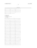 | 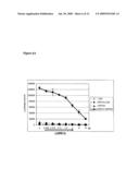 |
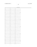 |  |
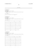 | 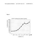 |
 |  |
 |  |
 |  |
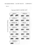 |  |
 |  |
 |  |
 |  |
 |  |
 |  |
 |  |
 |  |
 | 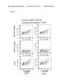 |
 |  |
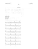 | 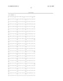 |
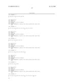 | 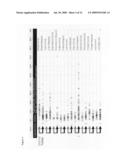 |
 | 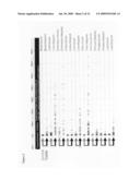 |
 |  |
 |  |
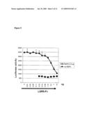 |  |
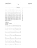 | 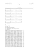 |
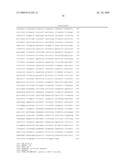 |
| Similar patent applications: | |
| Date | Title |
|---|---|
| 2011-12-01 | Chimeric therapeutics, compositions, and methods for using same |
| 2011-12-01 | Chimeric therapeutics, compositions, and methods for using same |
| 2011-12-01 | Modulation of gsk-3beta and method of treating proliferative disorders |
| 2011-12-01 | Feed composition comprising a mineral complex and methods of using the mineral complex |
| 2011-12-01 | Feed composition comprising a mineral complex and methods of using the mineral complex |
| New patent applications in this class: | |
| Date | Title |
|---|---|
| 2022-05-05 | Methods of treating locally advanced or metastatic breast cancers using pd-1 axis binding antagonists and taxanes |
| 2019-05-16 | Methods of treating diseases associated with ilc3 cells |
| 2018-01-25 | Monoclonal antibodies against her2 epitope |
| 2016-12-29 | Protein purification |
| 2016-09-01 | Dosage and administration of anti-egfr therapeutics |
| New patent applications from these inventors: | |
| Date | Title |
|---|---|
| 2016-06-30 | Compositions and methods for diagnosing and treating cancer |
| 2015-07-02 | Compositions and methods for diagnosing and treating cancer |
| 2014-11-27 | Compositions and methods for diagnosing and treating cancer |
| 2014-07-03 | Compositions and methods for diagnosing and treating cancer |
| 2014-01-30 | Compositions and methods for treating and diagnosing cancer |
| Top Inventors for class "Drug, bio-affecting and body treating compositions" | |
| Rank | Inventor's name |
|---|---|
| 1 | David M. Goldenberg |
| 2 | Hy Si Bui |
| 3 | Lowell L. Wood, Jr. |
| 4 | Roderick A. Hyde |
| 5 | Yat Sun Or |