Patent application title: Suppression of inflammation associated with transplantation using an epsilon PKC inhibitor
Inventors:
Daria D. Mochly-Rosen (Menlo Park, CA, US)
Daria D. Mochly-Rosen (Menlo Park, CA, US)
Tomoyoshi Koyanagi (Kobe, JP)
Koichi Inagaki (Otsu, JP)
Akifumi Ootani (Palo Alto, CA, US)
IPC8 Class: AA61K3810FI
USPC Class:
514 13
Class name: Designated organic active ingredient containing (doai) peptide containing (e.g., protein, peptones, fibrinogen, etc.) doai 16 to 24 peptide repeating units in known peptide chain
Publication date: 2009-05-14
Patent application number: 20090124553
Inventors list |
Agents list |
Assignees list |
List by place |
Classification tree browser |
Top 100 Inventors |
Top 100 Agents |
Top 100 Assignees |
Usenet FAQ Index |
Documents |
Other FAQs |
Patent application title: Suppression of inflammation associated with transplantation using an epsilon PKC inhibitor
Inventors:
Koichi Inagaki
Daria D. Mochly-Rosen
Tomoyoshi Koyanagi
Akifumi Ootani
Agents:
King & Spalding LLP
Assignees:
Origin: BELMONT, CA US
IPC8 Class: AA61K3810FI
USPC Class:
514 13
Abstract:
The described compositions and methods relate to the suppression of
inflammatory responses following allograft transplantation by
administering an inhibitor of epsilon protein kinases c (εPKC)
following transplantation.Claims:
1. A method for attenuating an inflammatory response due to allograft
transplantation, comprisingadministering an inhibitor of epsilon protein
kinase c (εPKC).
2. The method of claim 1, wherein the inhibitor is administered after allograft transplantation.
3. The method of claim 1, wherein the inhibitor consists of between about 5-30 contiguous amino acid residues from a conserved set of amino acid resides from the sequence identified herein as εPKC.
4. The method of claim 1, wherein the inhibitor is (i) EAVSLKPT (SEQ ID NO: 1) or (ii) a peptide having one or two conservative modifications to EAVSLKPT.
5. The method of claim 1, wherein the inhibitor is modified with a terminal amino acid residue that provides a reactive moiety.
6. The method of claim 5, wherein the inhibitor is modified with a cysteine residue.
7. The method of claim 5, wherein the reactive moiety is attached to a delivery peptide having activity to facilitate intracellular delivery of the inhibitor.
8. The method of claim 7, wherein the delivery peptide is selected from polyarginine, Drosophila Antennapedia homeodomain-derived sequence (RQIKIWFQNRRMKWKK; SEQ ID NO: 4), and Transactivating Regulatory Protein (Tat)-derived transport polypeptide (YGRKKRRQRRR; SEQ ID NO: 5) from the Human Immunodeficiency virus.
9. The method of claim 1, wherein the allograft transplantation involves a kidney, gall bladder, lung, liver, eye, blood, bone marrow, or vessel.
10. A method for enhancing survival of an allograft transplantation recipient, comprisingadministering an inhibitor of epsilon protein kinase c (εPKC).
11. A kit comprising an inhibitor of epsilon protein kinase c (εPKC) and instructions for use in administration to an allograft transplantation recipient.
12. The kit of claim 11, further comprising a delivery element for parenteral administration of the inhibitor.
Description:
PRIORITY
[0001]The present application claims priority to U.S. Provisional Application Ser. No. 60/927,580, filed on May 4, 2007, which is hereby incorporated by reference in its entirety.
TECHNICAL FIELD
[0003]The present subject matter relates to compositions and methods using an epsilon protein kinase C (εPKC) inhibitor for suppressing inflammation associated with transplantation.
BACKGROUND
[0004]Transplantation is currently the most effective treatment for end-stage organ failure. However, allograft survival is affected by acute and chronic rejections due to incomplete histocompatibility, which causes severe inflammatory responses in transplant recipients (Thom, T. et al. (2006) Circulation. 113:e85-151; Taylor, D. O. et al. (2003) J. Heart Lung Transplant. 22:616-24.) While the survival rate of transplant patients continues to increase, largely due to the wide availability of immunomodulatory drugs, chronic or late-stage allograft rejection still causes high mortality. In addition, immunomodulatory drugs have significant side-effects.
[0005]Ten protein kinase C (PKC) isozymes are encoded in the human genome and each isozyme mediates distinct roles in normal and disease states (Nishizuka, Y. (1995) Faseb J. 9: 484-96). Using pharmaceutical agents selective for particular PKC isozymes, it was shown that δPKC and εPKC isozymes have opposing roles in cardiac ischemia and reperfusion (Chen, L. et al. (2001) Proc. Natl. Acad. Sci. U.S.A. 98:11114-9; Inagaki. K. et al. (2003) Circulation 108:2304-7; Inagaki, K. et al. (2006) Cardiovasc. Res. 70:222-30). In addition, using both knockout mice and PKC peptide modulators, Levine and collaborators demonstrated that εPKC inhibition profoundly suppressed acute and chronic inflammatory pain response (Hucho, T. B. et al. (2005) J. Neurosci. 25:6119-26) and Aksoy et al. suggested that εPKC controls inflammation and immune-mediated disorders (Aksoy, E. et al. (2004) Int. J. Biochem. Cell. Biol. 36:183-8).
[0006]It would be desirable to further explore the role of εPKC on transplant inflammation and rejection and to determine whether the administration of εPKC offers beneficial therapeutic effect to transplant recipients.
REFERENCES
[0007]Thom, T. et al. (2006) Circulation. 113:e85-151. [0008]Taylor, D. O. et al. (2003) J Heart Lung Transplant. 22:616-24. [0009]Tanaka, M. et al. (2004) Circulation 110:II194-9. [0010]Tanaka, M. (2005) J. Thorac. Cardiovasc. Surg. 129:1160-7. [0011]Nishizuka, Y. (1995) Faseb J. 9: 484-96. [0012]Smith, B. L. and Mochly-Rosen, D. (1992) Biochem. Biophys. Res. Commun. 188:1235-40. [0013]Souroujon, M. C. and Mochly-Rosen, D. (1998) Nat. Biotechnol. 16: 919-24. [0014]Chen, L. et al. (2001) Proc. Natl. Acad. Sci. U.S.A. 98:11114-9. [0015]Inagaki. K. et al. (2003) Circulation 108:2304-7. [0016]Inagaki, K. et al. (2006) Cardiovasc. Res. 70:222-30. [0017]Hucho, T. B. et al. (2005) J. Neurosci. 25:6119-26. [0018]Aksoy, E. et al. (2004) Int. J. Biochem. Cell. Biol. 36:183-8. [0019]Tanaka, M. et al. (2005) J. Heart Lung Transplant. 24:446-53. [0020]Corry, R. J. et al. (1973) Transplantation 16:343-50. [0021]Chen, L. et al. (2001) Chem. Biol. 8:1123-9. [0022]Billingham, M. E. et al. (1990) J. Heart. Transplant. 9:587-93. [0023]Tanaka, M. et al. (1986) Br. Heart J. 55:575-81. [0024]Booz, G. W. et al. (1995) Cardiovasc. Res. 30:537-43. [0025]Crews, G. M. et al. (2005) Transplant. Proc. 37:1926-8. [0026]Aziz, T. M. et al. (2003) J. Heart Lung Transplant. 22:663-73. [0027]Tojima, Y. et al. (2000) Nature 404:778-82. [0028]Chen, L. Y. et al. (2005) J. Biol. Chem. 280:22497-501. [0029]Inagaki, K. et al. (2005) Circulation 111:44-50. [0030]Altschul et al. (1990) J. Mol. Biol. 215:403-410. [0031]Altschul et al. (1997) Nucleic Acids Res. 25:3389-3402. [0032]Chen, L. et al. (2001) Chem. Biol. 8:1123-9. [0033]Current Protocols in Molecular Biology (Ausubel et al., eds.), John Wiley & Sons. [0034]Dorn, G. W. et al. (1999) Proc. Natl. Acad. Sci. U.S.A. 96:12798-12803. [0035]Dorn, G. W. et al. (1999) Proc. Natl. Acad. Sci. U.S.A. 96:12798-12803. [0036]Freidinger, R. M. (2003) J. Med. Chem. 46:5553. [0037]Gellman, S. H. (1998) Acc. Chem. Res. 31:173. [0038]Goodman, M. et al. (1996) Pure Appl. Chem. 68:1303. [0039]Hagihara et al. (1992) J. Am. Chem. Soc. 114:6568. [0040]Hirschmann et al. (2000) J. Am. Chem. Soc. 122:11037. [0041]Johnson, J. A. and Mochly-Rosen, D. (1995) Circ Res. 76:654-63. [0042]Johnson, J. A. et al. (1996) Circ. Res. 79:1086. [0043]Liron, T. et al. (2007) J. Molecular and Cellular Cardiology 42:835-841 [0044]Karlin and Altschul (1990) Proc. Natl. Acad. Sci. USA 87:2264-2268. [0045]Karlin and Altschul (1993) Proc. Natl. Acad. Sci. USA 90:5873-5877. [0046]Kulesza, A. et al. (2003) Org. Letts. 5:1163. [0047]Martin, Robin, Protein Synthesis: Methods and Protocols, Humana Press, Totowa, N.J. (1998). [0048]Mitchell et al. (2000) J. Peptide Res. 56:318-325. [0049]Patani, G. A. and Lavoie, E. J. (1996) Chem. Rev. 96:3147-3176. [0050]Patil et al. (2003) J. Org. Chem. 68:7274-7280. [0051]Ripka, A. S. and Rich, D. H. (1998) Curr. Opin. Chem. Biol. 2:441. [0052]Rolhbard et al. (2000) Nature Med. 6:1253-1257. [0053]Ron, D. and Mochly-Rosen, D. (1995) Proc. Natl. Acad. Sci. U.S.A. 92:492-496; [0054]Ron, D. et al. (1994) Proc. Natl. Acad. Sci. USA 91:839-843. [0055]Sambrook et al., Molecular Cloning: A Laboratory Manual, Cold Springs Harbor laboratory, 2nd ed., Cold Springs Harbor, N.Y. (1989). [0056]Schechtman, D. et al. (2004) J. Biol. Chem. 279:15831-15840. [0057]Scott et al. (2004) Org. Letts. 6:1629-1632. [0058]Souroujon, M. C. and Mochly-Rosen, D. (1998) Nature Biotech. 16:919-924. [0059]Theodore, L. et al. (1995) J. Neurosci. 15:7158-7167. [0060]U.S. Pat. No. 5,804,604. [0061]U.S. Publication Nos. US2004-0009919A1, US2005-0209160A1, US2005-0164947A1, and US2006-0148700A1. [0062]Vives et al. (1997) J. Biol. Chem. 272:16010-17. [0063]Williams, Paul Lloyd, et al., (1997) Chemical Approaches to the Synthesis of Peptides and Proteins, CRC Press, Boca Raton, Fla. [0064]Wootton and Federhen (1993) Computers and Chemistry 17:149-163. [0065]Zega and Urleb (2002) Acta Chim. Slov. 49:649-662.
SUMMARY
[0066]In one aspect, a method for attenuating an inflammatory response due to allograft transplantation is provided, comprising administering an inhibitor of epsilon protein kinase C (εPKC).
[0067]In some embodiments, the inhibitor is administered after allograft transplantation.
[0068]In some embodiments, the inhibitor consists of between about 5-30 contiguous amino acid residues from a conserved set of amino acid resides from the sequence identified herein as εPKC.
[0069]In particular embodiments, the inhibitor is (i) EAVSLKPT (SEQ ID NO: 1) or (ii) a peptide having one or two conservative modifications to EAVSLKPT (SEQ ID NO: 1). In other embodiments, the inhibitor is β'-COP 285-292 (NNVALGYD; SEQ ID NO: 2). In yet another embodiment, the inhibitor is a mutated ψε-RACK peptide having, e.g., the amino acid sequence HNAPIGYD (SEQ ID NO: 3).
[0070]In some embodiments, the inhibitor is modified with a terminal amino acid residue that provides a reactive moiety. In particular embodiments, the inhibitor is modified with a cysteine residue.
[0071]In some embodiments, the reactive moiety is attached to a delivery peptide having activity to facilitate intracellular delivery of the inhibitor. In particular embodiments, the delivery peptide is selected from polyarginine, Drosophila Antennapedia homeodomain-derived sequence (RQIKIWFQNRRMKWKK; SEQ ID NO: 4), and Transactivating Regulatory Protein (Tat)-derived transport polypeptide (YGRKKRRQRRR; SEQ ID NO: 5) from the Human Immunodeficiency virus.
[0072]In some embodiments, the allograft transplantation involve the heart. In other embodiments, the allograft transplantation involves a kidney, gall bladder, lung, liver, eye, blood, bone marrow, or vessel.
[0073]In another aspect, a method for enhancing survival of an allograft transplantation recipient is provided, comprising
[0074]administering an inhibitor of εPKC.
[0075]In a further aspect, a kit comprising an inhibitor of εPKC and instructions for use in administration to an allograft transplantation recipient is provided. In some embodiments, the kit further comprises a delivery element for parenteral administration of the inhibitor.
BRIEF DESCRIPTION OF THE DRAWINGS
[0076]FIG. 1 shows the results of an experiment performed in support of the present methods. A timeline of events is shown above the graph. The beating score in the days following transplantation are shown in the graph. Animals were treated with TAT47-57-εV1-2 (n=9; solid line) or TAT47-57 alone as a control (n=8, dotted line; line graphs on the bottom).
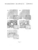
[0077]FIG. 2 shows histological features of the transplanted murine cardiac allografts treated with TAT (A and C-E) or εV1-2 (B and C'-E') based on hematoxylin and eosin (H&E) staining (A and B with enlarged picture in small panels) and 3-methyl-4-diazobenzene (DAB) staining for a macrophage peroxidase marker (F4/80; C and C'), a T-cell marker (CD3; D and D'), or a B-cell marker (CD45R/B220; E and E').
[0078]FIGS. 3A and 3B show histological staining of murine cardiac allografts treated with TAT (A) or εV1-2 (B) stained with Elastica van Gieson (EVG; scale bar=50 μm). FIG. 3C is a bar graph showing the amount of luminal narrowing in cardiac allografts treated with TAT or εV1-2.
[0079]FIGS. 4A and 4B show histological staining of murine cardiac allografts treated with TAT (A) or εV1-2 (B) stained with Masson's trichrome to assess cardiac fibrosis (blue). Scale bar=1 mm. For quantitative analysis, the fibrotic area in left coronary artery area, right coronary artery area and anterior side of septum were measured, averaged and indicated as percent of the total area. FIG. 4C is a graph summarizing the observed luminal narrowing.
[0080]FIGS. 5A-5C are bar graphs showing the local levels of TGF-β (A), PDGF-BB (B), and MCP-1 (C) in TAT or εV1-2-treated animals, as determined by ELISA. FIG. 5D shows the results of western blot analysis using RAW264.7 cells treated with 10 ng/ml TGF-β and incubated for 15 min (D). To observe the effect of εPKC on IκB polyubiquitination, εV1-2 or TAT control peptide (and MG132) were added to culture 10 min prior to TGF-β treatment.
DETAILED DESCRIPTION
I. Overview
[0081]The present methods relate to the administration of a selective εPKC inhibitor to suppress inflammation associated with allograft transplantation. The methods are based on the observation that the selective inhibition of εPKC with a peptide inhibitor prolonged allograft survival and improved functional recovery in a murine cardiac transplantation model. Inhibition of εPKC markedly attenuated the inflammatory response, resulting in reduced infiltration of macrophages and T-cells and a decrease in the attachment of mononuclear inflammatory cells to the arterial wall. εPKC administration further caused a substantial reduction in the extent of luminal narrowing and fibrosis in the allograft, preserving cardiac tissue architecture following transplantation.
[0082]To better understand the mechanism by which εPKC inhibitors suppress inflammatory response, the levels of several inflammatory cytokines, TGF-β, PDGF, and MCP-1 were measured before and after allograft transplantation. As expected, the process of transplantation significantly increased cytokine expression in control animals. Cytokine expression was attenuated/suppressed by εV1-2 treatment. The increase in MCP-1 and TGF-β that resulted from transplantation was abolished by εPKC inhibitor treatment, with the levels reduced to those observed in naive allografts. Treatment with an εPKC inhibitor also reduced the increased expression of PDGF associated with allograft transplantation, although it was not statistically significant.
[0083]Without being limited to a theory, it is believed that decreased MCP-1 levels may result from decreased macrophage invasion into the perivascular area, subsequently decreasing vasculitis. In support of this theory, macrophage and T-cell invasion of perivascular and parenchymal regions was drastically decreased 30 days after transplantation in εV1-2-treated animals. TGF-β is a stimulator of proliferation and differentiation for cardiac fibroblasts (Aziz, T. M. et al. (2003) J. Heart Lung Transplant. 22:663-73) and its induction correlated with the level of fibrosis observed by histological analysis. In addition to the role in fibrosis, a decrease in TGF-β levels was reported to associate with inhibition of chronic rejection in rat kidney grafts (Tojima, Y. et al. (2000) Nature 404:778-82) and TGF-β expression in human cardiac allografts correlates with impaired cardiac function (Chen, L. Y. et al. (2005) J. Biol. Chem. 280:22497-501).
[0084]Alternatively, the inhibition of cellular inflammatory responses, such as NF-κB activation and subsequent effects on cytokine expression, may be an important mechanism by which the εPKC inhibitor reduces inflammation in allograft transplantation. εPKC is reported to control NF-κB activation in human embryonic kidney cells and human peripheral blood monocytes (Booz, G. W. et al. (1995) Cardiovasc. Res. 30:537-43; Crews, G. M. et al. (2005) Transplant. Proc. 37:1926-8). These observations are consistent with reduced levels of IκB poly-ubiquitination following administration of a εPKC inhibitor (see below).
[0085]In a further alternative mechanism, the εPKC inhibitor may affect the Toll-like receptor 4 (TLR4) pathway, which drives the IL-12-dependent Th1 responses of the immune system (Aksoy, E. et al. (2004) Int. J. Biochem. Cell. Biol. 36:183-8). One skilled in the art will recognize that such mechanisms are not mutually excusive, and that several mechanisms may contribute to the effects observed with the present methods.
[0086]Previous studies showed that acute activation of εPKC prior to organ transplantation mimics ischemic preconditioning (Tanaka, M. et al. (2004) Circulation 110:II194-9; Tanaka, M. (2005) J. Thorac. Cardiovasc. Surg. 129:1160-7; Inagaki. K. et al. (2003) Circulation 108:2304-7; Inagaki, K. et al. (2006) Cardiovasc. Res. 70:222-30). The present methods are drawn to sustained delivery of a εPKC inhibitor following transplantation. Several different therapeutic windows are noted. For example, sustained administration of an εPKC inhibitor is effective in reducing inflammation when administration is initiated about three days following transplantation. While the exact number of days following transplantation is likely not critical, it is preferred that εPKC inhibitor administration be administered following transplantation, rather than before or during transplantation.
[0087]Moreover, the ability of sustained administration of an εPKC inhibitor to attenuate rejection following allograft transplantation may be distinct from the ability of an εPKC activator to improve cardiac function following a brief pretreatment of the organ In any case, the ability of the εPKC inhibitor to reduce inflammation when administered following transplantation was unexpected in view of previous results.
[0088]An additional benefit of the present εεPKC inhibitors is the absence of side-effects in other organs (i.e., endogenous organs in the recipient) observed both in the present methods and those previously reported (Inagaki, K. et al. (2005) Circulation 111:44-50).
[0089]Since coronary artery disease and immune rejection are the major and leading causes of cardiac allograft dysfunction (Taylor, D. O. et al. (2003) J Heart Lung Transplant. 22:616-24), the ability to inhibit parenchymal inflammation and vasculitis using an εεPKC inhibitor will increase the survival rate, average survival time, and quality of life for transplant recipients.
[0090]Moreover, since inflammation is generally associated with allograft transplantation, and not limited to cardiac transplantation, it is expected that εPKC inhibitors will be effective in suppressing inflammation associated with a wide variety of allograft transplantations, including but not limited to kidney, gall bladder, lung, liver, eye, blood, bone marrow, and other organ transplantation.
II. εPKC Inhibitors
[0091]The present methods feature the administration of an antagonist/inhibitor of εPKC. As used herein, an antagonist or inhibitor of εPKC is a compound that reduces εPKC expression, activity, activation, or stability, or reduces the effects of εPKC expression or activity, in a mammalian subject. An inhibitor of εPKC may be a compound that inactivates εPKC to form inactive εPKC, prevents εPKC from performing its biological functions, or otherwise antagonizes the activity of εPKC. The antagonist/inhibitor may be a competitive, non-competitive, or uncompetitive inhibitor of εPKC. In some embodiments, the inhibitor is a selective peptide inhibitor of εPKC, as opposed to an inhibitor of multiple PKC isozymes (e.g., α, β, δ, etc.).
[0092]As known in the art, εPKC is a serine/threonine kinase and is involved in a myriad of cellular process, including regulation of various physiological functions, such as the activation of various biological systems, including the nervous, endocrine, and exocrine systems.
[0093]The polypeptide sequences of murine, rat, and human εPKC are reproduced, below. The present compositions and methods contemplate the use of any one of these polypeptides, chimeric/hybrid polypeptides including sequence from one or more of these polypeptides, and/or fragments, variants, and derivatives, thereof.
TABLE-US-00001 εPKC (Mus musculus); gi:6755084; ACCESSION: NP_036234 XP_994572 XP_994601 XP_994628 (SEQ ID NO: 6): 1 MVVFNGLLKI KICEAVSLKP TAWSLRHAVG PRPQTFLLDP YIALNVDDSR IGQTATKQKT 61 NSPAWHDEFV TDVCNGRKIE LAVFHDAPIG YDDFVANCTI QFEELLQNGS RHFEDWIDLE 121 PEGKVYVIID LSGSSGEAPK DNEERVFRER MRPRKRQGAV RRRVHQVNGH KFMATYLRQP 181 TYCSHCRDFI WGVIGKQGYQ CQVCTCVVHK RCHELIITKC AGLKKQETPD EVGSQRFSVN 241 MPHKFGIHNY KVPTFCDHCG SLLWGLLRQG LQCKVCKMNV HRRCETNVAP NCGVDARGIA 301 KVLADLGVTP DKITNSGQRR KKLAAGAESP QPASGNSPSE DDRSKSAPTS PCDQELKELE 361 NNIRKALSFD NRGEEHRASS ATDGQLASPG ENGEVRPGQA KRLGLDEFNF IKVLGKGSFG 421 KVMLAELKGK DEVYAVKVLK KDVILQDDDV DCTMTEKRIL ALARKHPYLT QLYCCFQTKD 481 RLFFVMEYVN GGDLMFQIQR SRKFDEPRSR FYAAEVTSAL MFLHQHGVIY RDLKLDNILL 541 DAEGHCKLAD FGMCKEGIMN GVTTTTFCGT PDYIAPEILQ ELEYGPSVDW WALGVLMYEM 601 MAGQPPFEAD NEDDLFESIL HDDVLYPVWL SKEAVSILKA FMTKNPHKRL GCVAAQNGED 661 AIKQHPFFKE IDWVLLEQKK IKPPFKPRIK TKRDVNNFDQ DFTREEPILT LVDEAIIKQI 721 NQEEFKGFSY FGEDLMP εPKC (Rattus norvegicus); ACCESSION: NP_058867 XP_343013 (SEQ ID NO: 7): 1 MVVFNGLLKI KICEAVSLKP TAWSLRHAVG PRPQTFLLDP YIALNVDDSR IGQTATKQKT 61 NSPAWHDEFV TDVCNGRKIE LAVFHDAPIG YDDFVANCTI QFEELLQNGS RHFEDWIDLE 121 PEGKVYVIID LSGSSGEAPK DNEERVFRER MRPRKRQGAV RRRVHQVNGH KFMATYLRQP 181 TYCSHCRDFI WGVIGKQGYQ CQVCTCVVHK RCHELIITKC AGLKKQETPD EVGSQRFSVN 241 MPHKFGIHNY KVPTFCDHCG SLLWGLLRQG LQCKVCKMNV HRRCETNVAP NCGVDARGIA 301 KVLADLGVTP DKITNSGQRR KKLAAGAESP QPASGNSPSE DDRSKSAPTS PCDQELKELE 361 NNIRKALSFD NRGEEHRASS STDGQLASPG ENGEVRQGQA KRLGLDEFNF IKVLGKGSFG 421 KVMLAELKGK DEVYAVKVLK KDVILQDDDV DCTMTEKRIL ALARKHPYLT QLYCCFQTKD 481 RLFFVMEYVN GGDLMFQIQR SRKFDEPRSG FYAAEVTSAL MFLHQHGVIY RDLKLDNILL 541 DAEGHSKLAD FGMCKEGILN GVTTTTFCGT PDYIAPEILQ ELEYGPSVDW WALGVLMYEM 601 MAGQPPFEAD NEDDLFESIL HDDVLYPVWL SKEAVSILKA FMTKNPHKRL GCVAAQNGED 661 AIKQHPFFKE IDWVLLEQKK MKPPFKPRIK TKRDVNNFDQ DFTREEPILT LVDEAIVKQI 721 NQEEFKGFSY FGEDLMP εPKC (Homo sapiens); ACCESSION: NP_005391 (SEQ ID NO: 8): 1 mvvfngllki kiceavslkp tawslrhavg prpqtflldp yialnvddsr igqtatkqkt 61 nspawhdefv tdvcngrkie lavfhdapig yddfvancti qfeellqngs rhfedwidle 121 pegrvyviid lsgssgeapk dneervfrer mrprkrqgav rrrvhqvngh kfmatylrqp 181 tycshcrdfi wgvigkqgyq cqvctcvvhk rcheliitkc aglkkqetpd qvgsqrfsvn 241 mphkfgihny kvptfcdhcg sllwgllrqg lqckvckmnv hrrcetnvap ncgvdargia 301 kvladlgvtp dkitnsgqrr kkliagaesp qpasgsspse edrsksapts pcdqeikele 361 nnirkalsfd nrgeehraas spdgqlmspq engevrqgqa krlgldefnf ikvlgkgsfg 421 kvmlaelkgk devyavkvlk kdvilqdddv dctmtekril alarkhpylt qlyccfqtkd 481 rlffvmeyvn ggdlmfqiqr srkfdeprsr fyaaevtsal mflhqhgviy rdlkldnill 541 daeghcklad fgmckegiln gvttttfcgt pdyiapeilq eleygpsvdw walgvlmyem 601 magqppfead neddlfesil hddvlypvwl skeavsilka fmtknphkrl qcvasqnged 661 aikqhpffke idwvlleqkk ikppfkprik tkrdvnnfdq dftreepvlt lvdeaivkqi 721 nqeefkgfsy fgedlmp
[0094]The inhibitor may be a protein, or other organic or inorganic compound, including a peptidomimetic small-molecule.
[0095]Suitable small molecules that may act as an inhibitor of εPKC may be determined by methods known to the art. For example, such molecules may be identified by their ability to translocate εPKC to its subcellular location. Such assays may utilize, for example, fluorescently-labeled enzyme and fluorescent microscopy to determine whether a particular compound or agent may aid in the cellular translocation of εPKC. Such assays are described, for example, in Schechtman, D. et al. ((2004) J. Biol. Chem. 279:15831-15840) and include use of selected antibodies. Other assays to measure cellular translocation include Western blot analysis as described in Dorn, G. W. et al. ((1999) Proc. Natl. Acad. Sci. U.S.A. 96:12798-12803) and Johnson, J. A. and Mochly-Rosen, D. ((1995) Circ Res. 76:654-63).
[0096]In certain forms of the invention, a protein inhibitor of εPKC may be utilized. The protein inhibitor may be in the form of a peptide. Protein, peptide and polypeptide are used interchangeably herein and refer to a compound made up of a chain of amino acid monomers linked by peptide bonds. Unless otherwise stated, the individual sequence of the peptide is given in the order from the amino terminus to the carboxyl terminus. Typically, peptides of εPKC having activity as an inhibitor of the translocation of εPKC are preferred, and generally have between about 5-30 amino acid residues, more preferably between about 6-25 amino acid residues, and still more preferably between about 6-15, and even more preferably from 6-12 or 8-15 amino acid residues.
[0097]The inhibitor of εPKC may be obtained by methods known to the skilled artisan. For example, the protein inhibitor may be chemically synthesized using various solid phase synthetic technologies known to the art and as described in, for example, Williams, Paul Lloyd et al. ((1997) Chemical Approaches to the Synthesis of Peptides and Proteins, CRC Press, Boca Raton, Fla.).
[0098]Alternatively, the protein inhibitor may be produced by recombinant technology methods as known in the art and as described, for example, in Sambrook et al., Molecular Cloning: A Laboratory Manual, Cold Springs Harbor laboratory, 2nd ed., Cold Springs Harbor, N.Y. (1989); Martin, Robin, Protein Synthesis: Methods and Protocols, Humana Press, Totowa, N.J. (1998); and Current Protocols in Molecular Biology (Ausubel et al., eds.), John Wiley & Sons, which is regularly and periodically updated. For example, an expression vector may be used to produce the desired peptide inhibitor in an appropriate host cell and the product may then be isolated by known methods.
[0099]An exemplary εPKC inhibitor peptide is TAT47-57-εV1-2, which contains amino acid residues 47-57 of the HIV TAT transactivator protein (SEQ ID NO: 5), which directs entry into cells, and amino acid residues 14-21 of εPKC (i.e., EAVSLKPT; (SEQ ID NO: 1). This εPKC inhibitor is described in Chen, L. et al. ((2001) Chem. Biol. 8:1123-9) and in U.S. Publication Nos. US2004-0009919A1, US2005-0209160A1, US2005-0164947A1, US2006-0148700A1, which further describe the characterization of εPKC agonists and antagonists and which are incorporated by reference herein. Other εPKC inhibitor peptides may be used, including but not limited to peptides containing conservative amino acid substitutions and peptides having similarity to εPKC RACK amino acid residues, as described, below.
[0100]Polypeptides may be encoded by an expression vector, which may include, for example, the nucleotide sequence encoding the desired peptide wherein the nucleotide sequence is operably linked to a promoter sequence. As defined herein, a nucleotide sequence is "operably linked" to another nucleotide sequence when it is placed in a functional relationship with another nucleotide sequence. For example, if a coding sequence is operably linked to a promoter sequence, this generally means that the promoter may promote transcription of the coding sequence. Operably linked means that the DNA sequences being linked are typically contiguous and, where necessary to join two protein coding regions, contiguous and in reading frame. However, since enhancers may function when separated from the promoter by several kilobases and intronic sequences may be of variable length, some nucleotide sequences may be operably linked but not contiguous. Additionally, as defined herein, a nucleotide sequence is intended to refer to a natural or synthetic linear and sequential array of nucleotides and/or nucleosides, and derivatives thereof. The terms "encoding" and "coding" refer to the process by which a nucleotide sequence, through the mechanisms of transcription and translation, provides the information to a cell from which a series of amino acids can be assembled into a specific amino acid sequence to produce a polypeptide.
[0101]The εPKC inhibitor peptide may be capable of preventing activation signaling proteins, such as εPKC, that are activated in vivo by binding to a cognate polypeptide such as a receptor protein (RACK). Regions of homology between the εPKC signaling peptide and its RACK are termed "pseudo-RACK" sequences (ψ-RACK; Ron, D. et al. (1994) Proc. Natl. Acad. Sci. USA 91:839-843; Ron, D. and Mochly-Rosen, D. (1995) Proc. Natl. Acad. Sci. U.S.A. 92:492-496; Dorn, G. W. et al. (1999) Proc. Natl. Acad. Sci. U.S.A. 96:12798-12803; and Souroujon, M. C. and Mochly-Rosen, D. (1998) Nature Biotech. 16:919-924) and typically have a sequence similar to the PKC-binding region of the corresponding RACK.
[0102]In εPKC, the sequence HDAPIGYD (εPKC 85-92; Genbank Accession No. NP--058867; SEQ ID NO: 9), named ψεRACK, has 75% homology with a sequence in εRACK consisting of amino acids NNVALGYD (RACK 285-292; SEQ ID NO: 2). A peptide corresponding to the ψεRACK sequence functioned as a εPKC-selective agonist (Dorn, G. W. et al. (1999) Proc. Nat. Acad. Sci. U.S.A. 96:12798-12803), possibly by stabilizing the "open" form of εPKC. Mutating Asp-86 in the ωεRACK sequence of εPKC to an Asn (i.e., D→N) produced an enzyme that translocated more slowly than the wild-type enzyme, presumably due to increased intramolecular interaction between the εRACK and the mutated ψεRACK-binding site in εPKC, which stabilized the "closed form." Accordingly, the mutated ωεRACK peptide having the amino acid sequence HNAPIGYD (SEQ ID NO: 3), functioned as a εPKC antatgonist/inhibitor (Schechtman, D. et al. (2004) J. Biol. Chem. 279:15831-15840; Liron, T. et al. (2007) J. Molecular and Cellular Cardiology 42:835-841). Other mutated ψεRACK are expected to function as εPKC antatgonists/inhibitors. In addition, the polypeptide β'-COP has an εPKC binding motif (i.e., NNVALGYD; SEQ ID NO: 2), which is expected to function as an antagonist/inhibitor of εPKC (Dorn et al. (1999) Proc. Natl. Acad. Sci., USA; Schechtman et al (2004) J. Biol. Chem.).
[0103]Also included within these definitions, and in the scope of the invention, are variants of the peptides which function in reducing injury to a transplanted organ or tissue, modulating the activity and/or production of mediators of inflammation as described herein or modulating a pro-apoptotic event, or a combination thereof as described herein.
[0104]The peptides may include natural amino acids, such as the L-amino acids or non-natural amino acids, such as D-amino acids. The amino acids in the peptide may be linked by peptide bonds or, in modified peptides described herein, by non-peptide bonds. A wide variety of modifications to the amide bonds which link amino acids may be made and are known in the art. Such modifications are discussed in general reviews, including in Freidinger, R. M. ((2003) J. Med. Chem. 46:5553) and Ripka, A. S. and Rich, D. H. ((1998) Curr. Opin. Chem. Biol. 2:441). These modifications are designed to improve the properties of the peptide in one of two ways: (a) increase the potency of the peptide by restricting conformational flexibility; (b) increase the half-life of the peptide by introducing non-degradable moieties to the peptide chain.
[0105]Examples of strategy (a) include the placement of additional alkyl groups on the nitrogen or alpha-carbon of the amide bond, such as the peptoid strategy of Zuckerman et al, and the alpha modifications of, for example Goodman, M. et al. ((1996) Pure Appl. Chem. 68:1303). The amide nitrogen and alpha carbon may be linked together to provide additional constraint (Scott et al. (2004) Org. Letts. 6:1629-1632).
[0106]Examples of strategy (b) include replacement of the amide bond by, for instance, a urea residue (Patil et al. (2003) J. Org. Chem. 68:7274-7280) or an aza-peptide link (Zega and Urleb (2002) Acta Chim. Slov. 49:649-662). Other examples such as introducing an additional carbon "beta peptides" (Gellman, S. H. (1998) Acc. Chem. Res. 31:173) or ethene unit (Hagihara et al. (1992) J. Am. Chem. Soc. 114:6568) to the chain, or the use of hydroxyethylene moieties (Patani, G. A. and Lavoie, E. J. (1996) Chem. Rev. 96:3147-3176) are also well known. One or more amino acids may be replaced by an isosteric moiety such as, for example, the pyrrolinones of Hirschmann et al. (2000) J. Am. Chem. Soc. 122:11037), or tetrahydropyrans (Kulesza, A. et al. (2003) Org. Letts. 5:1163).
[0107]The εPKC inhibitors may be based on any mammalian polypeptide or polynucleotide sequence, including human sequences. Skilled artisans will recognize that, through the process of mutation and/or evolution, polypeptides of different lengths and having different constituents, e.g., with amino acid insertions, substitutions, deletions, and the like, may arise that are related to, or sufficiently similar to, a sequence set forth herein by virtue of amino acid sequence homology and advantageous functionality as described herein.
[0108]The peptide inhibitors described herein also encompass amino acid sequences similar to the amino acid sequences set forth herein that have at least about 50% identity thereto and function in reducing inflammation associated with allograft transplantation, modulating the activity of mediators of inflammation as described herein, modulating a pro-apoptotic event, or a combination thereof. Preferably, the amino acid sequences of the peptide inhibitors encompassed in the invention have at least about 60% identity, further at least about 70% identity, preferably at least about 80% identity, more preferably at least about 90% identity, and further preferably at least about 95% identity to the amino acid sequences of, e.g., εV1-2, β'-COP 285-292, and/or mutated ωεRACK, as set forth herein. Exemplary levels of sequence homology are 90%, 91%, 92%, 93%, 94%, 95%, 96%, 97%, 98%, and even 99%.
[0109]Percent identity may be determined, for example, by comparing sequence information using the advanced BLAST computer program, including version 2.2.9, available from the National Institutes of Health. The BLAST program is based on the alignment method of Karlin and Altschul ((1990) Proc. Natl. Acad. Sci. USA 87:2264-2268) and as discussed in (Altschul et al. (1990) J. Mol. Biol. 215:403-410; Karlin and Altschul (1993) Proc. Natl. Acad. Sci. USA 90:5873-5877; and Altschul et al. (1997) Nucleic Acids Res. 25:3389-3402. Briefly, the BLAST program defines identity as the number of identical aligned symbols (i.e., nucleotides or amino acids), divided by the total number of symbols in the shorter of the two sequences. The program may be used to determine percent identity over the entire length of the proteins being compared. Default parameters are provided to optimize searches with short query sequences in, for example, blastp with the program. The program also allows use of an SEG filter to mask-off segments of the query sequences as determined by the SEG program of Wootton and Federhen ((1993) Computers and Chemistry 17:149-163).
[0110]Accordingly, fragments or derivatives of peptide agonists described herein may also be advantageously utilized that include amino acid sequences having the specified percent identities to the amino acid sequences described herein to reduce inflammation associated with allograft transplantation, to modulate the activity and/or production of mediators of inflammation as described herein, to modulate a pro-apoptotic event, or a combination thereof.
[0111]Conservative amino acid substitutions may be made in the amino acid sequences described herein to obtain derivatives of the peptides that may advantageously be utilized in the present invention. Conservative amino acid substitutions, as known in the art and as referred to herein, involve substituting amino acids in a protein with amino acids having similar side chains in terms of, for example, structure, size and/or chemical properties. For example, the amino acids within each of the following groups may be interchanged with other amino acids in the same group: amino acids having aliphatic side chains, including glycine, alanine, valine, leucine and isoleucine; amino acids having non-aromatic, hydroxyl-containing side chains, such as serine and threonine; amino acids having acidic side chains, such as aspartic acid and glutamic acid; amino acids having amide side chains, including glutamine and asparagine; basic amino acids, including lysine, arginine and histidine; amino acids having aromatic ring side chains, including phenylalanine, tyrosine and tryptophan; and amino acids having sulfur-containing side chains, including cysteine and methionine. Additionally, amino acids having acidic side chains, such as aspartic acid and glutamic acid, are considered interchangeable herein with amino acids having amide side chains, such as asparagine and glutamine.
[0112]The derivatives include amino acid sequences where a given amino acid of one group (such as a non-polar amino acid, an uncharged polar amino acid, a charged polar amino acidic amino acid or a charged polar basic amino acid) is substituted with another amino acid from the same amino acid group. For example, it is know that the uncharged polar amino acid serine may be commonly substituted with the uncharged polar amino acid threonine in a peptide without substantially altering the functionality of the peptide. If one is unsure whether a given substitution will affect the functionality of the peptide, then this may be determined without undue experimentation using synthetic techniques and screening assays known in the art.
[0113]The εPKC inhibitor peptides described herein may be modified by being part of a fusion protein. The fusion protein may include a protein or peptide that functions to increase the cellular uptake of the peptide inhibitors, has another desired biological effect, such as a therapeutic effect, or may have both of these functions. For example, it may be desirable to conjugate, or otherwise attach, the εV1-2 peptide, an inhibitor derived from the ψεRACK peptide, or other εPKC inhibitor peptide, to a cytokine or other peptide that elicits a desired biological response. The fusion protein may be produced by methods known to the skilled artisan. The inhibitor peptide may be bound, or otherwise conjugated, to another peptide in a variety of ways known to the art. For example, inhibitor peptide may be bound to a carrier peptide or other peptide described herein by cross-linking wherein both peptides of the fusion protein retain their activity. As a further example, the inhibitor peptide may be linked or otherwise conjugated to each other by an amide bond from the C-terminal of one peptide to the N-terminal of the other peptide. The linkage between the transmembrane carrier or therapeutic peptide may be non-cleavable, with a peptide bond, or cleavable with, for example, an ester or other cleavable bond.
[0114]Furthermore, in other forms of the invention, the carrier protein or peptide that may increase cellular uptake of the peptide agonist or inhibitor may be, for example, a Drosophila melanogaster Antennapedia homeodomain-derived sequence (unmodified sequence may be found in Genbank Accession No. AAD19795; RQIKIWFQNRRMKWKK; SEQ ID NO: 4), and may be attached to the agonist or inhibitor by cross-linking via an N-terminal Cys-Cys bond as discussed in (Theodore, L. et al. (1995) J. Neurosci. 15:7158-7167; Johnson, J. A. et al. (1996) Circ. Res. 79:1086). The sequence may also be sought from Drosophila hydei and Drosophila virilis. Alternatively, the εPKC inhibitor may be modified by a Transactivating Regulatory Protein (Tat)-derived transport polypeptide (such as from amino acids 47-57 of Tat (i.e., YGRKKRRQRRR; SEQ ID NO: 5) from the Human Immunodeficiency Virus, Type 1, as described in Vives et al. ((1997) J. Biol. Chem. 272:16010-17); U.S. Pat. No. 5,804,604; as seen in Genbank Accession No. AAT48070; or with polyarginine, as described in Mitchell et al. ((2000) J. Peptide Res. 56:318-325) and Rothbard et al. ((2000) Nature Med. 6:1253-1257). The inhibitors may be modified by other methods known to the skilled artisan in order to increase the cellular uptake of the inhibitors.
III. Supporting Studies
[0115]In studies performed in support of the present methods, hearts of FVB mice (H-2q) were transplanted into C57BL/6 mice (H-2b) to provide an animal model for allograft transplantation. Following transplantation, an isozyme-specific εPKC inhibitor was administered to the transplant recipients. The particular inhibitor was TAT47-57-εV1-2 (i.e., εV1-2), which is described herein.
[0116]A. εPKC Inhibition Prolongs Allograft Survival
[0117]The εPKC-specific inhibitor, εV1-2, or the carrier/control peptide, TAT, was administered to the recipient animals using osmotic pumps to provide continuous treatment from day 3 following allograft transplantation until sacrifice on day 30. All animals were given 20 mg/kg/day of cyclosporine A (CyA) to ensure survival.
[0118]Animals treated with εV1-2-did not demonstrate toxic effects when compared to the TAT-treated group. Importantly, εV1-2 treatment significantly improved the beating score of cardiac allografts compared to TAT-peptide treatment (FIG. 1), suggesting that εV1-2 treatment augmented graft survival promoted by CyA without toxic side effects. εPKC inhibitor-treated animals had a similar beat score at 30 days as TAT-treated animals at 14 days (FIG. 1). Both groups showed normal behavior with no difference in body weight (TAT: 21.6±0.5 g; εV1-2; 21.7±0.9 g).
[0119]P values <0.05 were calculated for the beat scores on days 12-30. P=0.007 at day 15 and P<0.0001 at day 30. The overall p-value with repeated Anova test was <0.0001.
[0120]B. εPKC Inhibition Reduces Macrophages and T Cells Infiltration
[0121]FIG. 2 shows histological features of the allografts in recipient animals treated with TAT (FIGS. 2A and 2C-E) or εV1-2 (FIGS. 2B and 2C'-E') based on H&E staining for general tissue morphology; DAB staining for the macrophage marker, F4/80; antibody staining for the T-cell marker, CD3; or antibody staining of the B-cell marker, CD45R/B220. Diffused infiltration of inflammatory cells with severe multifocal myocyte damage, hemorrhage, and vasculitis were observed in control-treated (TAT) transplanted hearts (FIGS. 2A and 2C-E). In contrast, εV1-2 treatment markedly suppressed the inflammatory response in the transplant recipients (FIGS. 2B and 2C'-E'). Allografts from the εV1-2-treated animals showed only a few foci of perivascular mononuclear cell infiltratration. Parenchymal rejection (PR) scores were significantly lower in allografts treated with εV1-2 (2.3±0.3) compared to control animals (4.0±0.0; p<0.0001). Immunohistochemical analysis of tissue samples revealed that the infiltrating inflammatory cells were mainly macrophages and T-cells but not B-cells (FIGS. 2C-D).
[0122]Parenchymal rejection scores were significantly lower in allografts treated with εV1-2 (2.3±0.3) as compared to control (4.0±0.0; provide p<0.0001). Immunohistochemical analysis revealed those inflammatory cells were mainly macrophages and T cells but not B cells (FIG. 2C-G).
[0123]EVG staining to identify vascular structures demonstrated a significant decrease in the attachment of mononuclear inflammatory cells to the arterial walls in the allografts in the εV1-2-treated animals compared to the TAT-treated animals (FIGS. 3A and B). Luminal narrowing in middle-sized arteries was decreased by 30% in the εV1-2-treated animals compared to control animals (FIG. 4C). Furthermore, fibrosis, measured by Masson's Trichrome staining, was significantly decreased (40% of control) following εV1-2 treatment (FIG. 4).
[0124]C. εPKC Inhibition Reduces TGF-1 and MCP-1 Levels and Inflammatory Response in Culture
[0125]Organ transplantation is known to be associated with prolonged changes in the levels of a number of cytokines and chemokines. In particular, heterotopic cardiac transplantation significantly increases the levels of transforming growth factor-β (TGF-β), pletelet derived growth factor (PDGF), and monocyte chemoattractant protein-1 (MCP-1).
[0126]Administration of the εPKC inhibitor significantly reduced/suppressed the induction of TGF-β and MCP-1 (FIGS. 5A and C), demonstrating that the εPKC inhibitor suppressed the induction of inflammatory cytokines. However, administration of the εPKC inhibitor did not significantly suppress PDGF induction (FIG. 5B). Notably, the PDGF levels were below detection levels in untreated cardiac allografts.
[0127]Nuclear factor-κB (NF-κB) is one of the major transcription factors involved in inflammation. NF-κB is inhibited by inhibitors of NF-κB (IκB), which involves the ubiquitin-proteasome pathway. To determine whether the εPKC inhibitor affected the activation of NF-κB by εPKC, a macrophage derived cell line, RAW264.7, was treated with the inflammatory cytokine, tumor necrosis factor-α (TNF-α), in the presence of a proteasome inhibitor, MG132. The assay allowed measurement of poly-ubiquitination of IκB, while preventing the destruction (and inability to detect) poly-ubiquitinated IκB. As shown in FIG. 5D, the addition of the εPKC inhibitor, εV1-2, reduced induction of poly-ubiquitination of IκB by TNF-α to a greater extent than the TAT control peptide, indicating that NF-κB activity was down-regulated (i.e., reduced) by εV1-2 via poly-ubiquitination of IκB.
[0128]D. Summary of Results
[0129]Based on the results described herein, administration of an εPKC inhibitor following allograft transplantation significantly improved the beating score of a cardiac allograft throughout the treatment period. Infiltration of macrophages and T-cells into the allografts was reduced significantly and parenchymal fibrosis was decreased in animals treated with εV1-2 compared to control-treated animals. Finally, the increase in TGF-β and MCP-1 levels that accompanied allograft transplantation were almost abolished by εV1-2-administration. These data suggest that εPKC activity contributes to the chronic immune response in inflammation associated with allograft transplantation and that an εPKC-selective inhibitor, such as εV1-2, can augment current therapeutic strategies to suppress inflammation and prolong graft survival in humans.
[0130]All references cited herein are hereby incorporated by reference in their entirety. Modifications, substitutions, variations, and alternatives of the present inhibitors and methods will be apparent in view of the disclosure.
[0131]The following Examples are provide to illustrate the compositions and methods and are not intended to be limiting.
EXAMPLES
Example 1
Animals
[0132]Male εVB (H-2q) and C57BL/6J (H-2b) mice, 6-8 weeks old, were purchased from Jackson Laboratory (Bar Harbor, Me.) and housed at the animal facility at Stanford University Medical Center (Stanford, Calif.). The εVB mice were used as allograft donors and the C57BL/6J mice were used as recipients. All mice were kept under standard temperature, humidity and timed lighting conditions and were provided mouse chow and water ad libitum. Animals were treated in compliance with the "Guide for the Care and Use of Laboratory Animals" prepared by the Institute of Laboratory Animal Resources, National Research Council, and published by the National Academy Press (revised 1996).
Example 2
Heterotopic Cardiac Transplantation
[0133]Heterotopic cardiac transplantation was performed according to the method of Corry et al. (Corry, R. J. et al., (1973) Transplantation 16:343-50) with some modifications. Anesthesia was induced with 5% inhaled isoflurane (Halocarbon Laboratories, River Edge, N.J.). During surgery, the animals were maintained on 2.5% inhaled isoflurane. Donor animals were systemically heparinized (50 mg/kg) before heart procurement. The donor heart was rapidly excised after coronary perfusion with ice-cold saline. The procured hearts were kept in ice-cold saline for 20 minutes. Because standard graft implantation averages 30 minutes, the total ischemic time was 50 minutes.
Example 3
Drug Administration
[0134]The selective εPKC inhibitor εV1-2 (εPKC amino acids 14-21; EAVSLKPT) was synthesized and conjugated to TAT (carrier peptide, amino acids 47-57; YGRKKRRQRRR) via a disulfide bond between cysteine residues added on amino termini of each peptides by American Peptides (Sunnyvale, Calif.), as previously described (Chen, L. et al. (2001) Chem. Biol. 8:1123-9). Recipient mice were treated with εV1-2 (n=9, 20 mg/kg/day), or with TAT as a control (13 mg/kg/day; n=8) using 0.1 mL osmotic pumps (release rate; 0.25 μL/hour, 30 mM of each peptide in sterile saline, Alzet, Alza) implanted subcutaneously from 3 to 30 days after transplantation. Recipients in both the εV1-2-treated group and control group received daily CyA (20 mg/kg/day) by intraperitoneal injection.
[0135]Allograft recipient mice were monitored daily. Graft viability was assessed by direct abdominal palpation of the heterotopically transplanted hearts and expressed as beating score, assessed by the Stanford cardiac surgery lab graft scoring system (Tanaka, M. et al. (2004) Circulation 110:II194-9).
Example 4
Histological Analysis
[0136]For histological analysis, grafts from 30-day post-transplantation animals (n=8 for TAT and n=9 for εV1-2) were harvested, fixed with 10% buffered formalin and embedded in paraffin. Sections were prepared from each specimen and stained with hematoxylin and eosin (H&E), Masson's trichrome, elastica von Gieson (EVG), or DAB for immunohisotochemistry by anti-F4/80, CD3 (Abcam) or CD45R/B220 (BD pharmingen). Parenchymal rejection (PR) severity was graded with a scale modified from the International Society for Heart and Lung Transplantation (Billingham, M. E. et al. (1990) J. Heart. Transplant. 9:587-93). The area of cardiac fibrosis were measured by a point-counting method (Tanaka, M. et al. (1986) Br. Heart J. 55:575-81). Percent luminal narrowing was calculated as [(inside area of internal elastic lamina)-(area of lumen)/(inside area of internal elastic lamina)]×100. Mid-sized coronary arteries (diameter; 40-120 μm) from multiple sections of the middle of the heart were analyzed (5 arteries for each graft) using ImageJ 1.35s software (Rasband, W. S., NIH, Bethesda, Md.).
Example 5
ELISA Determination of Local Cytokine Level
[0137]Harvested transplanted hearts were homogenized in 1× phosphate-buffered saline containing protease inhibitor cocktail (Sigma, St Louis, Mo.) and centrifuged at 30,000×g for 20 min at 4° C. The protein concentration of the supernatant was measured using Protein Assay (Bio-Rad, Hercules, Calif.) and 1-50 μg of protein were used for each analysis. All ELISA kits were purchased from R&D systems (Minneapolis, Minn.) and assays were performed according to the manufacturer's instructions.
Example 6
Western Blot Analysis
[0138]RAW264.7 cells were maintained in DMEM (Invitrogen) with supplemental 10% fetal bovine serum and penicillin-streptomycin solution (Invitrogen). Equal amounts of whole cell lysates were loaded to 10% SDS-polyacrylamide gel electrophoresis and were transferred to immobilon-P transfer membrane (Millipore). Anti-IκB (Cell Signaling) or GAPDH (Santa Cruz Biotechnology) antibodies were used for immunoblotting followed by HRP-conjugated anti-mouse (GE Healthcare) or rabbit (Santa Cruz Biotechnology) IgG antibodies.
Example 7
Statistical Analysis
[0139]Values were expressed as Mean±SEM. Statistical analysis was assessed by 1-way factorial ANOVA with Fisher's test, 2-way repeated ANOVA or Student's t-test when appropriate. A probability value <0.05 was considered significant.
Sequence CWU
1
918PRTArtificial SequenceSynthetic peptide 1Glu Ala Val Ser Leu Lys Pro
Thr1 528PRTArtificial SequenceSynthetic peptide 2Asn Asn
Val Ala Leu Gly Tyr Asp1 538PRTArtificial SequenceSynthetic
peptide 3His Asn Ala Pro Ile Gly Tyr Asp1 5416PRTArtificial
SequenceSynthetic peptide 4Arg Gln Ile Lys Ile Trp Phe Gln Asn Arg Arg
Met Lys Trp Lys Lys1 5 10
15511PRTArtificial SequenceSynthetic polypeptide 5Tyr Gly Arg Lys Lys
Arg Arg Gln Arg Arg Arg1 5 106737PRTMus
musculus 6Met Val Val Phe Asn Gly Leu Leu Lys Ile Lys Ile Cys Glu Ala
Val1 5 10 15Ser Leu Lys
Pro Thr Ala Trp Ser Leu Arg His Ala Val Gly Pro Arg20 25
30Pro Gln Thr Phe Leu Leu Asp Pro Tyr Ile Ala Leu Asn
Val Asp Asp35 40 45Ser Arg Ile Gly Gln
Thr Ala Thr Lys Gln Lys Thr Asn Ser Pro Ala50 55
60Trp His Asp Glu Phe Val Thr Asp Val Cys Asn Gly Arg Lys Ile
Glu65 70 75 80Leu Ala
Val Phe His Asp Ala Pro Ile Gly Tyr Asp Asp Phe Val Ala85
90 95Asn Cys Thr Ile Gln Phe Glu Glu Leu Leu Gln Asn
Gly Ser Arg His100 105 110Phe Glu Asp Trp
Ile Asp Leu Glu Pro Glu Gly Lys Val Tyr Val Ile115 120
125Ile Asp Leu Ser Gly Ser Ser Gly Glu Ala Pro Lys Asp Asn
Glu Glu130 135 140Arg Val Phe Arg Glu Arg
Met Arg Pro Arg Lys Arg Gln Gly Ala Val145 150
155 160Arg Arg Arg Val His Gln Val Asn Gly His Lys
Phe Met Ala Thr Tyr165 170 175Leu Arg Gln
Pro Thr Tyr Cys Ser His Cys Arg Asp Phe Ile Trp Gly180
185 190Val Ile Gly Lys Gln Gly Tyr Gln Cys Gln Val Cys
Thr Cys Val Val195 200 205His Lys Arg Cys
His Glu Leu Ile Ile Thr Lys Cys Ala Gly Leu Lys210 215
220Lys Gln Glu Thr Pro Asp Glu Val Gly Ser Gln Arg Phe Ser
Val Asn225 230 235 240Met
Pro His Lys Phe Gly Ile His Asn Tyr Lys Val Pro Thr Phe Cys245
250 255Asp His Cys Gly Ser Leu Leu Trp Gly Leu Leu
Arg Gln Gly Leu Gln260 265 270Cys Lys Val
Cys Lys Met Asn Val His Arg Arg Cys Glu Thr Asn Val275
280 285Ala Pro Asn Cys Gly Val Asp Ala Arg Gly Ile Ala
Lys Val Leu Ala290 295 300Asp Leu Gly Val
Thr Pro Asp Lys Ile Thr Asn Ser Gly Gln Arg Arg305 310
315 320Lys Lys Leu Ala Ala Gly Ala Glu Ser
Pro Gln Pro Ala Ser Gly Asn325 330 335Ser
Pro Ser Glu Asp Asp Arg Ser Lys Ser Ala Pro Thr Ser Pro Cys340
345 350Asp Gln Glu Leu Lys Glu Leu Glu Asn Asn Ile
Arg Lys Ala Leu Ser355 360 365Phe Asp Asn
Arg Gly Glu Glu His Arg Ala Ser Ser Ala Thr Asp Gly370
375 380Gln Leu Ala Ser Pro Gly Glu Asn Gly Glu Val Arg
Pro Gly Gln Ala385 390 395
400Lys Arg Leu Gly Leu Asp Glu Phe Asn Phe Ile Lys Val Leu Gly Lys405
410 415Gly Ser Phe Gly Lys Val Met Leu Ala
Glu Leu Lys Gly Lys Asp Glu420 425 430Val
Tyr Ala Val Lys Val Leu Lys Lys Asp Val Ile Leu Gln Asp Asp435
440 445Asp Val Asp Cys Thr Met Thr Glu Lys Arg Ile
Leu Ala Leu Ala Arg450 455 460Lys His Pro
Tyr Leu Thr Gln Leu Tyr Cys Cys Phe Gln Thr Lys Asp465
470 475 480Arg Leu Phe Phe Val Met Glu
Tyr Val Asn Gly Gly Asp Leu Met Phe485 490
495Gln Ile Gln Arg Ser Arg Lys Phe Asp Glu Pro Arg Ser Arg Phe Tyr500
505 510Ala Ala Glu Val Thr Ser Ala Leu Met
Phe Leu His Gln His Gly Val515 520 525Ile
Tyr Arg Asp Leu Lys Leu Asp Asn Ile Leu Leu Asp Ala Glu Gly530
535 540His Cys Lys Leu Ala Asp Phe Gly Met Cys Lys
Glu Gly Ile Met Asn545 550 555
560Gly Val Thr Thr Thr Thr Phe Cys Gly Thr Pro Asp Tyr Ile Ala
Pro565 570 575Glu Ile Leu Gln Glu Leu Glu
Tyr Gly Pro Ser Val Asp Trp Trp Ala580 585
590Leu Gly Val Leu Met Tyr Glu Met Met Ala Gly Gln Pro Pro Phe Glu595
600 605Ala Asp Asn Glu Asp Asp Leu Phe Glu
Ser Ile Leu His Asp Asp Val610 615 620Leu
Tyr Pro Val Trp Leu Ser Lys Glu Ala Val Ser Ile Leu Lys Ala625
630 635 640Phe Met Thr Lys Asn Pro
His Lys Arg Leu Gly Cys Val Ala Ala Gln645 650
655Asn Gly Glu Asp Ala Ile Lys Gln His Pro Phe Phe Lys Glu Ile
Asp660 665 670Trp Val Leu Leu Glu Gln Lys
Lys Ile Lys Pro Pro Phe Lys Pro Arg675 680
685Ile Lys Thr Lys Arg Asp Val Asn Asn Phe Asp Gln Asp Phe Thr Arg690
695 700Glu Glu Pro Ile Leu Thr Leu Val Asp
Glu Ala Ile Ile Lys Gln Ile705 710 715
720Asn Gln Glu Glu Phe Lys Gly Phe Ser Tyr Phe Gly Glu Asp
Leu Met725 730 735Pro7737PRTRattus
norvegicus 7Met Val Val Phe Asn Gly Leu Leu Lys Ile Lys Ile Cys Glu Ala
Val1 5 10 15Ser Leu Lys
Pro Thr Ala Trp Ser Leu Arg His Ala Val Gly Pro Arg20 25
30Pro Gln Thr Phe Leu Leu Asp Pro Tyr Ile Ala Leu Asn
Val Asp Asp35 40 45Ser Arg Ile Gly Gln
Thr Ala Thr Lys Gln Lys Thr Asn Ser Pro Ala50 55
60Trp His Asp Glu Phe Val Thr Asp Val Cys Asn Gly Arg Lys Ile
Glu65 70 75 80Leu Ala
Val Phe His Asp Ala Pro Ile Gly Tyr Asp Asp Phe Val Ala85
90 95Asn Cys Thr Ile Gln Phe Glu Glu Leu Leu Gln Asn
Gly Ser Arg His100 105 110Phe Glu Asp Trp
Ile Asp Leu Glu Pro Glu Gly Lys Val Tyr Val Ile115 120
125Ile Asp Leu Ser Gly Ser Ser Gly Glu Ala Pro Lys Asp Asn
Glu Glu130 135 140Arg Val Phe Arg Glu Arg
Met Arg Pro Arg Lys Arg Gln Gly Ala Val145 150
155 160Arg Arg Arg Val His Gln Val Asn Gly His Lys
Phe Met Ala Thr Tyr165 170 175Leu Arg Gln
Pro Thr Tyr Cys Ser His Cys Arg Asp Phe Ile Trp Gly180
185 190Val Ile Gly Lys Gln Gly Tyr Gln Cys Gln Val Cys
Thr Cys Val Val195 200 205His Lys Arg Cys
His Glu Leu Ile Ile Thr Lys Cys Ala Gly Leu Lys210 215
220Lys Gln Glu Thr Pro Asp Glu Val Gly Ser Gln Arg Phe Ser
Val Asn225 230 235 240Met
Pro His Lys Phe Gly Ile His Asn Tyr Lys Val Pro Thr Phe Cys245
250 255Asp His Cys Gly Ser Leu Leu Trp Gly Leu Leu
Arg Gln Gly Leu Gln260 265 270Cys Lys Val
Cys Lys Met Asn Val His Arg Arg Cys Glu Thr Asn Val275
280 285Ala Pro Asn Cys Gly Val Asp Ala Arg Gly Ile Ala
Lys Val Leu Ala290 295 300Asp Leu Gly Val
Thr Pro Asp Lys Ile Thr Asn Ser Gly Gln Arg Arg305 310
315 320Lys Lys Leu Ala Ala Gly Ala Glu Ser
Pro Gln Pro Ala Ser Gly Asn325 330 335Ser
Pro Ser Glu Asp Asp Arg Ser Lys Ser Ala Pro Thr Ser Pro Cys340
345 350Asp Gln Glu Leu Lys Glu Leu Glu Asn Asn Ile
Arg Lys Ala Leu Ser355 360 365Phe Asp Asn
Arg Gly Glu Glu His Arg Ala Ser Ser Ser Thr Asp Gly370
375 380Gln Leu Ala Ser Pro Gly Glu Asn Gly Glu Val Arg
Gln Gly Gln Ala385 390 395
400Lys Arg Leu Gly Leu Asp Glu Phe Asn Phe Ile Lys Val Leu Gly Lys405
410 415Gly Ser Phe Gly Lys Val Met Leu Ala
Glu Leu Lys Gly Lys Asp Glu420 425 430Val
Tyr Ala Val Lys Val Leu Lys Lys Asp Val Ile Leu Gln Asp Asp435
440 445Asp Val Asp Cys Thr Met Thr Glu Lys Arg Ile
Leu Ala Leu Ala Arg450 455 460Lys His Pro
Tyr Leu Thr Gln Leu Tyr Cys Cys Phe Gln Thr Lys Asp465
470 475 480Arg Leu Phe Phe Val Met Glu
Tyr Val Asn Gly Gly Asp Leu Met Phe485 490
495Gln Ile Gln Arg Ser Arg Lys Phe Asp Glu Pro Arg Ser Gly Phe Tyr500
505 510Ala Ala Glu Val Thr Ser Ala Leu Met
Phe Leu His Gln His Gly Val515 520 525Ile
Tyr Arg Asp Leu Lys Leu Asp Asn Ile Leu Leu Asp Ala Glu Gly530
535 540His Ser Lys Leu Ala Asp Phe Gly Met Cys Lys
Glu Gly Ile Leu Asn545 550 555
560Gly Val Thr Thr Thr Thr Phe Cys Gly Thr Pro Asp Tyr Ile Ala
Pro565 570 575Glu Ile Leu Gln Glu Leu Glu
Tyr Gly Pro Ser Val Asp Trp Trp Ala580 585
590Leu Gly Val Leu Met Tyr Glu Met Met Ala Gly Gln Pro Pro Phe Glu595
600 605Ala Asp Asn Glu Asp Asp Leu Phe Glu
Ser Ile Leu His Asp Asp Val610 615 620Leu
Tyr Pro Val Trp Leu Ser Lys Glu Ala Val Ser Ile Leu Lys Ala625
630 635 640Phe Met Thr Lys Asn Pro
His Lys Arg Leu Gly Cys Val Ala Ala Gln645 650
655Asn Gly Glu Asp Ala Ile Lys Gln His Pro Phe Phe Lys Glu Ile
Asp660 665 670Trp Val Leu Leu Glu Gln Lys
Lys Met Lys Pro Pro Phe Lys Pro Arg675 680
685Ile Lys Thr Lys Arg Asp Val Asn Asn Phe Asp Gln Asp Phe Thr Arg690
695 700Glu Glu Pro Ile Leu Thr Leu Val Asp
Glu Ala Ile Val Lys Gln Ile705 710 715
720Asn Gln Glu Glu Phe Lys Gly Phe Ser Tyr Phe Gly Glu Asp
Leu Met725 730 735Pro8737PRTHomo sapiens
8Met Val Val Phe Asn Gly Leu Leu Lys Ile Lys Ile Cys Glu Ala Val1
5 10 15Ser Leu Lys Pro Thr Ala
Trp Ser Leu Arg His Ala Val Gly Pro Arg20 25
30Pro Gln Thr Phe Leu Leu Asp Pro Tyr Ile Ala Leu Asn Val Asp Asp35
40 45Ser Arg Ile Gly Gln Thr Ala Thr Lys
Gln Lys Thr Asn Ser Pro Ala50 55 60Trp
His Asp Glu Phe Val Thr Asp Val Cys Asn Gly Arg Lys Ile Glu65
70 75 80Leu Ala Val Phe His Asp
Ala Pro Ile Gly Tyr Asp Asp Phe Val Ala85 90
95Asn Cys Thr Ile Gln Phe Glu Glu Leu Leu Gln Asn Gly Ser Arg His100
105 110Phe Glu Asp Trp Ile Asp Leu Glu
Pro Glu Gly Arg Val Tyr Val Ile115 120
125Ile Asp Leu Ser Gly Ser Ser Gly Glu Ala Pro Lys Asp Asn Glu Glu130
135 140Arg Val Phe Arg Glu Arg Met Arg Pro
Arg Lys Arg Gln Gly Ala Val145 150 155
160Arg Arg Arg Val His Gln Val Asn Gly His Lys Phe Met Ala
Thr Tyr165 170 175Leu Arg Gln Pro Thr Tyr
Cys Ser His Cys Arg Asp Phe Ile Trp Gly180 185
190Val Ile Gly Lys Gln Gly Tyr Gln Cys Gln Val Cys Thr Cys Val
Val195 200 205His Lys Arg Cys His Glu Leu
Ile Ile Thr Lys Cys Ala Gly Leu Lys210 215
220Lys Gln Glu Thr Pro Asp Gln Val Gly Ser Gln Arg Phe Ser Val Asn225
230 235 240Met Pro His Lys
Phe Gly Ile His Asn Tyr Lys Val Pro Thr Phe Cys245 250
255Asp His Cys Gly Ser Leu Leu Trp Gly Leu Leu Arg Gln Gly
Leu Gln260 265 270Cys Lys Val Cys Lys Met
Asn Val His Arg Arg Cys Glu Thr Asn Val275 280
285Ala Pro Asn Cys Gly Val Asp Ala Arg Gly Ile Ala Lys Val Leu
Ala290 295 300Asp Leu Gly Val Thr Pro Asp
Lys Ile Thr Asn Ser Gly Gln Arg Arg305 310
315 320Lys Lys Leu Ile Ala Gly Ala Glu Ser Pro Gln Pro
Ala Ser Gly Ser325 330 335Ser Pro Ser Glu
Glu Asp Arg Ser Lys Ser Ala Pro Thr Ser Pro Cys340 345
350Asp Gln Glu Ile Lys Glu Leu Glu Asn Asn Ile Arg Lys Ala
Leu Ser355 360 365Phe Asp Asn Arg Gly Glu
Glu His Arg Ala Ala Ser Ser Pro Asp Gly370 375
380Gln Leu Met Ser Pro Gly Glu Asn Gly Glu Val Arg Gln Gly Gln
Ala385 390 395 400Lys Arg
Leu Gly Leu Asp Glu Phe Asn Phe Ile Lys Val Leu Gly Lys405
410 415Gly Ser Phe Gly Lys Val Met Leu Ala Glu Leu Lys
Gly Lys Asp Glu420 425 430Val Tyr Ala Val
Lys Val Leu Lys Lys Asp Val Ile Leu Gln Asp Asp435 440
445Asp Val Asp Cys Thr Met Thr Glu Lys Arg Ile Leu Ala Leu
Ala Arg450 455 460Lys His Pro Tyr Leu Thr
Gln Leu Tyr Cys Cys Phe Gln Thr Lys Asp465 470
475 480Arg Leu Phe Phe Val Met Glu Tyr Val Asn Gly
Gly Asp Leu Met Phe485 490 495Gln Ile Gln
Arg Ser Arg Lys Phe Asp Glu Pro Arg Ser Arg Phe Tyr500
505 510Ala Ala Glu Val Thr Ser Ala Leu Met Phe Leu His
Gln His Gly Val515 520 525Ile Tyr Arg Asp
Leu Lys Leu Asp Asn Ile Leu Leu Asp Ala Glu Gly530 535
540His Cys Lys Leu Ala Asp Phe Gly Met Cys Lys Glu Gly Ile
Leu Asn545 550 555 560Gly
Val Thr Thr Thr Thr Phe Cys Gly Thr Pro Asp Tyr Ile Ala Pro565
570 575Glu Ile Leu Gln Glu Leu Glu Tyr Gly Pro Ser
Val Asp Trp Trp Ala580 585 590Leu Gly Val
Leu Met Tyr Glu Met Met Ala Gly Gln Pro Pro Phe Glu595
600 605Ala Asp Asn Glu Asp Asp Leu Phe Glu Ser Ile Leu
His Asp Asp Val610 615 620Leu Tyr Pro Val
Trp Leu Ser Lys Glu Ala Val Ser Ile Leu Lys Ala625 630
635 640Phe Met Thr Lys Asn Pro His Lys Arg
Leu Gly Cys Val Ala Ser Gln645 650 655Asn
Gly Glu Asp Ala Ile Lys Gln His Pro Phe Phe Lys Glu Ile Asp660
665 670Trp Val Leu Leu Glu Gln Lys Lys Ile Lys Pro
Pro Phe Lys Pro Arg675 680 685Ile Lys Thr
Lys Arg Asp Val Asn Asn Phe Asp Gln Asp Phe Thr Arg690
695 700Glu Glu Pro Val Leu Thr Leu Val Asp Glu Ala Ile
Val Lys Gln Ile705 710 715
720Asn Gln Glu Glu Phe Lys Gly Phe Ser Tyr Phe Gly Glu Asp Leu Met725
730 735Pro98PRTArtificial SequenceSynthetic
peptide 9His Asp Ala Pro Ile Gly Tyr Asp1 5
User Contributions:
comments("1"); ?> comment_form("1"); ?>Inventors list |
Agents list |
Assignees list |
List by place |
Classification tree browser |
Top 100 Inventors |
Top 100 Agents |
Top 100 Assignees |
Usenet FAQ Index |
Documents |
Other FAQs |
User Contributions:
Comment about this patent or add new information about this topic:
 |  |
 |  |
 |  |
 |  |
 |  |
 |  |
 |  |
 |
| New patent applications in this class: | |
| Date | Title |
|---|---|
| 2010-10-28 | Highly potent full and partial agonists and antagonists of the nociceptin/orphanin fq receptor |
| 2010-09-30 | Human epo mimetic hinge core mimetibodies, compositions, methods and uses |
| 2010-09-16 | Methods and compositions for the treatment of gastrointestinal disorders |
| 2010-09-09 | Peptides and peptide mimetics to treat cancer |
| 2010-09-09 | Pharmaceutical peptides for the treatment of inflammatory diseases |
| New patent applications from these inventors: | |
| Date | Title |
|---|---|
| 2013-08-15 | Method of preventing progression of hypertension-induced heart failure with pkc peptides |
| 2011-06-16 | Inhibition of tumor metastases using protein kinase c (pkc) inhibitors |
| 2011-04-07 | Peptides derived from the c2 domain of epsilon pkc and methods of use, thereof |
| 2010-11-11 | Method of preventing progression of hypertension-induced heart failure with pkc peptides |
| 2009-06-18 | Peptide sequence for modulation of delta protein kinase c |
| Top Inventors for class "Drug, bio-affecting and body treating compositions" | |
| Rank | Inventor's name |
|---|---|
| 1 | Anthony W. Czarnik |
| 2 | Ulrike Wachendorff-Neumann |
| 3 | Ken Chow |
| 4 | John E. Donello |
| 5 | Rajinder Singh |