Patent application title: COMBINATION OF BLyS AND/OR APRIL INHIBITION AND IMMUNNOSUPPRESSANTS FOR TREATMENT OF AUTOIMMUNE DISEASE
Inventors:
Rafael A. Ponce (Seattle, WA, US)
Wayne J. Wallis (Seattle, WA, US)
Matthew S. Holdren (Seattle, WA, US)
Linda Zuckerman (Seattle, WA, US)
Alisa M. Littau (Woodinville, WA, US)
Kirk P. Van Ness (Bainbridge Island, WA, US)
Claudia Pena-Rossi (Geneva, CH)
Hans Otto Lennart Graffner (Helsingborg, SE)
IPC8 Class: AA61K39395FI
USPC Class:
4241341
Class name: Immunoglobulin, antiserum, antibody, or antibody fragment, except conjugate or complex of the same with nonimmunoglobulin material structurally-modified antibody, immunoglobulin, or fragment thereof (e.g., chimeric, humanized, cdr-grafted, mutated, etc.) antibody, immunoglobulin, or fragment thereof fused via peptide linkage to nonimmunoglobulin protein, polypeptide, or fragment thereof (i.e., antibody or immunoglobulin fusion protein or polypeptide)
Publication date: 2008-10-23
Patent application number: 20080260737
Inventors list |
Agents list |
Assignees list |
List by place |
Classification tree browser |
Top 100 Inventors |
Top 100 Agents |
Top 100 Assignees |
Usenet FAQ Index |
Documents |
Other FAQs |
Patent application title: COMBINATION OF BLyS AND/OR APRIL INHIBITION AND IMMUNNOSUPPRESSANTS FOR TREATMENT OF AUTOIMMUNE DISEASE
Inventors:
Rafael A. Ponce
Wayne J. Wallis
Matthew S. Holdren
Linda Zuckerman
Alisa M. Littau
Kirk P. Van Ness
Claudia Pena Rossi
Hans Otto Lennart Graffner
Agents:
ZYMOGENETICS, INC.;INTELLECTUAL PROPERTY DEPARTMENT
Assignees:
Origin: SEATTLE, WA US
IPC8 Class: AA61K39395FI
USPC Class:
4241341
Abstract:
The invention relates to novel combination therapies involving BLyS or
BLyS/APRIL inhibition and immunosuppressants for the treatment of
autoimmune diseases. One preferred method is where the BLyS and/or APRIL
antagonist is a Fc-fusion protein which can be a TACI-Fc-fusion protein
comprising the extracellular domain of TACI or a functional fragment
thereof, a BAFF-R-Fc-fusion protein comprising the extracellular domain
of BAFF-R or a functional fragment thereof, or a BCMA-Fc-fusion protein
comprising the extracellular domain of BCMA or a functional fragment
thereof. In the methods of the present invention some of the
immunosuppressive drugs contemplated include cyclophosphamide (CYC),
azathioprine (AZA), cyclosporine A (CSA), or mycophenolate mofetil (MMF),
although any drug that suppresses the immune system may be suitable. The
methods of the present invention reduce the levels of various
immunoglobulins in patients in need of such reduction, such as those
suffering from autoimmune diseases.Claims:
1. A method of reducing immunoglobulin levels in a mammal comprising
administering a BLyS antagonist and an immunosuppressive drug.
2. The method of claim 1 wherein the BLyS antagonist is a Fc-fusion protein.
3. The method of claim 2, wherein the Fc-fusion protein is selected from the group consisting of a TACI-Fc-fusion protein comprising the extracellular domain of TACI or a functional fragment thereof, a BAFF-R-Fc-fusion protein comprising the extracellular domain of BAFF-R or a functional fragment thereof, and a BCMA-Fc-fusion protein comprising the extracellular domain of BCMA or a functional fragment thereof.
4. The method of claim 3, wherein the Fc-fusion protein comprises the polypeptide sequence selected from the group consisting of SEQ ID NO: 19, SEQ ID NO: 21, SEQ ID NO: 23, and SEQ ID NO: 25.
5. The method of claim 3, wherein the Fc-fusion protein comprises the polypeptide sequence of SEQ ID NO: 26.
6. The method of claim 1, wherein the BLyS antagonist is a BLyS antibody.
7. The method of claim 6, wherein the BLyS antibody binds BLyS within a region comprising amino acids 162-275 of SEQ ID NO: 8.
8. The method of claim 6, wherein the BLyS antibody is LymphoStat-B.
9. The method of claim 1, wherein the BLyS antagonist is a TACI antibody.
10. The method of claim 1, wherein the TACI antibody binds TACI within a region comprising amino acids selected from the group consisting of 72-109 of SEQ ID NO:2 and 82-222 of SEQ ID NO:2.
11. The method of claim 10, wherein the TACI antibody binds both TACI and BCMA.
12. The method of claim 1, wherein the immunosuppressive drug is selected from the group consisting of cyclophosphamide (CYC), azathioprine (AZA), cyclosporine A (CSA), and mycophenolate mofetil (MMF).
13. The method of claim 12, wherein the immunosuppressive drug is mycophenolate mofetil (MMF).
14. The method of claim 1 wherein the BLyS antagonist and the immunosuppressive drug act synergistically to reduce immunoglobulin levels.
15. The method of claim 1 wherein the immunoglobulin level that is reduced is selected from the group consisting of IgM, IgG, IgA, IgD and IgE.
16. A method of alleviating a B-cell regulated autoimmune disorder comprising administering to a patient suffering from the disorder a therapeutically effective amount of a BLyS antagonist and an immunosuppressive drug.
17. The method of claim 16 wherein the BLyS antagonist is a Fc-fusion protein.
18. The method of claim 17, wherein the Fc-fusion protein is selected from the group consisting of a TACI-Fc-fusion protein comprising the extracellular domain of TACI or a functional fragment thereof, a BAFF-R-Fc-fusion protein comprising the extracellular domain of BAFF-R or a functional fragment thereof, and a BCMA-Fc-fusion protein comprising the extracellular domain of BCMA or a functional fragment thereof.
19. The method of claim 18, wherein the Fc-fusion protein comprises the polypeptide sequence selected from the group consisting of SEQ ID NO: 19, SEQ ID NO: 21, SEQ ID NO: 23, and SEQ ID NO: 25.
20. The method of claim 18, wherein the Fc-fusion protein comprises the polypeptide sequence of SEQ ID NO: 26.
21. The method of claim 16, wherein the BLyS antagonist is a BLyS antibody.
22. The method of claim 16, wherein the BLyS antibody binds BLyS within a region comprising amino acids 162-275 of SEQ ID NO: 8.
23. The method of claim 16, wherein the BLyS antibody is LymphoStat-B.
24. The method of claim 16, wherein the BLyS antagonist is a TACI antibody.
25. The method of claim 16, wherein the TACI antibody binds TACI within a region comprising amino acids selected from the group consisting of 72-109 of SEQ ID NO:2 and 82-222 of SEQ ID NO:2.
26. The method of claim 24, wherein the TACI antibody binds both TACI and BCMA.
27. The method of claim 16, wherein the immunosuppressive drug is selected from the group consisting of cyclophosphamide (CYC), azathioprine (AZA), cyclosporine A (CSA), and mycophenolate mofetil (MMF).
28. The method of claim 27, wherein the immunosuppressive drug is mycophenolate mofetil (MMF).
29. The method of claim 16 wherein the BLyS antagonist and the immunosuppressive drug act synergistically to reduce immunoglobulin levels.
30. The method of claim 16 wherein the immunoglobulin level that is reduced is selected from the group consisting of IgM, IgG, IgA, IgD and IgE.
31. The method of claim 16 wherein the autoimmune disease is selected from the group consisting of rheumatoid arthritis, juvenile rheumatoid arthritis, systemic lupus erythematosus (SLE), lupus nephritis (LN), Wegener's disease, inflammatory bowel disease, idiopathic thrombocytopenic purpuria (ITP), thrombotic thrombocytopenic purpura (TTP), autoimmune thrombocytopenia, multiple sclerosis, psoriasis, IgA nephropathy, IgM polyneuropathies, myasthenia gravis, vasculitis, diabetes, mellitus, Reynauld's syndrome, Sjorgen's syndrome, glomerulonephritis, autoimmune hepatitis, and autoimmune thyroiditis.
32. The method of claim 31 wherein the autoimmune disease is lupus nephritis.
33. The method of claim 16 the BLyS antagonist is administered at a dosage of about 1 to about 2.5 mg/kg and the immunosuppressive drug is administered at a dosage of about 1 to about 4 mg/kg.
34. The method of claim 16 wherein the BLyS antagonist and the immunosuppressive drug is administered in conjunction with therapy using a second immunosuppressive drug selected from the group consisting of nonsteroidal anti-inflammatory drugs (NSAIDs), glucocorticoid, prednisone, and disease-modifying antirheumatic drugs (DMARDs).
35. A composition comprising a BLyS antagonist and an immunosuppressive drug.
36. An article of manufacture comprising a BLyS antagonist and an immunosuppressive drug, and a label wherein the label indicates that the composition is for treating a B cell regulated autoimmune disorder.
37. The method of claim 1 wherein said BLyS antagonist is also an APRIL antagonist.
38. The method of claim 16 wherein said BLyS antagonist is also an APRIL antagonist.
39. The composition of claim 34 wherein said BLyS antagonist is also an APRIL antagonist.
40. The article of manufacture of claim 35 wherein said BLyS antagonist is also an APRIL antagonist.
41. A method of reducing immunoglobulin levels in a mammal comprising administering an APRIL antagonist and an immunosuppressive drug.
42. A method of alleviating a B-cell regulated autoimmune disorder comprising administering to a patient suffering from the disorder a therapeutically effective amount of an APRIL antagonist and an immunosuppressive drug.
43. A composition comprising an APRIL antagonist and an immunosuppressive drug.
44. An article of manufacture comprising an APRIL antagonist and an immunosuppressive drug, and a label wherein the label indicates that the composition is for treating a B cell regulated autoimmune disorder.
Description:
CROSS-REFERENCE TO RELATED APPLICATIONS
[0001]The present application claims the benefit of U.S. Provisional Patent Application Nos. 60/908,365, filed Mar. 27, 2007, which is herein incorporated by reference in its entirety.
FIELD OF THE INVENTION
[0002]The invention relates to novel combination therapies involving BLyS or BLyS/APRIL inhibition and immunosuppressants for the treatment of autoimmune diseases.
BACKGROUND OF THE INVENTION
[0003]Lymphocytes are one of several populations of white blood cells; they specifically recognize and respond to foreign antigen. The three major classes of lymphocytes are B lymphocytes (B cells), T lymphocytes (T cells) and natural killer (NK) cells. B lymphocytes are the cells responsible for antibody production and provide humoral immunity. B cells mature within the bone marrow and leave the marrow expressing an antigen-binding antibody on their cell surface. When a naive B cell first encounters the antigen for which its membrane-bound antibody is specific, the cell begins to divide rapidly and its progeny differentiate into memory B cells and effector cells called plasma cells. Memory B cells have a longer life span and continue to express membrane-bound antibody with the same specificity as the original parent cell. Plasma cells do not produce membrane-bound antibody but instead produce secreted form of the antibody. Secreted antibodies are the major effector molecules of humoral immunity.
[0004]A group of tumor necrosis factor (TNF) receptors found on the surface of B cells under various conditions are among the cellular regulators of B cell function in the immune system. In particular, three TNF receptors: transmembrane activator and CAML interactor (TACI), B cell activator belonging to the TNF family receptor (BAFF-R), and B cell maturation protein (BCMA) are known to bind one or both TNF ligands--B Lymphocyte stimulator (BLyS also known as BAFF, TALL-1, ztnf4 and THANK) and a proliferation-inducing ligand (APRIL). Specifically, TACI and BCMA are known to bind both BLyS and APRIL and BAFF-R binds only BLyS.
[0005]A number of BLyS and/or APRIL antagonists have been developed in order to block the various functions of BLyS, which include but should not be limited to B cell co-stimulation, plasmablast and plasma cell survival, Ig class switching, enhanced B-cell antigen presenting cell function, survival of malignant B cells, development of B-1 cell function, B cell development beyond the T-1 stage, and complete germinal centre formation Some of these molecules can also bind to and block the effect of APRIL on B cells and other components of the immune system (Dillon et al. (2006) Nat. Rev. Drug Dis. 5, 235-246). Molecules that have been developed to affect B cell function by interfering with BLyS and/or APRIL binding include BLyS antibodies such as Lymphostat-B (Belimumab) (Baker et al, (2003) Arthritis Rheum, 48, 3253-3265 and WO 02/02641); receptor-extracellular domain/Fc domain fusions proteins such as TACI-Ig, including one particular embodiment, atacicept (U.S. Patent Application No. 20060034852), BAFF-R-Fc (WO 05/0000351), and BCMA-Ig or other fusion proteins utilizing receptor extracellular domains. A further class of BLyS and/or APRIL antagonists include other molecules relying on BLyS binding ability to block binding to its receptors such as AMG 623, receptor antibodies, and other molecules disclosed in WO 03/035846 and WO 02/16312
[0006]The current approach for the treatment of autoimmune diseases is suppression of the unwanted immune reaction. For example, in the treatment of lupus nephritis (LN), a serious complication involving the kidney of patients suffering from systemic lupus nephritis (SLE), several immunosuppressive drugs have proven beneficial. These drugs include cyclophosphamide (CYC), azathioprine (AZA), cyclosporine A (CSA), and mycophenolate mofetil (MMF) (for general reviews see, Mok et al. (2003) Ann Rheum Dis 62, 799-804 and Iaccarino et al. (2007) Autoimmunity Reviews 6, 190-195). Although these drugs are beneficial, there remains a need in the art to improve the response to immune suppressive drugs in order to effectively treat autoimmune disease.
SUMMARY OF THE INVENTION
[0007]One aspect of the present invention is a method of reducing immunoglobulin levels in a mammal comprising administering a BLyS and/or APRIL antagonist and an immunosuppressive drug. One preferred method is where the BLyS and/or APRIL antagonist is a Fc-fusion protein which can be a TACI-Fc-fusion protein comprising the extracellular domain of TACI or a functional fragment thereof, a BAFF-R-Fc-fusion protein comprising the extracellular domain of BAFF-R or a functional fragment thereof, or a BCMA-Fc-fusion protein comprising the extracellular domain of BCMA or a functional fragment thereof. In particular, the Fc-fusion protein comprises the polypeptide sequences of SEQ ID NO: 19, SEQ ID NO: 21, SEQ ID NO: 23, SEQ ID NO: 25, or SEQ ID NO: 26.
[0008]In another embodiment, the BLyS and/or APRIL antagonist is a BLyS antibody, preferably that binds BLyS within a region comprising amino acids 162-275 of SEQ ID NO: 8, or the BLyS antibody known as LymphoStat-B. In further embodiment, the BLyS and/or APRIL antagonist is a TACI antibody, preferably that binds TACI within a region comprising 72-109 of SEQ ID NO:2 or 82-222 of SEQ ID NO:2.
[0009]In the methods of the present invention some of the immunosuppressive drugs contemplated include cyclophosphamide (CYC), azathioprine (AZA), cyclosporine A (CSA), or mycophenolate mofetil (MMF), although any drug that suppresses the immune system may be suitable. The immunoglobulin level that is reduced by the present method can be IgM, IgG, IgA, IgD or IgE or combinations thereof
[0010]The present invention also encompasses a method of alleviating a B-cell regulated autoimmune disorders comprising administering to a patient suffering from the disorder, a therapeutically effective amount of an immunosuppressive drug and of a BLyS and/or APRIL antagonist. In one embodiment, the autoimmune disorder is selected from the group consisting of rheumatoid arthritis, juvenile rheumatoid arthritis, systemic lupus erythematosus (SLE), lupus nephritis (LN), Wegener's disease, inflammatory bowel disease, idiopathic thrombocytopenic purpura (ITP), thrombotic throbocytopenic purpura (TTP), autoimmune thrombocytopenia, multiple sclerosis, psoriasis, IgA nephropathy, IgM polyneuropathies, myasthenia gravis, vasculitis, diabetes mellitus, Reynaud's syndrome, Sjorgen's syndrome and glomerulonephritis. One disease specifically treated in this manner is lupus nephritis.
[0011]Particularly when the autoimmune disorder is rheumatoid arthritis, systemic lupus erythematosus, or lupus nephritis, in one embodiment, the BLyS and/or APRIL antagonist and the immunosuppressive drug can be administered in further conjunction with therapy using a second immunosuppressive drug such as nonsteroidal anti-inflammatory drugs (NSAIDs), glucocorticoid, prednisone, and disease-modifying antirheumatic drug (DMARD).
[0012]The methods of the present invention utilize immunosuppressive drugs which have been shown to be effective in treating autoimmune disease in combination with the BLyS and/or APRIL antagonist. Among the immunosuppressive drugs contemplated for use in the methods of the present invention are cyclophosphamide (CYC), azathioprine (AZA), cyclosporine A (CSA), and mycophenolate mofetil (MMF).
[0013]In any of the methods of treatment or alleviation of a disorder where the immunosuppressive drug and the BLyS and/or APRIL antagonist are administered to a patient, the immunosuppressive drug and BLyS and/or APRIL antagonist can be administered concurrently or sequentially. In a specific embodiment, the immunosuppressive agent is administered before BLyS and/or APRIL antagonist
[0014]A composition comprising an immunosuppressive agent and a BLyS and/or APRIL antagonist is also provided.
[0015]Further provided by the invention is an article of manufacture comprising an immunosuppressive agent, a BLyS and/or APRIL antagonist, and a label wherein the label indicates that the composition is for treating a B cell regulated autoimmune disorder.
[0016]In any of the embodiments of the methods, compositions and articles of manufacture of the invention, the BLyS and/or APRIL antagonist, if an antibody, includes chimeric and humanized antibodies.
[0017]In any of the embodiments of the methods, compositions and articles of manufacture of the invention, the BLyS and/or APRIL antagonist, in one embodiment, is a fusion protein between the extracellular domain of a receptor that binds BLyS and the Fc domain of an immunoglobulin, or an Fc-fusion protein. In specific embodiments, the Fc-fusion protein selected from the group consisting of TACI Fc-fusion protein comprising the extracellular domain of TACI, in particular atacicept, BAFF-R Fc-fusion protein comprising the extracellular domain of BAFF-R, in particular BR3-Ig, and BCMA Fc-fusion protein comprising the extracellular domain of BCMA. In other embodiments, the BLyS and/or APRIL antagonist is a BLyS antibody, in particular, a BLyS antibody that binds BLyS within a region of BLyS comprising residues 162-275, in particular Lymphostat B. In another embodiment, the BLyS and/or APRIL antagonist is a BAFF-R antibody including one that binds in a region comprising residues 23-38 of human BAFF-R. In another embodiment, the BLyS and/or APRIL antagonist is a TACI antibody, a BCMA antibody, or an antibody that binds both molecules as described in U.S. Patent Application No. 2003-0012783.
BRIEF DESCRIPTION OF THE FIGURES
[0018]FIG. 1 graphs the mean value (±standard deviation) of IgG for the four treatment groups--vehicle, MMF only, atacicept only, and atacicept and MMF. All pairwise comparisons of these values were statistically significant (p<0.05) using log-transformed data other than vehicle vs. MMF alone.
[0019]FIG. 2 graphs the mean value (±standard deviation) of IgM for the four treatment groups--vehicle, MMF only, atacicept only, and atacicept and MMF. All pairwise comparisons of these values were statistically significant (p<0.05) using log-transformed data other than vehicle vs. MMF alone.
[0020]FIG. 3 graphs the mean value (±standard deviation) of IgA for the four treatment groups--vehicle, MMF only, atacicept only, and atacicept and MMF. All pairwise comparisons of these values were statistically significant (p<0.05) using log-transformed data other than vehicle vs. MMF alone.
DETAILED DESCRIPTION OF THE PREFERRED EMBODIMENTS
[0021]While immunosuppressive treatment appears useful in the treatment of autoimmune disease, it was discovered from the experiments described herein that administration of a combination of immunosuppressives with a BLyS and/or APRIL antagonist is a method of treatment that will block multiple signal pathways in B cells believed responsible for the production of antibodies directed at self-antigens, thereby triggering and/or perpetuating the autoimmune condition. This results in a reduction of circulating immunoglobulin in a mammal undergoing such treatment. As such circulating immunoglobulin is believed at least partially responsible for triggering the negative symptoms of autoimmune disease, the combination of immunesuppressive drugs and therapies directed against the BLyS pathway therefore provides a novel method of treating B cell-mediated diseases such as B cell-based autoimmune diseases. The combination therapy of immunesuppressive drugs with a BLyS and/or APRIL antagonist may offer more effective alternatives to existing treatments for certain diseases, e.g., lupus nephritis.
[0022]An "autoimmune disease" herein is any non-malignant disease or disorder arising from antibodies that are produced directed against an individual's own (self) antigens and/or tissues.
[0023]As used herein, "B cell depletion" refers to a reduction in B cell levels in an animal or human after drug or antibody treatment, as compared to the level before treatment. B cell levels are measurable using well known assays such as by getting a complete blood count, by FACS analysis staining for known B cell markers, and by methods such as described in the Experimental Examples. B cell depletion can be partial or complete. In a patient receiving a B cell depleting drug, B cells are generally depleted for the duration of time when the drug is circulating in the patient's body and the time for recovery of B cells.
[0024]Immunosuppressive drugs" are any molecules that interfere with the immune system and blunt its response to foreign or self antigens. Cyclophosphamide (CYC) and mycophenolate mofetil (MMF) are two such kinds of molecules. This term is intended to encompass any drug or molecule useful as a therapeutic agent in downregulating the immune system. This method particularly contemplates drugs that have been used to treat autoimmune diseases such as rheumatoid arthritis, juvenile rheumatoid arthritis, systemic lupus erythematosus (SLE), lupus nephritis (LN), Wegener's disease, inflammatory bowel disease, idiopathic thrombocytopenic purpura (ITP), thrombotic throbocytopenic purpura (TTP), autoimmune thrombocytopenia, multiple sclerosis, psoriasis, IgA nephropathy, IgM polyneuropathies, myasthenia gravis, vasculitis, diabetes mellitus, Reynaud's syndrome, Sjorgen's syndrome and glomerulonephritis.
[0025]The terms "BLyS" or "BLyS polypeptide," "TALL-1" or "TALL-1 polypeptide," or "BAFF" or "BAFF polypeptide" when used herein encompass "native sequence BLyS polypeptides" and "BLyS variants." "BLyS" is a designation given to those polypeptides which are encoded by the Human BLyS sequence (SEQ ID NO: 7) or the Mouse BLyS sequence (SEQ ID NO: 9). Polypeptides which show BLyS biological activity are encompassed within this designation as well. For example, a biologically active BLyS potentiates any one or combination of the following events in vitro or in vivo: an increased survival of B cells, an increased level of IgGand/or IgM, an increased numbers of plasma cells, and processing of NF-Kb2/100 to p 52NF-Kb in splenic B cells (e.g., Batten, M et al., (2000) J. Exp. Med. 192: 1453-1465; Moore, et al., (1999) Science 285: 260-263; Kayagaki, et al., (2002) 10: 515-524). Several assays useful for testing BLyS and/or APRIL antagonists such as the B cell proliferation assay described in WO 00/40716 among others are well known to one of ordinary skill in the art.
[0026]Briefly, human B cells are isolated from peripheral blood mononuclear cells using CD19 magnetic beads and the VarioMacs magnetic separation system (Miltenyi Biotec Auburn, Calif.) according to the manufacturer's instructions. Purified B cells are mixed with soluble BLyS (25 ng/ml) and recombinant human IL-4 (10 ng/ml Pharmingen), and the cells are plated onto round bottom 96 well plates at 1×10⁵ cells per well. The BLyS and/or APRIL antagonist to be tested can be diluted from about 5 μg/ml to about 6 ng/ml, and incubated with the B cells for five days, pulsing overnight on day four with 1 μCi 3H-thymidine per well. As a control, BLyS and/or APRIL antagonist can also be incubated with B cells and IL-4 without BLyS. Plates are harvested using Packard plate harvester, and counted using the Packard reader.
[0027]A "native sequence" polypeptide comprises a polypeptide having the same amino acid sequence as the corresponding polypeptide derived from nature. Such native sequence polypeptides can be isolated from nature or can be produced by recombinant and/or synthetic means. The term "native sequence" specifically encompasses naturally-occurring truncated, soluble or secreted forms (e.g., an extracellular domain sequence), naturally-occurring variant forms (e.g., alternatively spliced forms) and naturally-occurring allelic variants of the polypeptide.
[0028]In general, "variant" polypeptides for any of the polypeptides disclosed in the present specification include polypeptides wherein one or more amino acid residues are added or deleted at the N- and/or C-terminus, as well as within one or more internal domains, of the full-length or "native sequence" amino acid sequence. When discussing extracellular domains of receptors, fragments that bind a native sequence BlyS polypeptide are also contemplated. Conversely, when discussing BLyS fragments, fragments that bind any one or more of the three BLyS receptors are contemplated. Ordinarily, a variant polypeptide will have at least about 80% amino acid sequence identity, more preferably at least about 81% amino acid sequence identity, more preferably at least about 82% amino acid sequence identity, more preferably at least about 83% amino acid sequence identity, more preferably at least about 84% amino acid sequence identity, more preferably at least about 85% amino acid sequence identity, more preferably at least about 86% amino acid sequence identity, more preferably at least about 87% amino acid sequence identity, more preferably at least about 88% amino acid sequence identity, more preferably at least about 89% amino acid sequence identity, more preferably at least about 90% amino acid sequence identity, more preferably at least about 91% amino acid sequence identity, more preferably at least about 92% amino acid sequence identity, more preferably at least about 93% amino acid sequence identity, more preferably at least about 94% amino acid sequence identity, more preferably at least about 95% amino acid sequence identity, more preferably at least about 96% amino acid sequence identity, more preferably at least about 97% amino acid sequence identity, more preferably at least about 98% amino acid sequence identity and yet more preferably at least about 99% amino acid sequence identity with the polypeptide or a specified fragment thereof. Generally, variant polypeptides do not encompass the native polypeptide sequence. Ordinarily, variant polypeptides are at least about 10 amino acids in length, often at least about 20 amino acids in length, more often at least about 30 amino acids in length, more often at least about 40 amino acids in length, more often at least about 50 amino acids in length, more often at least about 60 amino acids in length, more often at least about 70 amino acids in length, more often at least about 80 amino acids in length, more often at least about 90 amino acids in length, more often at least about 100 amino acids in length, more often at least about 150 amino acids in length, more often at least about 200 amino acids in length, more often at least about 250 amino acids in length, more often at least about 300 amino acids in length, or more.
[0029]As mentioned above, a BLyS and/or APRIL antagonist can function in a direct or indirect manner to partially or fully block, inhibit or neutralize BLyS signaling, in vitro or in vivo. For instance, the BLyS and/or APRIL antagonist can directly bind BLyS. For example, a direct binder is a polypeptide comprising the extracellular domain (ECD) of a BLyS receptor such as TACI, BAFF-R, and BCMA.
[0030]The BLyS receptors involved in the present invention can be described as follows. The TACI polypeptides of the invention include TACI polypeptides comprising or consisting of amino acids 1-246 of SEQ ID NO: 2. The general term "TACI" includes the TACI polypeptides described in WO 98/39361, WO 00/40716, WO 01/85782, WO 01/87979, WO 01/81417, and WO 02/094852. The TACI polypeptides of the invention can be isolated from a variety of sources, such as from human tissue types or from another source, or prepared by recombinant and/or synthetic methods. The BAFF-R polypeptides of the invention include the BAFF-R polypeptide comprising or consisting of the contiguous sequence of amino acid residues 1 to 184 of SEQ ID NO:4. The general term "BAFF-R" includes the BAFF-R polypeptides described in WO 02/24909 and WO 03/14294. The BAFF-R polypeptides of the invention can be isolated from a variety of sources, such as from human tissue types or from another source, or prepared by recombinant and/or synthetic methods. The BCMA polypeptide of the invention include BCMA polypeptides comprising or consisting of amino acid residues 1-184 of SEQ ID NO:6. The general term "BCMA" includes the BCMA polypeptides described in Laabi et al., EMBO J., 11: 3897-3904 (1992); Laabi et al., Nucleic Acids Res., 22: 1147-1154 (1994); Gras et al., Int. Immunology, 7: 1093-1106 (1995); and Madry et al., Int. Immunology, 10: 1693-1702 (1998). The BCMA polypeptides of the invention can be isolated from a variety of sources, such as from human tissue types or from another source, or prepared by recombinant and/or synthetic methods.
[0031]For the purposes of functioning as a BLyS and/or APRIL antagonist, the ECD of these receptors is a polypeptide essentially free of the transmembrane or cytoplasmic domains that generally retains the ability to bind BLyS. Specifically, the extracellular domain of TACI can comprise amino acids 1 to 154 of the TACI polypeptide sequence (SEQ ID NO: 2). Additionally, the ECD can be fragments or variants of this sequence, such as ECD forms of TACI as described in von Bulow et al., supra, WO 98/39361, WO 00/40716, WO 01/85782, WO 01/87979, and WO 01/81417. In particular, these ECD forms can comprise amino acids 1-106 of SEQ ID NO:2, amino acids 1-142 of SEQ ID NO:2, amino acids 30-154 of SEQ ID NO:2, amino acids 30-106 of SEQ ID NO:2, amino acids 30-110 of SEQ ID NO:2, amino acids 30-119 of SEQ ID NO:2, amino acids 1-166 of SEQ ID NO:2, amino acids 1-165 of SEQ ID NO:2, amino acids 1-114 of SEQ ID NO: 2, amino acids 1-119 of SEQ ID NO:2, amino acids 1-120 of SEQ ID NO:2, and amino acids 1-126 of SEQ ID NO:2. In addition, the TACI ECD can comprise those molecules having only one cysteine rich domain
[0032]ECD forms of BAFF-R include those comprising amino acids 1-71 of the BAFF-R polypeptide sequence (SEQ ID NO: 4). Additionally, the ECD can be fragments or variants of this sequence such as ECD forms of BAFF-R as described in WO 02/24909, WO 03/14294, and WO 02/38766. In particular, these ECD forms can comprise amino acids 1-77 of SEQ ID NO: 4, amino acids 7-77 of SEQ ID NO:4, amino acids 1-69 of SEQ ID NO:4, amino acids 7-69 of SEQ ID NO:4, amino acids 2-62 of SEQ ID NO:4, amino acids 2-71 of SEQ ID NO:4, amino acids 1-61 of SEQ ID NO:4 and amino acids 2-63 of SEQ ID NO:4, amino acids 1-45 of SEQ ID NO:4, amino acids 1-39 of SEQ ID NO:4, amino acids 7-39 of SEQ ID NO:4, amino acids 1-17 of SEQ ID NO:4, amino acids 39-64 of SEQ ID NO:4, amino acids 19-35 of SEQ ID NO:4, and amino acids 17-42 of SEQ ID NO:4. In addition, the BAFF-R ECD can comprise those molecules having a cysteine rich domain.
[0033]ECD forms of BCMA include those comprising amino acids 1-48 of the BCMA polypeptide sequence (SEQ ID NO: 6). Additionally, the ECD can be fragments or variants of this sequence, such as ECD forms of BCMA as described in WO 00/40716 and WO 05/075511. In particular, these ECD forms can comprise amino acids 1-150 of SEQ ID NO:6, amino acids 1-48 of SEQ ID NO:6, amino acids 1-41 of SEQ ID NO:6, amino acids 8-41 of SEQ ID NO:6, amino acids 8-37 of SEQ ID NO:6, amino acids 8-88 of SEQ ID NO:6, amino acids 41-88 of SEQ ID NO:6, amino acids 1-54 of SEQ ID NO:6, amino acids 4-55 of SEQ ID NO:6, amino acids 4-51 of SEQ ID NO:6, and amino acids 21-53 of SEQ ID NO:6. In addition, the BCMA ECD can comprise those molecules having only a partial cysteine rich domain.
[0034]In a further embodiment, the BLyS binding region of a BLyS receptor (e.g., an extracellular domain or fragment thereof of BAFF-R, BCMA or TACI) can be fused to an Fc portion of an immunoglobulin molecule to facilitate its solubility in vivo. According to one embodiment, the BLyS and/or APRIL antagonist binds to a BLyS polypeptide with a binding affinity of 100 nM or less. According to another embodiment, the BLyS and/or APRIL antagonist binds to a BLyS polypeptide with a binding affinity of 10 nM or less. According to yet another embodiment, the BLyS and/or APRIL antagonist binds to a BLyS polypeptide with a binding affinity of 1 nM or less.
[0035]In another example, BLyS and/or APRIL antagonists include BLyS binding polypeptides that are not native sequences or varients thereof. Some examples of such polypeptides are those having the sequence of Formula I, Formula II, Formula III as described in WO 05/000351. In particular, some binding polypeptides include ECFDLLVRAWVPCSVLK (SEQ ID NO:13), ECFDLLVRHWVPCGLLR (SEQ ID NO:14), ECFDLLVRRWVPCEMLG (SEQ ID NO:15), ECFDLLVRSWVPCHMLR (SEQ ID NO:16), ECFDLLVRHWVACGLLR (SEQ ID NO:17), or sequences listed in FIG. 32 of WO 05/000351.
[0036]Alternatively, the BLyS and/or APRIL antagonist can bind an extracellular domain of native sequence TACI, BAFF-R, or BCMA at its BLyS binding region to partially or fully block, inhibit or neutralize BLyS binding in vitro, in situ, or in vivo. For example, such indirect antagonist is a TACI antibody that binds in a region of TACI such that the binding of BLyS is sterically hindered. For example, binding at amino acids 72-109 or a neighboring region is believed to block BLyS binding. It could also be advantageous to block APRIL binding to this molecule, which is believed to occur in the region of amino acids 82-222. Another BLyS and/or APRIL antagonist is a BAFF-R antibody that binds in a region of BAFF-R such that binding of human BAFF-R to BLyS is sterically hindered. For example, binding at amino acids 23-38 or amino acids 17-42 or a neighboring region is believed to block BLyS binding. Finally, a further indirect antagonist would be a BCMA antibody that binds in a region of BCMA such that the binding of BLyS is sterically hindered. For example, binding at amino acids 5-43 or a neighboring region is believed to block BLyS (or APRIL) binding.
[0037]In some embodiments, a BLyS and/or APRIL antagonist according to this invention includes BLyS antibodies. The term "antibody" when referring to is used in the broadest sense and specifically covers, for example, monoclonal antibodies, polyclonal antibodies, antibodies with polyepitopic specificity, single chain antibodies, and fragments of antibodies. According to some embodiments, a polypeptide of this invention is fused into an antibody framework, for example, in the variable region or in a CDR such that the antibody can bind to and inhibit BLyS binding to TACI, BAFF-R, or BCMA or inhibits BLyS signaling. The antibodies comprising a polypeptide of this invention can be chimeric, humanized, or human. The antibodies comprising a polypeptide of this invention can be an antibody fragment. Alternatively, an antibody of this invention can be produced by immunizing an animal with a polypeptide of this invention. Thus, an antibody directed against a polypeptide of this invention is contemplated.
[0038]In particular, antibodies specific for BLyS that bind within a region of human BLyS (SEQ ID NO: 8) comprising residues 162-275 and/or a neighboring amino acid of amino acids selected from the group consisting of 162, 163, 206, 211, 231, 233, 264 and 265 of human BLyS are contemplated. The binding of the antibodies are such that the antibody sterically hinders BLyS binding to one or more of its receptors. Such antibodies are described in WO 02/02641 and WO 03/055979. A particularly preferred antibody is the one described as Lyphostat-B (Baker et al. (2003) Arthritis Rheum, 48, 3253-3265).
[0039]The term "monoclonal antibody" as used herein refers to an antibody obtained from a population of substantially homogeneous antibodies, i.e., the individual antibodies comprising the population are identical except for possible naturally occurring mutations that can be present in minor amounts.
[0040]Monoclonal antibodies are highly specific, being directed against a single antigenic site. Furthermore, in contrast to conventional (polyclonal) antibody preparations which typically include different antibodies directed against different determinants (epitopes), each monoclonal antibody is directed against a single determinant on the antigen. In addition to their specificity, the monoclonal antibodies are advantageous in that they are synthesized by the hybridoma culture, uncontaminated by other immunoglobulins. The modifier "monoclonal" indicates the character of the antibody as being obtained from a substantially homogeneous population of antibodies, and is not to be construed as requiring production of the antibody by any particular method. For example, the monoclonal antibodies to be used in accordance with the present invention may be made by the hybridoma method first described by Kohler et al., Nature, 256: 495 (1975), or may be made by recombinant DNA methods (see, e.g., U.S. Pat. No. 4,816,567). The "monoclonal antibodies" may also be isolated from phage antibody libraries using the techniques described in Clackson et al., Nature, 352: 624-628 (1991) and Marks et al., J. Mol. Biol., 222: 581-597 (1991), for example.
[0041]The monoclonal antibodies herein specifically include "chimeric" antibodies (immunoglobulins) in which a portion of the heavy and/or light chain is identical with or homologous to corresponding sequences in antibodies derived from a particular species or belonging to a particular antibody class or subclass, while the remainder of the chain(s) is identical with or homologous to corresponding sequences in antibodies derived from another species or belonging to another antibody class or subclass, as well as fragments of such antibodies, so long as they exhibit the desired biological activity (U.S. Pat. No. 4,816,567; Morrison et al., Proc. Natl. Acad. Sci. USA, 81: 6851-6855 (1984)). Methods of making chimeric antibodies are known in the art.
[0042]Humanized" forms of non-human (e.g., murine) antibodies are chimeric immunoglobulins, immunoglobulin chains or fragments thereof (such as Fv, Fab, Fab', F (ab') 2 or other antigen-binding subsequences of antibodies) which contain minimal sequence derived from non-human immunoglobulin.
[0043]For the most part, humanized antibodies are human immunoglobulins (recipient antibody) in which residues from a complementarity-determining region (CDR) of the recipient are replaced by residues from a CDR of a non-human species (donor antibody) such as mouse, rat or rabbit having the desired specificity, affinity, and capacity. In some instances, Fv framework region (FR) residues of the human immunoglobulin are replaced by corresponding non-human residues. Furthermore, humanized antibodies may comprise residues which are found neither in the recipient antibody nor in the imported CDR or framework sequences. These modifications are made to further refine and maximize antibody performance. In general, the humanized antibody will comprise substantially all of at least one, and typically two, variable domains, in which all or substantially all of the hypervariable loops correspond to those of a non-human immunoglobulin and all or substantially all of the FR regions are those of a human immunoglobulin sequence although the FR regions may include one or more amino acid substitutions that improve binding affinity. The number of these amino acid substitutions in the FR are typically no more than 6 in the H chain, and in the L chain, no more than 3. The humanized antibody optimally also will comprise at least a portion of an immunoglobulin constant region (Fc), typically that of a human immunoglobulin. For further details, see Jones et al., Nature, 321: 522-525 (1986); Reichmann et al., Nature, 332: 323-329 (1988); and Presta, Curr. Op. Struct. Biol., 2: 593-596 (1992). The humanized antibody includes a PRIMATIZED antibody wherein the antigen-binding region of the antibody is derived from an antibody produced by, e.g., immunizing macaque monkeys with the antigen of interest. Methods of making humanized antibodies are known in the art.
[0044]Human antibodies can also be produced using various techniques known in the art, including phage-display libraries. Hoogenboom and Winter, J. Mol. Biol., 227: 381 (1991); Marks et al., J. Mol. Biol., 222: 581 (1991). The techniques of Cole et al. and Boerner et al. are also available for the preparation of human monoclonal antibodies. Cole et al., Monoclonal Antibodies and Cancer Therapy, Alan R. Liss, p. 77 (1985); Boerner et al., J. Immunol., 147(1): 86-95 (1991).
[0045]Functional fragments" of the binding antibodies of the invention are those fragments that retain binding to BLyS, TACI, BAFF-R, or BCMA with substantially the same affinity as the intact full chain molecule from which they are derived and may be able to deplete B cells as measured by in vitro or in vivo assays such as those described herein.
[0046]Antibody "effector functions" refer to those biological activities attributable to the Fc region (a native sequence Fc region or amino acid sequence variant Fc region) of an antibody, and vary with the antibody isotype. Examples of antibody effector functions include: Clq binding and complement dependent cytotoxicity; Fc receptor binding; antibody-dependent cell-mediated cytotoxicity (ADCC); phagocytosis; down regulation of cell surface receptors (e.g. B cell receptor); and B cell activation.
[0047]Antibody-dependent cell-mediated cytotoxicity" or "ADCC" refers to a form of cytotoxicity in which secreted Ig bound onto Fc receptors (FcRs) present on certain cytotoxic cells (e.g. Natural Killer (NK) cells, neutrophils, and macrophages) enable these cytotoxic effector cells to bind specifically to an antigen-bearing-target cell and subsequently kill the-target cell with cytotoxins. The antibodies--"arm" the cytotoxic cells and are absolutely required for such killing. The primary cells for mediating ADCC, NK cells, express FcyRIII only, whereas monocytes express FcyRI, FcyRII and FcyRIII. FcR expression on hematopoietic cells is summarized in Table 3 on page 464 of Ravetch and Kinet, Ann. Rev. Immunol 9: 457-92 (1991). To assess ADCC activity of a molecule of interest, an in vitro ADCC assay, such as that described in U.S. Pat. No. 5,500,362 or 5,821,337 may be performed. Useful effector cells for such assays include peripheral blood mononuclear cells (PBMC) and Natural Killer (NK) cells. Alternatively, or additionally, ADCC activity of the molecule of interest may be assessed in vivo, e.g., in a animal model such as that disclosed in Clynes et al. PNAS (USA) 95: 652-656 (1998).
[0048]Complement dependent cytotoxicity" or "CDC" refers to the lysis of a target cell in the presence of complement. Activation of the classical complement pathway is initiated by the binding of the first component of the complement system (Clq) to antibodies (of the appropriate subclass) which are bound to their cognate antigen. To assess complement activation, a CDC assay, e.g. as described in Gazzano-Santoro et al., J. Immunol. Methods 202: 163 (1996), may be performed.
[0049]An "isolated" antibody is one which has been identified and separated and/or recovered from a component of its natural environment. Contaminant components of its natural environment are materials which would interfere with diagnostic or therapeutic uses for the antibody, and may include enzymes, hormones, and other proteinaceous or nonproteinaceous solutes. In preferred embodiments, the antibody will be purified (1) to greater than 95% by weight of antibody as determined by the Lowry method, and most preferably more than 99% by weight, (2) to a degree sufficient to obtain at least 15 residues of N-terminal or internal amino acid sequence by use of a spinning cup sequenator, or (3) to homogeneity by SDS-PAGE under reducing or nonreducing conditions using Coomassie blue or, preferably, silver stain.
[0050]Isolated antibody includes the antibody insitu within recombinant cells since at least one component of the antibody's natural environment will not be present. Ordinarily, however, isolated antibody will be prepared by at least one purification step.
[0051]Amino acids may be grouped according to similarities in the properties of their side chains (in A. L. Lehninger, in Biochemistry, second ed., pp. 73-75, Worth Publishers, New York (1975)): (1) non-polar: Ala (A), Val (V), Leu (L), Ile (I), Pro (P), Phe (F), Trp (W), Met (M) (2) uncharged polar: Gly (G), Ser (S), Thr (T), Cys (C), Tyr (Y), Asn (N) Gln (O) (3) acidic: Asp (D), Glu (E) (4) basic: Lys(K), Arg (R), His (H--) Alternatively, naturally occurring residues may be divided into groups based on common side-chain properties: (1) hydrophobic: Norleucine, Met, Ala, Val, Leu, Ile; (2) neutral hydrophilic: Cys, Ser, Thr, Asn, Gln; (3) acidic: Asp, Glu; (4) basic: H is, Lys, Arg; (5) residues that influence chain orientation: Gly, Pro; (6) aromatic: Trp, Tyr, Phe.
[0052]The term "conservative" amino acid substitution as used within this invention is meant to refer to amino acid substitutions which substitute functionally equivalent amino acids. Conservative amino acid changes result in silent changes in the amino acid sequence of the resulting peptide. For example, one or more amino acids of a similar polarity act as functional equivalents and result in a silent alteration within the amino acid sequence of the peptide. In general, substitutions within a group may be considered conservative with respect to structure and function. However, the skilled artisan will recognize that the role of a particular residue is determined by its context within the three-dimensional structure of the molecule in which it occurs. For example, Cys residues may occur in the oxidized (disulfide) form, which is less polar than the reduced (thiol) form. The long aliphatic portion of the Arg side chain may constitute a critical feature of its structural or functional role, and this may be best conserved by substitution of a nonpolar, rather than another basic residue. Also, it will be recognized that side chains containing aromatic groups (Trp, Tyr, and Phe) can participate in ionic-aromatic or "cation-pi" interactions. In these cases, substitution of one of these side chains with a member of the acidic or uncharged polar group may be conservative with respect to structure and function. Residues such as Pro, Gly, and Cys (disulfide form) can have direct effects on the main chain conformation, and often may not be substituted without structural distortions.
[0053]Percent (%) amino acid sequence identity" with respect to the ligand or receptor polypeptide sequences identified herein is defined as the percentage of amino acid residues in a candidate sequence that are identical with the amino acid residues in such a ligand or receptor sequence identified herein, after aligning the sequences and introducing gaps, if necessary, to achieve the maximum percent sequence identity, and not considering any conservative substitutions as part of the sequence identity. Alignment for purposes of determining percent amino acid sequence identity can be achieved in various ways that are within the skill in the art, for instance, using publicly available computer software such as BLAST, BLAST-2, ALIGN, ALIGN-2 or Megalign (DNASTAR) software. Those skilled in the art can determine appropriate parameters for measuring alignment, including any algorithms needed to achieve maximal alignment over the full-length of the sequences being compared. For purposes herein, however, % amino acid sequence identity values are obtained as described below by using the sequence comparison computer program ALIGN-2, wherein the complete source code for the ALIGN-2 program is provided in the table below. The ALIGN-2 sequence comparison computer program was authored by Genentech, Inc. and the source code shown in the table below has been filed with user documentation in the U.S. Copyright Office, Washington D.C., 20559, where it is registered under U.S. Copyright Registration No. TXU510087. The ALIGN-2 program is-publicly available through Genentech, Inc., South San Francisco, Calif. or can be compiled from the source code provided in the table below. The ALIGN-2 program should be compiled for use on a UNIX operating system, preferably digital UNIX V4.0D. All sequence comparison parameters are set by the ALIGN-2 program and do not vary.
[0054]A useful method for identification of certain residues or regions in a protein that are preferred locations for mutagenesis is called "alanine scanning mutagenesis" as described by Cunningham and Wells Science, 244: 1081-1085 (1989). A residue or group of target residues are identified (e.g., charged residues such as arg, asp, his, lys, and glu) and replaced by a neutral or negatively charged amino acid (most preferably alanine or polyalanine) to affect the interaction of the amino acids with a binding target. Those amino acid locations demonstrating functional sensitivity to the substitutions then are refined by introducing further or other variants at, or for, the sites of substitution. Thus, while the site for introducing an amino acid sequence variation is predetermined, the nature of the mutation per se need not be predetermined. For example, to analyze the performance of a mutation at a given site, ala scanning or random mutagenesis is conducted at the target codon or region and the expressed variants are screened for the desired activity.
[0055]The term, "dihedral angle" refers to a rotation about a bond. See e.g., Creighton, T. E., (1993) Protein: Structures and Molecular Properties, 2 ed., W. H. Freeman and Company, New York, N.Y. The term, "phi," is a dihedral angle that denotes a rotation about the N--C bond of an amino acid. See e.g., Creighton, T. E., (1993) Protein: Structures and Molecular Properties, 2 ed., W. H. Freeman and Company, New York, N.Y. Type I beta turns are described in Hutchinson, E. G. & Thornton, J. M. (1994) A revised set of potentials for beta turn formation in proteins. Protein Science 3, 2207-2216.
[0056]A "fusion protein" and a "fusion polypeptide" refer to a polypeptide having two portions covalently linked together, where each of the portions is a polypeptide having a different property. The property may be a biological property, such as activity in vitro or in vivo. The property may also be a simple chemical or physical property, such as binding to a target molecule, catalysis of a reaction, etc. The two portions may be linked directly by a single peptide bond or through a peptide linker containing one or more amino acid residues. Generally, the two portions and the linker will be in reading frame with each other.
[0057]A "conjugate" refers to any hybrid molecule, including fusion proteins and as well as molecules that contain both amino acid or protein portions and non-protein portions. Conjugates may be synthesized by a variety of techniques known in the art including, for example, recombinant DNA techniques, solid phase synthesis, solution phase synthesis, organic chemical synthetic techniques or a combination of these techniques. The choice of synthesis will depend upon the particular molecule to be generated. For example, a hybrid molecule not entirely "protein" in nature may be synthesized by a combination of recombinant techniques and solution phase techniques.
[0058]As used herein, the term "Fc-fusion protein" designates antibody-like molecules which combine the binding specificity of a heterologous protein with the effector functions of immunoglobulin constant domains. Structurally, the Fc-fusion proteins comprise a fusion of an amino acid sequence with the desired binding specificity which is other than the antigen recognition and binding site of an antibody (i.e., is "heterologous"), and an immunoglobulin constant domain sequence. The Fc-fusion protein molecule typically includes a contiguous amino acid sequence comprising at least the binding site of a receptor or a ligand. The immunoglobulin constant domain sequence in the Fc-fusion protein can be obtained from any immunoglobulin, such as IgG-1, IgG-2, IgG-3, or IgG-4 subtypes, IgA (including IgA-1 and IgA-2), IgE, IgD or IgM. For example, useful Fc-fusion proteins according to this invention are polypeptides that comprise the BLyS binding portions of a BLyS receptor without the transmembrane or cytoplasmic sequences of the BLyS receptor. In one embodiment, the extracellular domain of BAFF-R, TACI or BCMA is fused to a constant domain of an immunoglobulin sequence.
[0059]The term "mammal" refers to any animal classified as a mammal, including humans, domestic and farm animals, and zoo, sports, or pet animals, such as dogs, horses, cats, cows, etc. Preferably, the mammal herein is human.
[0060]The term "therapeutically effective amount" refers to an amount of an antibody or a antagonist drug effective to "alleviate" or "treat" a disease or disorder in a subject or mammal. In the case of cancer, the therapeutically effective amount of the drug may reduce the number of cancer cells; reduce the tumor size; inhibit (i.e., slow to some extent and preferably stop) cancer cell infiltration into peripheral organs; inhibit (i.e., slow to some extent and preferably stop) tumor metastasis; inhibit, to some extent, tumor growth; and/or relieve to some extent one or more of the symptoms associated with the cancer. See the definition of "treated" below. To the extent the drug may prevent growth and/or kill existing cancer cells, it may be cytostatic and/or cytotoxic.
[0061]The BLyS or BLyS receptor antibodies of the invention can be produced by transient or stable transfection eukaryotic host cells such as CHO cells.
[0062]Carriers" as used herein include pharmaceutically acceptable carriers, excipients, or stabilizers which are nontoxic to the cell or mammal being exposed thereto at the dosages and concentrations employed. Often the physiologically acceptable carrier is an aqueous pH buffered solution. Examples of physiologically acceptable carriers include buffers such as phosphate, citrate, and other organic acids; antioxidants including ascorbic acid; low molecular weight (less than about 10 residues) polypeptide; proteins, such as serum albumin, gelatin, or immunoglobulins; hydrophilic polymers such as polyvinylpyrrolidone; amino acids such as glycine, glutamine, asparagine, arginine or lysine; monosaccharides, disaccharides, and other carbohydrates including glucose, mannose, or dextrins; chelating agents such as EDTA; sugar alcohols such as mannitol or sorbitol; salt-forming counterions such as sodium; and/or nonionic surfactants such as TWEEN polyethylene glycol(PEG), and PLURONICS®.
Polynucleotides, Vectors, Host Cells
[0063]According to a number of embodiments disclosed herein, the BLyS and/or APRIL antagonist can comprise specific polypeptides that are produced using specific polynucleotides in specific vectors and using specific host cells. The various types of polypeptides of the present invention can be broadly described and are selected from the group consisting of receptor-based sequences, antibody-based sequences, and artificial (i.e., non-native) binding sequences. Examples of the receptor-based sequences are those sequences that bind BLyS that were isolated from or derived from domains of the receptors that bind BLyS in vivo, such as TACI, BAFF-R, or BCMA. Antibody-based sequences are those that are produced using antibody development technology and maintain the general structure of an antibody molecule. Examples of antibody-based sequences are LymphoStat-B, or antibodies to receptors of BLyS. Examples of the artificial binding sequences include the 17mer peptides described herein, polypeptides incorporating one or more 17mer peptides as core regions, and covalently modified forms of the 17 mer peptides and polypeptides (e.g., Fc-fusion proteins, labeled polypeptides, protected polypeptides, conjugated polypeptides, fusion proteins, etc.). Various techniques that are employed for making these forms of polypeptides are described herein. Methods for labeling polypeptides and conjugating molecules to polypeptides are known in the art.
[0064]Compositions of the invention can be prepared using recombinant techniques known in the art.
[0065]The description below relates to methods of producing such specific polypeptides by culturing host cells transformed or transfected with a vector containing the encoding nucleic acid and recovering the polypeptide from the cell culture. (See, e.g., Sambrook et al., Molecular Cloning: A Laboratory Manual (New York: Cold Spring Harbor Laboratory Press, 1989); Dieffenbach et al., PCR Primer: A Laboratory Manual (Cold Spring Harbor Laboratory Press, 1995)).
[0066]The nucleic acid (e.g., cDNA orgenomic DNA) encoding the desired polypeptide may be inserted into a replicable vector for further cloning (amplification of the DNA) or for expression. Various vectors are publicly available. The vector components generally include, but are not limited to, one or more of the following: a signal sequence, an origin of replication, one or more marker genes, an enhancer element, a promoter, and a transcription termination sequence, each of which is described below. Optional signal sequences, origins of replication, marker genes, enhancer elements and transcription terminator sequences that may be employed are known in the art and described in further detail in WO 97/25428.
[0067]Expression and cloning vectors usually contain a promoter that is recognized by the host organism and is operably linked to the encoding nucleic acid sequence. Promoters are untranslated sequences located upstream (5') to the start codon of a structural gene (generally within about 100 to 1000 bp) that control the transcription and translation of a particular nucleic acid sequence, to which they are operably linked. Such promoters typically fall into two classes, inducible and constitutive. Inducible promoters are promoters that initiate increased levels of transcription from DNA under their control in response to some change in culture conditions, e.g., the presence or absence of a nutrient or a change in temperature. At this time a large number of promoters recognized by a variety of potential host cells are well known. These promoters are operably linked to the encoding DNA by removing the promoter from the source DNA by restriction enzyme digestion and inserting the isolated promoter sequence into the vector.
[0068]Construction of suitable vectors containing one or more of the above-listed components employs standard ligation techniques. Isolated plasmids or DNA fragments are cleaved, tailored, and re-ligated in the form desired to generate the plasmids required. For analysis to confirm correct sequences in plasmids constructed, the ligation mixtures can be used to transform E. coli K12 strain 294 (ATCC 31,446) and successful transformants selected by ampicillin or tetracycline resistance where appropriate. Plasmids from the transformants are prepared, analyzed by restriction endonuclease digestion, and/or sequenced using standard techniques known in the art. [See, e.g., Messing et al., Nucleic Acids Res., 9: 309 (1981); Maxam et al., Methods in Enzymology, 65: 499 (1980)].
[0069]Expression vectors that provide for the transient expression in mammalian cells of the encoding DNA may be employed. In general, transient expression involves the use of an expression vector that is able to replicate efficiently in a host cell, such that the host cell accumulates many copies of the expression vector and, in turn, synthesizes high levels of a desired polypeptide encoded by the expression vector [Sambrook et al., supra]. Transient expression systems, comprising a suitable expression vector and a host cell, allow for the convenient positive identification of polypeptides encoded by cloned DNAs, as well as for the rapid screening of such polypeptides for desired biological or physiological properties.
[0070]Other methods, vectors, and host cells suitable for adaptation to the synthesis of the desired polypeptide in recombinant vertebrate cell culture are described in Gething et al., Nature, 293: 620-625 (1981); Mantei et al., Nature, 281: 40-46 (1979); EP 117,060; and EP 117,058.
[0071]Suitable host cells for cloning or expressing the DNA in the vectors herein include prokaryote, yeast, or higher eukaryote cells. Suitable prokaryotes for this purpose include but are not limited to eubacteria, such as Gram-negative or Gram-positive organisms, for-example, Enterobacteriaceae such as Escherichia, e.g., E. coli, Enterobacter, Erwinia, Klebsiella, Proteus, Salmonella, e.g., Salmonella typhimurium, Serratia, e.g., Serratia marcescans, and Shigella, as well as Bacilli such as B. subtilis and B. licheniformis (e.g., B. licheniformis 41P disclosed in DD 266,710 published 12 Apr. 1989), Pseudomonas such as P. aeruginosa, and Streptomyces. Preferably, the host cell should secrete minimal amounts of proteolytic enzymes.
[0072]In addition to prokaryotes, eukaryotic microbes such as filamentous fungi or yeast are suitable cloning or expression hosts for vectors. Suitable host cells for the expression of glycosylated polypeptide are derived from multicellular organisms. Examples of all such host cells are described further in WO97/25428.
[0073]Host cells are transfected and preferably transformed with the above-described expression or cloning vectors and cultured in nutrient media modified as appropriate for inducing promoters, selecting transformants, or amplifying the genes encoding the desired sequences.
[0074]Transfection refers to the taking up of an expression vector by a host cell whether or not any coding sequences are in fact expressed. Numerous methods of transfection are known to the ordinarily skilled artisan, for example, CaP04 and electroporation. Successful transfection is generally recognized when any indication of the operation of this vector occurs within the host cell.
[0075]Transformation means introducing DNA into an organism so that the DNA is replicable, either as an extrachromosomal element or by chromosomal integrant. Depending on the host cell used, transformation is done using standard techniques appropriate to such cells. The calcium treatment employing calcium chloride, as described in Sambrook et al., supra, or electroporation is generally used for prokaryotes or other cells that contain substantial cell-wall barriers. Infection with Agrobacterium tumefaciens is used for transformation of certain plant cells, as described by Shaw et al., Gene, 23: 315 (1983) and WO 89/05859 published 29 Jun. 1989. In addition, plants may be transfected using ultrasound treatment as described in WO 91/00358 published 10 Jan. 1991.
[0076]For mammalian cells without such cell walls, the calcium phosphate precipitation method of Graham and van der Eb, Virology, 52: 456-457 (1978) may be employed. General aspects of mammalian cell host system transformations have been described in U.S. Pat. No. 4,399,216. Transformations into yeast are typically carried out according to the method of Van Solingen et al., J. Bact., 130: 946 (1977) and Hsiao et al., Proc. Natl. Acad. Sci. (USA), 76: 3829 (1979). However, other methods for introducing DNA into cells, such as by nuclear microinjection, electroporation, bacterial protoplast fusion with intact cells, or polycations, e.g., polybrene, polyornithine, may also be used. For various techniques for transforming mammalian cells, see Keown et al., Methods in Enzymology, 185: 527-537 (1990) and Mansour et al., Nature, 336: 348-352 (1988).
[0077]Prokaryotic cells can be cultured in suitable culture media as described generally in Sambrook et al., supra. Examples of commercially available culture media include Ham's F10 (Sigma), Minimal Essential Medium ("MEM", Sigma), RPMI-1640 (Sigma), and Dulbecco's Modified Eagle's Medium ("DMEM", Sigma). Any such media may be supplemented as necessary with hormones and/or other growth factors (such as insulin, transferrin, or epidermal growth factor), salts (such as sodium chloride, calcium, magnesium, and phosphate), buffers (such as HEPES), nucleosides (such as adenosine and thymidine), antibiotics (such as gentamycin), -trace elements (defined as inorganic compounds usually present at final concentrations in the micromolar range), and glucose or an equivalent energy source. Any other necessary supplements may also be included at appropriate concentrations that would be known to those skilled in the art. The culture conditions, such as temperature, pH, and the like, Ore those previously used with the host cell selected for expression, and will be apparent to the ordinarily skilled artisan.
[0078]In general, principles, protocols, and practical techniques for maximizing the productivity of mammalian cell cultures can be found in Mammalian Cell Biotechnology: A Practical Approach, M. Butler, ed. (IRL Press, 1991).
[0079]The expressed polypeptides may be recovered from the culture medium as a secreted polypeptide, although may also be recovered from host cell lysates when directly produced without a secretory signal. If the polypeptide is membrane-bound, it can be released from the membrane using a suitable detergent solution (e.g. Triton-X 100) or its extracellular region may be released by enzymatic cleavage.
[0080]When the polypeptide is produced in a recombinant cell other than one of human origin, it is free of proteins or polypeptides of human origin. However, it is usually necessary to recover or purify the polypeptide from recombinant cell proteins or polypeptides to obtain preparations that are substantially homogeneous. As a first step, the culture medium or lysate may be centrifuged to remove particulate cell debris. The following are procedures exemplary of suitable purification procedures: by fractionation on an ion-exchange column; ethanol precipitation; reverse phase HPLC; chromatography on silica or on a cation-exchange resin such as DEAE; chromatofocusing; SDS-PAGE; ammonium sulfate precipitation; gel filtration using, for example, Sephadex G-75; and protein A Sepharose columns to remove contaminants such as IgG.
Phage Display
[0081]According to some embodiments, the polypeptides of this invention selected from the group consisting of: Formula I, Formula II, Formula III, ECFDLLVRAWVPCSVLK (SEQ ID NO:13), ECFDLLVRHWVPCGLLR (SEQ ID NO:14), ECFDLLVRRWVPCEMLG (SEQ ID NO:15), ECFDLLVRSWVPCHMLR (SEQ ID NO:16), ECFDLLVRHWVACGLLR (SEQ ID NO:17), and sequences listed in FIG. 32 of WO 05/000351, may utilized in phage display.
[0082]Using the techniques of phage display allows the generation of large libraries of protein variants which can be rapidly sorted for those sequences that bind to a target molecule with high affinity. Nucleic acids encoding variant polypeptides are fused to a nucleic acid sequence encoding a viral coat protein, such as the gene III protein or the gene VIII protein. Monovalent phage display systems where the nucleic acid sequence encoding the protein or polypeptide is fused to a nucleic acid sequence encoding a portion of the gene III protein have been developed. (Bass, S., Proteins, 8: 309 (1990); Lowman and Wells, Methods: A Companion to Methods in Enzymology, 3: 205 (1991)). In a monovalent phage display system, the gene fusion is expressed at low levels and wild type gene III proteins are also expressed so that infectivity of the particles is retained. Methods of generating peptide libraries and screening those libraries have been disclosed in many patents (e.g. U.S. Pat. No. 5,723,286; U.S. Pat. No. 5,432,018; U.S. Pat. No. 5,580,717; U.S. Pat. No. 5,427,908; and U.S. Pat. No. 5,498,530).
[0083]In some embodiments, Formula I, Formula II or Formula III are expressed as peptide libraries on phage. The phage expressing the library of polypeptides of Formula I, Formula II or Formula III are then subjected to selection based on BLyS binding. In some embodiments, the selection process involves allowing some phage bind to biotinylated BLyS which is subsequently bound to a neutravidin plate. Phage bound to the plate through the BLyS-biotin-neutravidin binding are recovered and propogated. In some embodiments, the phage are subject to several rounds of selection. In some embodiments, the phage is incubated with BLyS-biotin, followed by the addition of unbiotinylated BLyS as a competitive binder.
[0084]Additional guidance of use of phage display in the context of the present invention is provided in the Examples.
Polypeptides Fused or Conjugated to Heterologous Polypeptides
[0085]Fc-fusion protein molecules comprising the polypeptides of this invention are further contemplated for use in the methods herein. In some embodiments, the molecule comprises a fusion of a polypeptide of this invention with an immunoglobulin or a particular region of an immunoglobulin. For a bivalent form of the Fc-fusion protein, such a fusion usefully comprises the Fc region of an IgG molecule. In a further embodiment, the Fc region is from a human IgG1 molecule. In some embodiments, the immunoglobulin fusion includes the hinge, CH2 and CH3, or the hinge, CH1, CH2 and CH3 regions of anIgG1 molecule.
[0086]For the production of immunoglobulin fusions, see also U.S. Pat. No. 5,428,130, U.S. Pat. No. 5,843,725, U.S. Pat. No. 6,018,026, and Chamow et al., TIBTECH, 14: 52-60 (1996).
[0087]The simplest and most straightforward Fc-fusion protein design often combines the binding domain(s) of an antagonist polypeptide of this invention, preferably a native sequence, with the Fc region of an immunoglobulin heavy chain. In another embodiment, the polypeptide can be artificial, such as a polypeptide comprising a sequence of Formula I, Formula II, Formula III, ECFDLLVRAWVPCSVLK (SEQ ID NO: 13), ECFDLLVRHWVPCGLLR (SEQ ID NO: 14), ECFDLLVRRWVPCEMLG (SEQ ID NO: 15), ECFDLLVRSWVPCHMLR (SEQ ID NO: 16), ECFDLLVRHWVACGLLR (SEQ ID NO: 17), or sequences listed in FIG. 32 can be covalently linked to an Fc portion of an immunoglobulin. In addition, one or more of these polypeptides can be linked to one another and linked to an Fe portion of an immunoglobulin.
[0088]Ordinarily, when preparing the Fc-fusion proteins of the present invention, nucleic acid encoding the binding domain will be fused C-terminally to nucleic acid encoding the N-terminus of an immunoglobulin constant domain sequence, however N-terminal fusions are also possible.
[0089]Typically, in such fusions the encoded chimeric polypeptide will retain at least functionally active hinge, CH2 and CH3 domains of the constant region of an immunoglobulin heavy chain. Fusions are also made to the C-terminus of the Fc portion of a constant domain, or immediately N-terminal to the CH1 of the heavy chain or the corresponding region of the light chain. The precise site at which the fusion is made is not critical; particular sites are well known and may be selected in order to optimize the biological activity, secretion, or binding characteristics of the Fc-fusion protein.
[0090]In a preferred embodiment, the binding domain sequence is fused to the N-terminus of the Fc region of immunoglobulin Gl(IgG1). It is possible to fuse the entire heavy chain constant region to the binding domain sequence. However, more preferably, a sequence beginning in the hinge region just upstream of the papain cleavage site which defines IgG Fe chemically (i.e. residue 216, taking the first residue of heavy chain constant region to be 114), or analogous sites of other immunoglobulins is used in the fusion. In a particularly preferred embodiment, the binding domain amino acid sequence is fused to (a) the hinge region and CH2 and CH3 or (b) the CH1, hinge, CH2 and CH3 domains, of an IgG heavy chain.
[0091]For bispecific Fc-fusion proteins, the Fc-fusion proteins are assembled as multimers, and particularly as heterodimers or heterotetramers. Generally, these assembled immunoglobulins will have known unit structures. A basic four chain structural unit is the form in which IgG, IgD, and IgE exist. A four chain unit is repeated in the higher molecular weight immunoglobulins; IgM generally exists as a pentamer of four basic units held together by disulfide bonds. IgA globulin, and occasionally IgG globulin, may also exist in multimeric form in serum. In the case of multimer, each of the four units may be the same or different.
[0092]Various exemplary assembled Fc-fusion proteins within the scope herein are schematically diagrammed below: (a) ACL-ACL; (b) ACH- (ACH, ACL-ACH, ACL-VHCH, or VLCL-ACH); (c) ACL-ACH- (ACL-ACH, ACL-VHCH, VLCL-ACH, or VLCL-VHCH) (d) ACL-VHCH- (ACH, or ACL-VHCH, or VLCL-ACH); (e) VLCL-ACH- (ACL-VHCH, or VLCL-ACH); and (f) (A-Y) n- (VLCL-VHCH) 2, wherein each A represents identical or different polypeptides comprising an amino acid sequence of sequences derived from BLyS receptor domains, sequences derived from antibodies to BLyS or to receptors to BLyS, or artificial sequences such as Formula I, Formula II, Formula III, ECFDLLVRAWVPCSVLK (SEQ ID NO 5), ECFDLLVRHWVPCGLLR (SEQ ID NO 6), ECFDLLVRRWVPCEMLG (SEQ ID NO 7), ECFDLLVRSWVPCHMLR (SEQ ID NO 8), ECFDLLVRHWVACGLLR (SEQ ID NO 9), or sequences listed in FIG. 32 or combinations thereof;
[0093]VL is an immunoglobulin light chain variable domain; VH is an immunoglobulin heavy chain variable domain; CL is an immunoglobulin light chain constant domain; CH is an immunoglobulin heavy chain constant domain; n is an integer greater than 1; Y designates the residue of a covalent cross-linking agent.
[0094]In the interests of brevity, the foregoing structures only show key features; they do not indicate joining (J) or other domains of the immunoglobulins, nor are disulfide bonds shown. However, where such domains are required for binding activity, they shall be constructed to be present in the ordinary locations which they occupy in the immunoglobulin molecules.
[0095]Alternatively, the Fc sequences can be inserted between immunoglobulin heavy chain and light chain sequences, such that an immunoglobulin comprising a chimeric heavy chain is obtained. In this embodiment, the Fc sequences are fused to the 3' end of an immunoglobulin heavy chain in each arm of an immunoglobulin, either between the hinge and the CH2 domain, or between the CH2 and CH3 domains. Similar constructs have been reported by Hoogenboom et al., Mol. Immunol., 28: 1027-1037 (1991).
[0096]Although the presence of an immunoglobulin light chain is not required in the Fc-fusion proteins of the present invention, an immunoglobulin light chain might be present either covalently associated to an binding domain-immunoglobulin heavy chain fusion polypeptide, or directly fused to the bdining domain. In the former case, DNA encoding an immunoglobulin light chain is typically coexpressed with the DNA encoding the binding domain-immunoglobulin heavy chain fusion protein. Upon secretion, the hybrid heavy chain and the light chain will be covalently associated to provide an immunoglobulin-like structure comprising two disulfide-linked immunoglobulin heavy chain-light chain pairs. Methods suitable for the preparation of such structures are, for example, disclosed in U.S. Pat. No. 4,816,567, issued 28 Mar. 1989.
[0097]Fc-fusion proteins are most conveniently constructed by fusing the cDNA sequence encoding the binding domain portion in-frame to an immunoglobulin cDNA sequence. However, fusion to genomic immunoglobulin fragments can also be used (see, e.g. Aruffo et al., Cell, 61: 1303-1313 (1990); and Stamenkovic et al., Cell, 66: 1133-1144 (1991)). The latter type of fusion requires the presence of Ig regulatory sequences for expression. cDNAs encoding IgG heavy-chain constant regions can be isolated based on published sequences from cDNA libraries derived from spleen or peripheral blood lymphocytes, by hybridization or by polymerase chain reaction (PCR) techniques. The cDNAs encoding the binding domain and the immunoglobulin parts of the Fc-fusion protein are inserted in tandem into a plasmid vector that directs efficient expression in the chosen host cells.
[0098]Particular modifications have been made to produce Fc sequences useful for creating fusion molecules such as the BLyS and/or APRIL antagonist Fc fusion molecules for use in the present invention. Specifically, six versions of a modified human IgG1 Fc were generated for creating Fc fusion proteins and are named Fc-488, as well as Fc4, Fc5, Fc6, Fc7, and Fc8. Fc-488 (SEQ ID NO: 39) was designed for convenient cloning of a fusion protein containing the human γ1 Fc region, and it was constructed using the wild-type human immunoglobulin γ1 constant region as a template. Concern about potential deleterious effects due to an unpaired cysteine residue led to the decision to replace the cysteine that normally disulfide bonds with the immunoglobulin light chain constant region with a serine residue. An additional change was introduced at the codon encoding EU index position 218 to introduce a BglII restriction enzyme recognition site for ease of future DNA manipulations. These changes were introduced into the PCR product encoded on the PCR primers. Due to the location of the BglII site and in order to complete the Fc hinge region, codons for EU index positions 216 and 217 were incorporated in the fusion protein partner sequences.
[0099]Fc4, Fc5, and Fc6 contain mutations to reduce effector functions mediated by the Fc by reducing FcγRI binding and complement Clq binding. Fc4 contains the same amino acid substitutions that were introduced into Fc-488. Additional amino acid substitutions were introduced to reduce potential Fc mediated effector functions. Specifically, three amino acid substitutions were introduced to reduce FcγRI binding. These are the substitutions at EU index position 234, 235, and 237. Substitutions at these positions have been shown to reduce binding to FcγRI (Duncan et al., Nature 332:563 (1988)). These amino acid substitutions may also reduce FcγRIIa binding, as well as FcγRIII binding (Sondermann et al., Nature 406:267 (2000); Wines et al., J. Immunol. 164:5313 (2000)).
[0100]Several groups have described the relevance of EU index positions 330 and 331 (amino acid residues 134 and 135 of SEQ ID NO:6) in complement Clq binding and subsequent complement fixation (Canfield and Morrison, J. Exp. Med. 173:1483 (1991); Tao et al., J. Exp. Med. 178:661 (1993)). Amino acid substitutions at these positions were introduced in Fc4 to reduce complement fixation. The CH3 domain of Fc4 is identical to that found in the corresponding wild-type polypeptide, except for the stop codon, which was changed from TGA to TAA to eliminate a potential dam methylation site when the cloned DNA is grown in dam plus strains of E. coli.
[0101]In Fc5, the arginine residue at EU index position 218 was mutated back to a lysine, because the BglII cloning scheme was not used in fusion proteins containing this particular Fc. The remainder of the Fc5 sequence matches the above description for Fc4.
[0102]Fc6 is identical to Fc5 except that the carboxyl terminal lysine codon has been eliminated. The C-terminal lysine of mature immunoglobulins is often removed from mature immunoglobulins post-translationally prior to secretion from B-cells, or removed during serum circulation. Consequently, the C-terminal lysine residue is typically not found on circulating antibodies. As in Fc4 and Fc5 above, the stop codon in the Fc6 sequence was changed to TAA.
[0103]Fc7 is identical to the wild-type γ1 Fc except for an amino acid substitution at EU index position 297 located in the CH2 domain. EU index position Asn-297 is a site of N-linked carbohydrate attachment. N-linked carbohydrate introduces a potential source of variability in a recombinantly expressed protein due to potential batch-to-batch variations in the carbohydrate structure. In an attempt to eliminate this potential variability, Asn-297 was mutated to a glutamine residue to prevent the attachment of N-linked carbohydrate at that residue position. The carbohydrate at residue 297 is also involved in Fc binding to the FcRIII (Sondermann et al., Nature 406:267 (2000)). Therefore, removal of the carbohydrate should decrease binding of recombinant Fc7 containing fusion proteins to the FcγRs in general. As above, the stop codon in the Fc7 sequence was mutated to TAA.
[0104]Fc8 is identical to the wild-type immunoglobulin γ1 region shown in SEQ ID NO:6, except that the cysteine residue at EU index position 220 was replaced with a serine residue. This mutation eliminated the cysteine residue that normally disulfide bonds with the immunoglobulin light chain constant region. The use of any of these specific Fc domains for formation of the BLyS and/or APRIL antagonist is within the scope of the present invention.
[0105]Leucine zipper forms of these molecules are also contemplated by the invention. "Leucine zipper" is a term in the art used to refer to a leucine rich sequence that enhances, promotes, or drives dimerization ortrimerization of its fusion partner (e.g., the sequence or molecule to which the leucine zipper is fused or linked to). Various leucine zipper polypeptides have been described in the art. See, e.g., Landschulz et al., Science, 240: 1759 (1988); U.S. Pat. No. 5,716,805; WO 94/10308; Hoppe et al., FEBS Letters, 344: 1991 (1994); Maniatis et al., Nature, 341: 24 (1989). Those skilled in the art will appreciate that a leucine zipper sequence may be fused at either the 5' or 3' end of the polypeptide of this invention.
[0106]The polypeptides of the present invention can also be modified in a way to form chimeric molecules by fusing the polypeptide to another, heterologous polypeptide or amino acid sequence.
[0107]According to some embodiments, such heterologous polypeptide or amino acid sequence is one which acts to oligimerize the chimeric molecule. In some embodiments, such a chimeric molecule comprises a fusion of the polypeptide with a tag polypeptide which provides an epitope to which a tag antibody can selectively bind. The epitope tag is generally placed at the amino- or carboxyl-terminus of the polypeptide.
[0108]The presence of such epitope-tagged forms of the polypeptide can be detected using an antibody against the tag polypeptide. Also, provision of the epitope tag enables the polypeptide to be readily purified by affinity purification using a tag antibody or another type of affinity matrix that binds to the epitope tag.
[0109]Various tag polypeptides and their respective antibodies are well known in the art. Examples include poly-histidine (poly-his) or poly-histidine-glycine (poly-his-gly) tags; the flu HA tag polypeptide and its antibody 12CA5 [Field et al., Mol. Cell. Biol., 8: 2159-2165 (1988)]; the c-myc tag and the 8F9, 3C7, 6E10, G4, B7 and 9E10 antibodies thereto [Evan et al., Molecular and Cellular Biology, 5: 3610-3616 (1985)]; and the Herpes Simplex virus glycoprotein D (gD) tag and its antibody [Paborsky et al., Protein Engineering, 3 (6): 547-553 (1990)]. Other tag polypeptides include the Flag-peptide [Hopp et al., BioTechnology, 6: 1204-1210 (1988)]; the KT3 epitope peptide [Martin et al., Science, 255: 192-194 (1992)]; an''-tubulin epitope peptide [Skinner et al., J. Biol. Chem., 266: 15163-15166 (1991)]; and the T7 gene 10 protein peptide tag [Lutz-Freyermuth et al., Proc. Natl. Acad. Sci: USA, 87: 6393-6397 (1990)].
Construction of Peptide-Polymer Conjugates
[0110]In some embodiments the strategy for the conjugation of a polymer, (e.g, PEGylation) of synthetic peptides consists of combining, through forming a conjugate linkage in solution, a peptide and a PEG moiety, each bearing a special functionality that is mutually reactive toward the other. The peptides can be easily prepared with conventional solid phase synthesis. The peptides are "preactivated" with an appropriate functional group at a specific, site. The precursors are purified and fully characterized prior to reacting with the PEG moiety. Ligation of the peptide with PEG usually takes place in aqueous phase and can be easily monitored by reverse phase analytical HPLC. The PEGylated peptides can be easily purified by preparative HPLC and characterized by analytical HPLC, amino acid analysis and laser desorption mass spectrometry.
[0111]In some embodiments, a peptide is covalently bonded via one or more of the amino acid residues of the peptide to a terminal reactive group on the polymer, depending mainly on the reaction conditions, the molecular weight of the polymer, etc. The polymer with the reactive group(s) is designated herein as activated polymer. The reactive group selectively reacts with free amino or other reactive groups on the peptide. Potential reactive sites include: N-terminal amino group, epsilon amino groups on lysine residues, as well as other amino, imino, carboxyl, sulfhydryl, hydroxyl, and other hydrophilic groups. It will be understood, however, that the type and amount of the reactive group chosen, as well as the type of polymer employed, to obtain optimum results, will depend on the particular peptide employed to avoid having the reactive group react with too many particularly active groups on the peptide. In some embodiments, a reactive residue, (e.g., lysine (K), a modified, non-natural amino acid, or other small molecule) may be substituted at a position suitable for conjugation.
[0112]In some embodiments, the peptide comprises the sequence of Formula I, Formula II, Formula III, ECFDLLVRAWVPCSVLK (SEQ ID NO 5), ECFDLLVRHWVPCGLLR (SEQ ID NO 6), ECFDLLVRRWVPCEMLG (SEQ ID NO 7), ECFDLLVRSWVPCHMLR (SEQ ID NO 8), ECFDLLVRHWVACGLLR (SEQ ID NO 9), or sequences listed in FIG. 32 of WO 05/000351 have a terminal reactive group.
[0113]In some embodiments, the peptide comprises at least one and more preferably, more than one of a polypeptide comprising a sequence of Formula I, Formula II, Formula III, ECFDLLVRAWVPCSVLK (SEQ ID NO 5), ECFDLLVRHWVPCGLLR (SEQ ID NO 6), ECFDLLVRRWVPCEMLG (SEQ ID NO 7), ECFDLLVRSWVPCHMLR (SEQ ID NO 8), ECFDLLVRHWVACGLLR (SEQ ID NO 9), or sequences listed in FIG. 32 of WO 05/000351. The polypeptides that are linked together can have the same sequence or have different sequences and a terminal reactive group. In some embodiments, these polypeptides can be joined to one another, optionally, through the use of a linker.
[0114]While conjugation may occur at any reactive amino acid on the polypeptide, in some embodiments, the reactive amino acid is lysine, which is linked to the reactive group of the activated polymer through its free epsilon-amino group, or glutamic oraspartic acid, which is linked to the polymer through an amide bond. In some embodiments, the reactive amino acids of the peptide are not cysteine residues at positions X2 and X1z.
[0115]The degree of polymer conjugation with each peptide will vary depending upon the number of reactive sites on the peptide, the molecular weight, hydrophilicity and other characteristics of the polymer, and the particular peptide derivatization sites chosen. In some embodiments, the conjugate has a final molar ratio of 1 to 10 polymer molecules per peptide molecule, but greater numbers of polymer molecules attached to the peptides of the invention are also contemplated. In some embodiments, each conjugate contains one polymer molecule. The desired amount of derivatization is easily achieved by using an experimental matrix in which the time, temperature and other reaction conditions are varied to change the degree of substitution, after which the level of polymer substitution of the conjugates is determined by size exclusion chromatography or other means known in the art.
[0116]In some embodiments, the polymer contains only a single group which is reactive. This helps to avoid cross-linking of protein molecules. However, it is within the scope herein to maximize reaction conditions to reduce cross-linking, or to purify the reaction products through gel filtration or ion exchange chromatography to recover substantially homogenous derivatives. In other embodiments, the polymer contains two or more reactive groups for the purpose of linking multiple peptides to the polymer backbone.
[0117]Again, gel filtration or ion exchange chromatography can be used to recover the desired derivative in substantially homogeneous form. In some embodiments, the polymer is covalently bonded directly to the peptide without the use of a multifunctional (ordinarily bifunctional) crosslinking agent. In some embodiments, there is a 1:1 molar ratio of PEG chain to peptide.
[0118]The covalent modification reaction may take place by any appropriate method generally used for reacting biologically active materials with inert polymers, preferably at about pH 5-9, more preferably 7-9 if the reactive groups on the peptide are lysine groups. Generally, the process involves preparing an activated polymer (the polymer typically having at least one terminal hydroxyl group to be activated), preparing an active substrate from this polymer, and thereafter reacting the peptide with the active substrate to produce the peptide suitable for formulation. The above modification reaction can be performed by several methods, which may involve one or more steps. Examples of modifying agents that can be used to produce the activated polymer in a one-step reaction include cyanuric acid chloride (2,4,6-trichloro-S-triazine) and cyanuric acid fluoride.
[0119]In some embodiments, the modification reaction takes place in two steps wherein the polymer is reacted first with an acid anhydride such as succinic or glutaric anhydride to form a carboxylic acid, and the carboxylic acid is then reacted with a compound capable of reacting with the carboxylic acid to form an activated polymer with a reactive ester group that is capable of reacting with the peptide. Examples of such compounds include N-hydroxysuccinimide, 4-hydroxy-3-nitrobenzene sulfonic acid, and the like, and preferably N-hydroxysuccinimide or 4-hydroxy-3-nitrobenzene sulfonic acid is used. For example, monomethyl substituted PEG may be reacted at elevated temperatures, preferably about 100-110 C for four hours, with glutaric anhydride. The monomethyl PEG-glutaric acid thus produced is then reacted with N-hydroxysuccinimide in the presence of a carbodiimide reagent such as dicyclohexyl or isopropyl carbodiimide to produce the activated polymer, methoxypolyethylene glycolyl-N-succinimidyl glutarate, which can then be reacted with the GH. This method is described in detail in Abuchowski et al., Cancer Biochem. Biophys., 7: 175-186 (1984). In another example, the monomethyl substituted PEG may be reacted with glutaric anhydride followed by reaction with 4-hydroxy-3-nitrobenzene sulfonic acid (HNSA) in the presence of dicyclohexyl carbodiimide to produce the activated polymer. HNSA is described by Bhatnagar et al., Peptides:Synthesis-Structure-Func-tion. Proceedings of the Seventh American Peptide Symposium, Rich et al. (eds.) (Pierce Chemical Co., RockfordIll., 1981), p. 97-100, and in Nitecki et al., High-Technology Route to Virus Vaccines (American Society for Microbiology: 1986) entitled "Novel Agent for Coupling Synthetic Peptides to Carriers and Its Applications."
[0120]In some embodiments, covalent binding to amino groups is accomplished by known chemistries based upon cyanuric chloride, carbonyl diimidazole, aldehyde reactive groups (PEG alkoxide plus diethyl acetal of bromoacetaldehyde; PEG plus DMSO and acetic anhydride, or PEG chloride plus the phenoxide of 4-hydroxybenzaldehyde, activated succinimidyl esters, activated dithiocarbonate PEG, 2,4,5-trichlorophenylcloroformate or P-nitrophenylcloroformate activated PEG. Carboxyl groups are derivatized by coupling PEG-amine using carbodiimide. Sulfhydryl groups are derivatized by coupling to maleimido-substituted PEG (e.g. alkoxy-PEG amine plus sulfosuccinimidyl 4-(N-maleimidomethyl) cyclohexane-1-carboxylate) as described in WO97/10847 published Mar. 27, 1997, or PEG-maleimide commercially available from Nektar Technologies, San Carlos, Calif. (formerly Shearwater Polymers, Inc.). Alternatively, free amino groups on the peptide (e.g. epsilon amino groups on lysine residues) may be coupled to N-hydroxysucciminidyl substituted PEG (PEG-NHS available from Nektar Technologies;) or can be thiolated with 2-imino-thiolane (Traut's reagent) and then coupled to maleimide-containing derivatives of PEG as described in Pedley et al., Br. J. Cancer, 70: 1126-1130 (1994).
[0121]Many inert polymers, including but not limited to PEG, are suitable for use in pharmaceuticals. See, e.g., Davis et al., Biomedical Polymers: Polymeric Materials and Pharmaceuticals for Biomedical Use, pp. 441-451 (1980). In some embodiments of the invention, a non-proteinaceous polymer is used. The nonproteinaceous polymer is typicially a hydrophilic synthetic polymer, i.e., a polymer not otherwise found in nature. However, polymers which exist in nature and are produced by recombinant or in vitro methods are also useful, as are polymers which are isolated from native sources. Hydrophilic polyvinyl polymers fall within the scope of this invention, e.g. polyvinylalcohol and polyvinylpyrrolidone. Particularly useful are polyalkylene ethers such as polyethylene glycol (PEG); polyoxyalkylenes such as polyoxyethylene, polyoxypropylene, and block copolymers of polyoxyethylene and polyoxypropylene (Pluronics); polymethacrylates; carbomers; branched or unbranched polysaccharides which comprise the saccharide monomers D-mannose, D- and L-galactose, fucose, fructose, D-xylose, L-arabinose, D-glucuronic acid, sialic acid, D-galacturonic acid, D-mannuronic acid (e.g. polymannuronic acid, or alginic acid), D-glucosamine, D-galactosamine, D-glucose and neuraminic acid including homopolysaccharides and heteropolysaccharides such as lactose, amylopectin, starch, hydroxyethyl starch, amylose, dextran sulfate, dextran, dextrins, glycogen, or the polysaccharide subunit of acid mucopolysaccharides, e.g. hyaluronic acid; polymers of sugar alcohols such as polysorbitol and polymannitol; heparin orheparon.
[0122]The polymer prior to conjugation need not be, but preferably is, water soluble, but the final conjugate is preferably water-soluble. Preferably, the conjugate exhibits a water solubility of at least about 0.01 mg/ml, and more preferably at least about 0.1 mg/ml, and still more preferably at least about 1 mg/ml.
[0123]In addition, the polymer should not be highly immunogenic in the conjugate form, nor should it possess viscosity that is incompatible with intravenous infusion, injection, or inhalation if the conjugate is intended to be administered by such routes.
[0124]The molecular weight of the polymer can range up to about 100,000 D, and preferably is at least about 500 D, or at least about 1,000 D, or at least about 5,000 D. In some embodiments, the PEG or other polymer has a molecular weight in the range of 5000 to 20,000 D. The molecular weight chosen can depend upon the effective size of the conjugate to be achieved, the nature (e.g. structure, such as linear or branched) of the polymer, and the degree of derivatization, i.e. the number of polymer molecules per peptide, and the polymer attachment site or sites on the peptide. In some embodiments, branched PEG's may used to induce a large increase in effective size of the peptides. PEG or other polymer conjugates may be utilized to increase half-life, increase solubility, stabilize against proteolytic attack, and reduce immunogenicity.
[0125]Functionalized PEG polymers to modify the peptides of the invention are available from Nektar Technologies of San Carlos, Calif. (formerly Shearwater Polymers, Inc.). Such commercially available PEG derivatives include, but are not limited to, amino-PEG, PEG amino acid esters. PEG-N-hydroxysuccinamide chemistry HS), PEG-hydrazide, PEG-thiol, PEG-succinate, carboxymethylated PEG, PEG-propionic acid, PEG amino acids, PEG succinimidyl succinate, PEG succinimidyl propionate, succinimidyl ester of carboxymethylated PEG, succinimidyl carbonate of PEG, succinimidyl esters of amino acid PEGs, PEG-xycarbonylimidazole, PEG-nitrophenyl carbonate, PEG tresylate, PEG-glycidyl ether, PEG-aldehyde, PEG vinylsulfone, PEG-maleimide, PEG-orthopyridyl-disulfide, hetero functional PEGs, PEG vinyl derivatives, PEG silanes, and PEG phospholides. The reaction conditions for coupling these PEG derivatives will vary depending on the protein, the desired degree of PEGylation, and the PEG derivative utilized. Some factors involved in the choice of PEG derivatives include: the desired point of attachment (such as lysine or cysteine R-groups), hydrolytic stability and reactivity of the derivatives, stability, toxicity and antigenicity of the linkage, suitability for analysis, etc. Specific instructions for the use of any particular derivative are available from the manufacturer.
[0126]The conjugates may be characterized by SDS-PAGE, gel filtration, NMR, tryptic mapping, liquid chromatrography-mass spectrophotometry, and in vitro biological assays. For example, the extent of PEG conjugation may be shown by SDS-PAGE and gel filtration, and then analyzed by NMR, which has a specific resonance peak for the methylene hydrogens of PEG. The number of PEG groups on each molecule can be calculated from the NMR spectrum or mass spectrometry. Polyacrylamide gelelectrophoresis in 10% SDS is appropriately run in 10 mMTris-HCI pH 8.0, 100 mMNaCI as elution buffer. To demonstrate which residue is PEGylated, tryptic mapping can be performed. Thus, PEGylated peptides are digested with trypsin at the protein/enzyme ratio of 100 to 1 in mg basis at 37 C for 4 hours in 100 mM sodium acetate, 10 mMTris-HCl, 1 mM calcium chloride, pH 8.3, and acidified to pH<4 to stop digestion before separating on HPLC NucleosilC-18 (4.6 mm. times. 150 mm, 5. mu., 100 A). The chromatogram is compared to that of non-PEGylated starting material. Each peak can then be analyzed by mass spectrometry to verify the size of the fragment in the peak. The fragment (s) that carried PEG groups are usually not retained on the HPLC column after injection and disappear from the chromatograph. Such disappearance from the chromatograph is an indication of PEGylation on that particular fragment that should contain at least one lysine residue. PEGylated peptides may then be assayed for ability to bind to the BLyS by conventional methods.
[0127]In some embodiments, conjugates are purified by ion-exchange chromatography, (e.g, ion-exchange HPLC. The chemistry of many of the electrophilically activated PEG's results in a reduction of amino group charge of the PEGylated product. Thus, high resolution ion exchange chromatography can be used to separate the free and conjugated proteins, and to resolvespecies-', with different levels of PEGylation.
[0128]In fact, the resolution of different species (e.g. containing one or two PEG residues) is also possible due to the difference in the ionic properties of the unreacted amino acids. In one embodiment, species with difference levels of PEGylation are resolved according to the methods described in WO 96/34015 (International Application No. PCT/US96/05550 published Oct. 31, 1996). Heterologous species of the conjugates are purified from one another in the same fashion.
[0129]In some embodiments, PEG-N-hydroxysuccinamide (NHS) reacts with a primary amine (e.g. lysines and the N-terminus). In some embodiments, PEG-NHS reacts with a C-terminal lysine (K) of the polypeptide. In some embodiments, the lysine residue is added to the C-terminus of the 17-mer polypeptide, while in other embodiments, Xi is substituted with lysine. In some embodiments, the polymer reacts with the N-terminus. In a preferred embodiment, the conjugate is generated by utilizing the derivatization and purification methods described in the Examples below.
[0130]In one aspect, the invention provides any of the above-described conjugates formed by its component parts, i.e. one or more peptide(s) covalently attached to one or more polymer molecule (s), without any extraneous matter in the covalent molecular structure of the conjugate.
[0131]The methods and articles of manufacture of the present invention use, or incorporate, an antibody which binds to BLyS or one or more of its three receptors. Accordingly, methods for generating such antibodies will be described here.
[0132]The BLyS or BLyS receptor to be used for production of, or screening for, antibodies may be, e.g., a soluble form of the antigen or a portion thereof, containing the desired epitope. As described above, the BLyS sequence and the sequence of the BLyS receptors are known as are the boundaries of the various domains of these polypeptides. Peptide fragments of the extracellular domain (ECD) can be used as immunogens. Based on these known sequences and domain delineations, one of skill in the art can express the BLyS or BLyS receptors polypeptides and fragments thereof for use to produce antibodies.
[0133]To generate antibodies to BLyS or its receptors, full length polypeptides or peptide fragments of 6 or greater residues in length can be used as immunogens to raise antibodies in rodents including mice, hamsters, and rats, in rabbit, goat, or other suitable animal. Soluble BLyS or BLyS receptor polypeptide or immunogenic fragments thereof can be expressed is suitable host cells such as bacteria or eukaryotic cells. In one embodiment, human and murine detergent-solubilized full-length BLyS are produced in E. coli and used to immunize and screen for hybridomas producing BLyS antibodies.
[0134]Alternatively, or additionally, B cells or cell lines expressing BLyS or BLyS receptors at their cell surface can be used to generate, and/or screen for, antibodies. Other forms of BLyS useful for generating antibodies will be apparent to those skilled in the art, such as phage display methodology can also be used to produce BLyS binding antibody. The antibodies that bind BLyS or the BLyS receptors may be chimeric, humanized, or human. Such antibodies and methods of generating them are described in more detail below.
[0135]Polyclonal antibodies are preferably raised in animals by multiple subcutaneous (sc) or intraperitoneal (ip) injections of the relevant antigen and an adjuvant. It may be useful to conjugate the relevant antigen to a protein that is immunogenic in the species to be immunized, e.g., keyhole limpet hemocyanin, serum albumin, bovine thyroglobulin, or soybean trypsin inhibitor using a bifunctional or derivatizing agent, for example, maleimidobenzoyl sulfosuccinimide ester (conjugation through cysteine residues), N-hydroxysuccinimide (through lysine residues), glutaraldehyde, succinic anhydride, SOC12, or R1N═C═NR, where R and R' are different alkyl groups.
[0136]Animals are immunized against the antigen, immunogenic conjugates, or derivatives by combining, e.g., 100 wu or 5 g of the protein or conjugate (for rabbits or mice, respectively) with 3 volumes of Freund's complete adjuvant and injecting the solution intradermally at multiple sites. One month later the animals are boosted with 1/5 to 1/10 the original amount of peptide or conjugate in Freund's complete adjuvant by subcutaneous injection at multiple sites. Seven to 14 days later the animals are bled and the serum is assayed for antibody titer. Animals are boosted until the titer plateaus. Preferably, the animal is boosted with the conjugate of the same antigen, but conjugated to a different protein and/or through a different cross-linking reagent. Conjugates also can be made in recombinant cell culture as protein fusions. Also, aggregating agents such as alum are suitably used to enhance the immune response.
[0137]Monoclonal antibodies are obtained from a population of substantially homogeneous antibodies, i.e., the individual antibodies comprising the population are identical except for possible naturally occurring mutations that may be present in minor amounts. Thus, the modifier "monoclonal" indicates the character of the antibody as not being a mixture of discrete antibodies.
[0138]For example, the monoclonal antibodies may be made using the hybridoma method first described by Kohler et al., Nature, 256: 495 (1975), or may be made by recombinant DNA methods (U.S. Pat. No. 4,816,567). In the hybridoma method, a mouse or other appropriate host animal, such as a hamster, is immunized as hereinabove described to elicit lymphocytes that produce or are capable of producing antibodies that will specifically bind to the protein used for immunization. Alternatively, lymphocytes may be immunized in vitro. Lymphocytes then are-fused with myeloma cells using a suitable fusing agent, such as polyethylene glycol, to form a hybridoma cell (Goding, Monoclonal Antibodies: Principles and Practice, pp. 59-103 (Academic Press, 1986)).
[0139]The hybridoma cells thus prepared are seeded and grown in a suitable culture medium that preferably contains one or more substances that inhibit the growth or survival of the unfused, parental myeloma cells. For example, if the parental myeloma cells lack the enzyme hypoxanthine guanine phosphoribosyl transferase (HGPRT or HPRT), the culture medium for the hybridomas typically will include hypoxanthine, aminopterin, and thymidine (HAT medium), which substances prevent the growth of HGPRT-deficient cells.
[0140]Preferred myeloma cells are those that fuse efficiently, support stable high-level production of antibody by the selected antibody-producing cells, and are sensitive to a medium such as HAT medium.
[0141]Among these, preferred myeloma cell lines are murine myeloma lines, such as those derived from MOPC-21 and MPC-11 mouse tumors available from the Salk Institute Cell Distribution Center, San Diego, Calif. USA, and SP-2 or X63-Ag8-653 cells available from the American Type Culture Collection, Rockville, Md. USA. Human myeloma and mouse-human heteromyeloma cell lines also have been described for the production of human monoclonal antibodies (Kozbor, J. Immunol., 133: 3001 (1984); Brodeur et al., Monoclonal Antibody Production Techniques and Applications, pp. 51-63 (Marcel Dekker, Inc., New York, 1987)).
[0142]Culture medium in which hybridoma cells are growing is assayed for production of monoclonal antibodies directed against the antigen. Preferably, the binding specificity of monoclonal antibodies produced by hybridoma cells is determined by immunoprecipitation or by an in vitro binding assay, such as radioimmunoassay (RIA) or enzyme-linked immunoabsorbent assay (ELISA). The binding affinity of the monoclonal antibody can, for example, be determined by the Scatchard analysis of Munson et al., Anal. Biochem., 107: 220 (1980).
[0143]After hybridoma cells are identified that produce antibodies of the desired specificity, affinity, and/or activity, the clones may be subcloned by limiting dilution procedures and grown by standard methods (Goding, Monoclonal Antibodies Principles and Practice, pp. 59-103 (Academic Press, 1986)). Suitable culture media for this purpose include, for example, D-MEM or RPMI-1640 medium. In addition, the hybridoma cells may be grown in vivo as ascites tumors in an animal.
[0144]The monoclonal antibodies secreted by the subclones are suitably separated from the culture medium, ascites fluid, or serum by conventional immunoglobulin purification procedures such as, for example, protein A-Sepharose, hydroxylapatite chromatography, gel electrophoresis, dialysis, or affinity chromatography. DNA encoding the monoclonal antibodies is readily isolated and sequenced using conventional procedures (e.g., by using oligonucleotide probes that are capable of binding specifically to genes encoding the heavy and light chains of murine antibodies). The hybridoma cells serve as a preferred source of such DNA. Once isolated, the DNA may be placed into expression vectors, which are then transfected into host cells such as E. coli cells, simian COS cells, Chinese Hamster Ovary (CHO) cells, or myeloma cells that do not otherwise produce immunoglobulin protein, to obtain the synthesis of monoclonal antibodies in the recombinant host cells. Review articles on recombinant expression in bacteria of DNA encoding the antibody include Skerra et al., Curr. Opinion in Immunol., 5: 256-262 (1993) and Pluckthun, Immunol Revs., 130: 151-188 (1992).
[0145]In a further embodiment, antibodies or antibody fragments can be isolated from antibody phage libraries generated using the techniques described in McCafferty et al., Nature, 348:552-554 (1990).
[0146]Clackson et al., Nature, 352: 624-628 (1991) and Marks et al., J. Mol. Biol., 222: 581-597 (1991) describe the isolation of murine and human antibodies, respectively, using phage libraries. Subsequent publications describe the production of high affinity (nM range) human antibodies by chain shuffling (Marks et al., BiolZ'echraology, 10: 779-783 (1992)), as well as combinatorial infection and in vivo recombination as a strategy for constructing very large phage libraries (Waterhouse et aL, Nuc. Acids. Res., 21: 2265-2266 (1993)). Thus, these techniques are viable alternatives to traditional monoclonal antibody hybridoma techniques for isolation of monoclonal antibodies.
[0147]The DNA also may be modified, for example, by substituting the coding sequence for human heavy- and light-chain constant domains in place of the homologous murine sequences (U.S. Pat. No. 4,816,567; Morrison, et al., Proc. Natl. Acad. Sci. USA, 81: 6851 (1984)), or by covalently joining to the immunoglobulin coding sequence all or part of the coding sequence for a non-immunoglobulin polypeptide.
[0148]Typically such non-immunoglobulin polypeptides are substituted for the constant domains of an antibody, or they are substituted for the variable domains of one antigen-combining site of an antibody to create a chimeric bivalent antibody comprising one antigen-combining site having specificity for an antigen and another antigen-combining site having specificity for a different antigen.
[0149]Methods for humanizing non-human antibodies have been described in the art. Preferably, a humanized antibody has one or more amino acid residues introduced into it from a source which is non-human. These non-human amino acid residues are often referred to as "import" residues, which are typically taken from an "import" variable domain. Humanization can be essentially performed following the method of Winter and co-workers (Jones et al., Nature, 321: 522-525 (1986); Riechmann et al., Nature, 332: 323-327 (1988); Verhoeyen et al., Science, 239: 1534-1536 (1988)), by substituting hypervariable region sequences for the corresponding sequences of a human antibody. Accordingly, such "humanized" antibodies are chimeric antibodies (U.S. Pat. No. 4,816,567) wherein substantially less than an intact human variable domain has been substituted by the corresponding sequence from a non-human species. In practice, humanized antibodies are typically human antibodies in which some hypervariable region residues and possibly some FR residues are substituted by residues from analogous sites in rodent antibodies.
[0150]The choice of human variable domains, both light and heavy, to be used in making the humanized antibodies is very important to reduce antigenicity. According to the so-called "best-fit" method, the sequence of the variable domain of a rodent antibody is screened against the entire library of known human variable-domain sequences. The human sequence which is closest to that of the rodent is then accepted as the human framework region (FR) for the humanized antibody (Sims et al., J. Immunol., 151: 2296 (1993); Chothia et al., J. Mol. Biol., 196: 901 (1987)). Another method uses a particular framework region derived from the consensus sequence of all human antibodies of a particular subgroup of light or heavy chains. The same framework may be used for several different humanized antibodies (Carter et al., Proc. Natl. Acad. Sci. USA, 89: 4285 (1992); Presta et al., J. Immunol., 15-1:2623 (1993)).
[0151]It is further important that antibodies be humanized with retention of high affinity for the antigen and other favorable biological properties. To achieve this goal, according to a preferred method, humanized antibodies are prepared by a process of analysis of the parental sequences and various conceptual humanized products using three-dimensional models of the parental and humanized sequences.
[0152]Three-dimensional immunoglobulin models are commonly available and are familiar to those skilled in the art. Computer programs are available which illustrate and display probable three-dimensional conformational structures of selected candidate immunoglobulin sequences. Inspection of these displays permits analysis of the likely role of the residues in the functioning of the candidate immunoglobulin sequence, i.e., the analysis of residues that influence the ability of the candidate immunoglobulin to bind its antigen. In this way, FR residues can be selected and combined from the recipient and import sequences so that the desired antibody characteristic, such as increased affinity for the target antigen (s), is achieved. In general, the hypervariable region residues are directly and most substantially involved in influencing antigen binding.
[0153]As an alternative to humanization, human antibodies can be generated. For example, it is now possible to produce transgenic animals (e.g., mice) that are capable, upon immunization, of producing a full repertoire of human antibodies in the absence of endogenous immunoglobulin production. For example, it has been described that the homozygous deletion of the antibody heavy-chain joining region (JH) gene in chimeric and germ-line mutant mice results in complete inhibition of endogenous antibody production.
[0154]Transfer of the human germ-line immunoglobulin gene array in such germ-line mutant mice will result in the production of human antibodies upon antigen challenge. See, e.g., Jakobovits et al., Proc. Natl. Acad. Sci. USA, 90: 2551 (1993); Jakobovits et al., Nature, 362: 255-258 (1993); Bruggermann et al., Year in Immuno., 7: 33 (1993); and U.S. Pat. Nos. 5,591,669, 5, 589,369 and 5,545,807.
[0155]Alternatively, phage display technology (McCafferty et al., Nature 348: 552-553 (1990)) can be used to produce human antibodies and antibody fragments in vitro, from immunoglobulin variable (V) domain gene repertoires from unimmunized donors. According to this technique, antibody V domain genes are cloned in-frame into either a major or minor coat protein gene of a filamentous bacteriophage, such as M13 or fd, and displayed as functional antibody fragments on the surface of the phage particle. Because the filamentous particle contains a single-stranded DNA copy of the phage genome, selections based on the functional properties of the antibody also result in selection of the gene encoding the antibody exhibiting those properties. Thus, the phage mimics some of the properties of the B cell.
[0156]Phage display can be performed in a variety of formats; for their review see, e.g., Johnson, Kevin S, and Chiswell, David J., Current Opinion in Structural Biology 3: 564-571 (1993). Several sources of V-gene segments can be used for phage display. Clackson et al., Nature, 352: 624-628 (1991) isolated a diverse array of anti-oxazolone antibodies from a small random combinatorial library of V genes derived from the spleens of immunized mice. A repertoire of V genes from unimmunized human donors can be constructed and antibodies to a diverse array of antigens (including self-antigens) can be isolated essentially following the techniques described by Marks et al., J. Mol. Biol. 222: 581-597 (1991), or Griffith et al., EMBO J. 12: 725-734 (1993). See, also, U.S. Pat. Nos. 5,565,332 and 5,573,905. Human antibodies may also be generated by in vitro activated B cells (see U.S. Pat. Nos. 5,567,610 and 5,229,275).
[0157]Various techniques have been developed for the production of antibody fragments. Traditionally, these fragments were derived via proteolytic digestion of intact antibodies (see, e.g., Morimoto et al., Journal of Biochemical and Biophysical Methods 24: 107-117 (1992) and Brennan et al., Science, 229: 81 (1985)). However, these fragments can now be produced directly by recombinant host cells. For example, the antibody fragments can be isolated from the antibody phage libraries discussed above. Alternatively, Fab'-SH fragments can be directly recovered from E. coli and chemically coupled to form F (ab') 2 fragments (Carter et al., Bio/Technology 10: 163-167 (1992)). According to another approach, F (ab')2 fragments can be isolated directly from recombinant host cell culture. Other techniques for the production of antibody fragments will be apparent to the skilled practitioner. In other embodiments, the antibody of choice is a single chain Fv fragment (scFv). See WO 93/16185; U.S. Pat. No. 5,571,894; and U.S. Pat. No. 5,587,458. The antibody fragment may also be a "linear antibody,", e.g., as described in U.S. Pat. No. 5,641,870 for example. Such linear antibody fragments may be monospecific or bispecific.
[0158]Bispecific antibodies are antibodies that have binding specificities for at least two different epitopes. Exemplary bispecific antibodies may bind to two different epitopes of the B cell surface marker. Other such antibodies may bind a first B cell marker and further bind a second B cell surface marker. Alternatively, an anti-B cell marker binding arm may be combined with an arm which binds to a triggering molecule on a leukocyte such as a T-cell receptor molecule (e.g. CD2 or CD3), or Fc receptors for IgG (FcγR), such as FcyRI (CD64), FcyRII (CD32) and FcyRIII (CD16) so as to focus cellular defense mechanisms to the B cell. Bispecific antibodies may also be used to localize cytotoxic agents to the B cell.
[0159]These antibodies possess a B cell marker-binding arm and an arm which binds the cytotoxic agent (e.g. saporin, anti-interferon-, vinca alkaloid, ricin A chain, methotrexate or radioactive isotope hapten).
[0160]Bispecific antibodies can be prepared as full length antibodies or antibody fragments (e.g. F (ab') 2 bispecific antibodies). Methods for making bispecific antibodies are known in the art. Traditional production of full length bispecific antibodies is based on the coexpression of two immunoglobulin heavy chain-light chain pairs, where the two chains have different specificities (Millstein et al., Nature, 305: 537-539 (1983)).
[0161]Because of the random assortment of immunoglobulin heavy and light chains, these hybridomas (quadromas) produce a potential mixture of 10 different antibody molecules, of which only one has the correct bispecific structure. Purification of the correct molecule, which is usually done by affinity chromatography steps, is rather cumbersome, and the product yields are low. Similar procedures are disclosed in WO93/08829, and in Traunecker et al., EMBO J., 10: 3655-3659 (1991).
[0162]According to a different approach, antibody variable domains with the desired binding specificities (antibody-antigen combining sites) are fused to immunoglobulin constant domain sequences. The fusion preferably is with an immunoglobulin heavy chain constant domain, comprising at least part of the hinge, CH2, and CH3 regions. It is preferred to have the first heavy-chain constant region(CH1) containing the site necessary for light chain binding, present in at least one of the fusions. DNAs-encoding the immunoglobulin heavy chain fusions and, if desired, the immunoglobulin light chain, are inserted into separate expression vectors, and are co-transfected into a suitable host organism. This provides for great flexibility in adjusting the mutual proportions of the three polypeptide fragments in embodiments when unequal ratios of the three polypeptide chains used in the construction provide the optimum yields. It is, however, possible to insert the coding sequences for two or all three polypeptide chains in one expression vector when the expression of at least two polypeptide chains in equal ratios results in high yields or when the ratios are of no particular significance.
[0163]In a preferred embodiment of this approach, the bispecific antibodies are composed of a hybrid immunoglobulin heavy chain with a first binding specificity in one arm, and a hybrid immunoglobulin heavy chain-light chain pair (providing a second binding specificity) in the other arm. It was found that this asymmetric structure facilitates the separation of the desired bispecific compound from unwanted immunoglobulin chain combinations, as the presence of an immunoglobulin light chain in only one half of the bispecific molecule provides for a facile way of separation. This approach is disclosed in WO 94/04690. For further details of generating bispecific antibodies see, for example, Suresh et al., Methods in Enzymology, 121: 210 (1986). According to another approach described in U.S. Pat. No. 5,731,168, the interface between a pair of antibody molecules can be engineered to maximize the percentage of heterodimers which are recovered from recombinant cell culture. The preferred interface comprises at least a part of the CH3 domain of an antibody constant domain. In this method, one or more small amino acid side chains from the interface of the first antibody molecule are replaced with larger side chains (e.g. tyrosine or tryptophan). Compensatory "cavities" of identical or similar size to the large side chain(s) are created on the interface of the second antibody molecule by replacing large amino acid side chains with smaller ones (e.g. alanine or threonine). This provides a mechanism for increasing the yield of the heterodimer over other unwanted end-products such as homodimers.
[0164]Bispecific antibodies include cross-linked or "heteroconjugate" antibodies. For example, one of the antibodies in the heteroconjugate can be coupled to avidin, the other to biotin. Such antibodies have, for example, been proposed to target immune system cells to unwanted cells (U.S. Pat. No. 4,676,980), and for treatment of HIV infection (WO 91/00360, WO 92/200373, and EP 03089). Heteroconjugate antibodies may be made using any convenient cross-linking methods. Suitable cross-linking agents are well known in the art, and are disclosed in U.S. Pat. No. 4,676,980, along with a number of cross-linking techniques.
[0165]Techniques for generating bispecific antibodies from antibody fragments have also been described in the literature. For example, bispecific antibodies can be prepared using chemical linkage. Brennan et al, Science, 229: 81 (1985) describe a procedure wherein intact antibodies are proteolytically cleaved to generate F (ab') 2 fragments. These fragments are reduced in the presence of the dithiol complexing agent sodium arsenite to stabilize vicinal dithiols and prevent intermolecular disulfide formation. The Fab' fragments generated are then converted to thionitrobenzoate (TNB) derivatives. One of the Fab'-TNB derivatives is then reconverted to the Fab'-thiol by reduction with mercaptoethylamine and is mixed with an equimolar amount of the other Fab'-TNB derivative to form the bispecific antibody. The bispecific antibodies produced can be used as agents for the selective immobilization of enzymes.
[0166]Recent progress has facilitated the direct recovery of Fab'-SH fragments from E. coli, which can be chemically coupled to form bispecific antibodies. Shalaby et al., J. Exp. Med., 175: 217-225 (1992) describe the production of a fully humanized bispecific antibody F (ab') 2 molecule. Each Fab' fragment was separately secreted from E. coli and subjected to directed chemical coupling in vitro to form the bispecific antibody. The bispecific antibody thus formed was able to bind to cells overexpressing the ErbB2 receptor and normal human T cells, as well as trigger the lytic activity of human cytotoxic lymphocytes against human breast tumor targets.
[0167]Various techniques for making and isolating bispecific antibody fragments directly from recombinant cell culture have also been described. For example, bispecific antibodies have been produced using leucine zippers. Kostelny et al., J. Immunol., 148 (5): 1547-1553 (1992). The leucine zipper peptides from the Fos and Jun proteins were linked to the Fab'portions of two different antibodies by gene fusion.
[0168]The antibody homodimers were reduced at the hinge region to form monomers and then re-oxidized to form the antibody heterodimers. This method can also be utilized for the production of antibody homodimers.
[0169]The "diabody" technology described by Hollinger et al, Proc. Natl. Acad. Sci. USA, 90: 6444-6448 (1993) has provided an alternative mechanism for making bispecific antibody fragments. The fragments comprise a heavy-chain variable domain (VH) connected to a light-chain variable domain (VL) by a linker which is too short to allow pairing between the two domains on the same chain. Accordingly, the VH and VL domains of one fragment are forced to pair with the complementary VL and VH domains of another fragment, thereby forming two antigen-binding sites. Another strategy for making bispecific antibody fragments by the use of single-chain Fv (sFv) dimers has also been reported. See Gruber et al., J. Immunol., 152: 5368 (1994).
[0170]Antibodies with more than two valencies are contemplated. For example, trispecific antibodies can be prepared. Tutt et al., J. Immunol. 147: 60 (1991).
[0171]Amino acid sequence modification(s) of protein or peptide antagonists and antibodies described herein are contemplated. For example, it may be desirable to improve the binding affinity and/or other biological properties of the BLYS binding antibody or antagonist. Amino acid sequence variants of the antagonist are prepared by introducing appropriate nucleotide changes into the antagonist nucleic acid, or by peptide synthesis. Such modifications include, for example, deletions from, and/or insertions into and/or substitutions of, residues within the amino acid sequences of the antagonist. Any combination of deletion, insertion, and substitution is made to arrive at the final construct, provided that the final construct possesses the desired characteristics. The amino acid changes also may alter post-translational processes of the antagonist, such as changing the number or position of glycosylation sites.
[0172]A useful method for identification of certain residues or regions of the antagonist that are preferred locations for mutagenesis is called "alanine scanning mutagenesis" as described by Cunningham and Wells Science, 244: 1081-1085 (1989). Here, a residue or group of target residues are identified (e.g., charged residues such as arg, asp, his, lys, and glu) and replaced by a neutral or negatively charged amino acid (most preferably alanine or polyalanine) to affect the interaction of the amino acids with antigen. Those amino acid locations demonstrating functional sensitivity to the substitutions then are refined by introducing further or other variants at, or for, -the sites of substitution. Thus, while the site for introducing an amino acid sequence variation is predetermined, the nature of the mutation per se need not be predetermined. For example, to analyze the performance of a mutation at a given site, tala scanning or random mutagenesis is conducted at the target codon or region and the expressed antagonist variants are screened for the desired activity.
[0173]Amino acid sequence insertions include amino- and/or carboxyl-terminal fusions ranging in length from one residue to polypeptides containing a hundred or more residues, as well as intrasequence insertions of single or multiple amino acid residues. Examples of terminal insertions include an antagonist with an N-terminal methionyl residue or the antagonist fused to a cytotoxic polypeptide. Other insertional variants of the antagonist molecule include the fusion to the N- or C-terminus of the antagonist of an enzyme, or a polypeptide which increases the serum half-life of the antagonist.
[0174]Another type of variant is an amino acid substitution variant. These variants have at least one amino acid residue in the antagonist molecule replaced by different residue. The sites of greatest interest for substitutional mutagenesis of antibody antagonists include the hypervariable regions, but FR alterations are also contemplated. Conservative substitutions are shown in Table 1 under the heading of "preferred substitutions". If such substitutions result in a change in biological activity, then more substantial changes, denominated "exemplary substitutions" in Table 1, or as further described below in reference to amino acid classes, may be introduced and the products screened.
[0175]Substantial modifications in the biological properties of the antagonist are accomplished by selecting substitutions that differ significantly in their effect on maintaining (a) the structure of the polypeptide backbone in the area of the substitution, for example, as a sheet or helical conformation, (b) the charge or hydrophobicity of the molecule at the target site, or (c) the bulk of the side chain. Naturally occurring residues are divided into groups based on common side-chain properties: (1) hydrophobic: norleucine, met, ala, val, leu, ile; (2) neutral hydrophilic: cys, ser, thr; (3) acidic: asp, glu; (4) basic: asn, gln, his, lys, arg; (5) residues that influence chain orientation: gly, pro; and (6) aromatic: trp, tyr, phe. Non-conservative substitutions will entail exchanging a member of one of these classes for another class.
[0176]Any cysteine residue not involved in maintaining the proper conformation of the antagonist also may be substituted, generally with serine, to improve the oxidative stability of the molecule and prevent aberrant crosslinking. Conversely, cysteine bond(s) may be added to the antagonist to improve its stability (particularly where the antagonist is an antibody fragment such as an Fv fragment).
[0177]A particularly preferred type of substitutional variant involves substituting one or more hypervariable region residues of a parent antibody. Generally, the resulting variant(s) selected for further development will have improved biological properties relative to the parent antibody from which they are generated. A convenient way for generating such substitutional variants is affinity maturation using phage display. Briefly, several hypervariable region sites (e.g. 6-7 sites) are mutated to generate all possible amino substitutions at each site. The antibody variants thus generated are displayed in a monovalent fashion from filamentous phage particles as fusions to the gene III product of M13 packaged within each particle. The phage-displayed variants are then screened for their biological activity (e.g. binding affinity) as herein disclosed. In order to identify candidate hypervariable region sites for modification, alanine scanning mutagenesis can be performed to identify hypervariable region residues contributing significantly to antigen binding. Alternatively, or in additionally, it may be beneficial to analyze a crystal structure of the antigen-antibody complex to identify contact points between the antibody and antigen. Such contact residues and neighboring residues are candidates for substitution-according to the techniques elaborated herein. Once such variants are generated, the panel of variants is subjected to screening as described herein and antibodies with superior properties in one or more relevant assays may be selected for further development.
[0178]Another type of amino acid variant of the antagonist alters the original glycosylation pattern of the antagonist. By altering is meant deleting one or more carbohydrate moieties found in the antagonist, and/or adding one or more glycosylation sites that are not present in the antagonist.
[0179]Glycosylation of polypeptides is typically either N-linked or 0-linked. N-linked refers to the attachment of the carbohydrate moiety to the side chain of an asparagine residue. The tripeptide sequences asparagine-X-serine and asparagine-X-threonine, where X is any amino acid except proline, are the recognition sequences for enzymatic attachment of the carbohydrate moiety to the asparagine side chain. Thus, the presence of either of these tripeptide sequences in a polypeptide creates a potential glycosylation site. 0-linked glycosylation refers to the attachment of one of the sugars N-aceylgalactosamine, galactose, or xylose to a hydroxyamino acid, most commonly serine or threonine, although 5-hydroxyproline or 5-hydroxylysine may also be used.
[0180]Addition of glycosylation sites to the antagonist is conveniently accomplished by altering the amino acid sequence such that it contains one or more of the above-described tripeptide sequences (for N-linked glycosylation sites). The alteration may also be made by the addition of, or substitution by, one or more serine or threonine residues to the sequence of the original antagonist (for 0-linked glycosylation sites).
[0181]Nucleic acid molecules encoding amino acid sequence variants of the antagonist are prepared by a variety of methods known in the art. These methods include, but are not limited to, isolation from a natural source (in the case of naturally occurring amino acid sequence variants) or preparation by oligonucleotide-mediated (or site-directed) mutagenesis, PCR mutagenesis, and cassette mutagenesis of an earlier prepared variant or a non-variant version of the antagonist.
[0182]It may be desirable to modify the antagonist of the invention with respect to effector function, e.g. so as to enhance antigen-dependent cell-mediated cyotoxicity (ADCC) and/or complement dependent cytotoxicity (CDC) of the antagonist. This may be achieved by introducing one or more amino acid substitutions in an Fc region of an antibody antagonist. Alternatively or additionally, cysteine residue (s) may be introduced in the Fc region, thereby allowing interchain disulfide bond formation in this region.
[0183]The homodimeric antibody thus generated may have improved internalization capability and/or increased complement-mediated cell killing and antibody-dependent cellular cytotoxicity (ADCC). See Caron et al., J. Exp Med. 176: 1191-1195 (1992) and Shopes, B. J. Immunol. 148: 2918-2922 (1992). Homodimeric antibodies with enhanced anti-tumor activity may also be prepared using heterobifunctional cross-linkers as described in Wolff et al. Cancer Research 53: 2560-2565 (1993). Alternatively, an antibody can be engineered which has dual Fc regions and may thereby have enhanced complement lysis and ADCC capabilities. See Stevenson et al. Anti-Cancer Drug Design 3: 219-230 (1989).
[0184]To increase the serum half life of the antagonist, one may incorporate a salvage receptor binding epitope into the antagonist (especially an antibody fragment) as described in U.S. Pat. No. 5,739,277, for example. As used herein, the term "salvage receptor binding epitope" refers to an epitope of the Fc region of an IgG molecule (e.g., -IgG1, IgG2, IgG3, or IgG4) that-is responsible for increasing the in vivo serum half-life of the IgG molecule.
Assays
[0185]Peripheral B-cell concentrations are determined by a FACS method that count CD3-/CD40+ cells.
[0186]The percent of CD3-CD40+B cells of total lymphocytes in samples can be obtained by the following gating strategy. The lymphocyte population is marked on the forward scatter/side scatter scattergram to define Region 1(Ri). Using events in RI, fluorescence intensity dot plots are displayed for CD40 and CD3 markers. Fluorescently labeled isotype controls are used to determine respective cutoff points for CD40 and CD3 positivity.
FACS Analysis
[0187]Half million cells are washed and resuspended in 1001 of FACS buffer, which is phosphate buffered saline with 1% BSA, containing 5, ul of staining or control antibody. All the staining antibodies, including isotype controls, are obtained from PharMingen, San Diego, Calif. Human BLYS expression is assessed by staining with Rituxan@ along with FITC-conjugated anti-human IgG1 secondary antibody.
[0188]FACS analysis is conducted using FACScan and Cell Quest (Becton Dickinson Immunocytometry Systems, San Jose, Calif.). All the lymphocytes are defined in the forward and side light scatterings, while all the B lymphocytes are defined with the expression of B220 on the cell surface.
[0189]B cell depletion and recovery are assessed by analyzing peripheral B cell counts and analysis of hBLYS+B cells by FACS in the spleen, lymph node and bone marrow on a daily basis for the first week after injection and thereafter on a weekly basis. Serum levels of the injected 2H7 variant antibody are monitored.
Pharmaceutical Formulations
[0190]Therapeutic formulations of the BLyS and/or APRIL antagonists such as BLyS-binding antibodies used in accordance with the present invention are prepared for storage by mixing an antibody having the desired degree of purity with optional pharmaceutically acceptable carriers, excipients or stabilizers (Remitgtorz's Phamamaceutical Science 16th edition, Osol, A. Ed. (1980)), in the form of lyophilized formulations or aqueous solutions. Acceptable carriers, excipients, or stabilizers are nontoxic to recipients at the dosages and concentrations employed, and include buffers such as phosphate, citrate, and other organic acids; antioxidants including ascorbic acid and methionine; preservatives (such as octadecyldimethylbenzyl ammonium chloride; hexamethonium chloride; benzalkonium chloride, benzethonium chloride; phenol, butyl or benzyl alcohol; alkyl parabens such as methyl or propyl paraben; catechol; resorcinol; cyclohexanol; 3-pentanol; and m-cresol); low molecular weight (less than about 10 residues) polypeptides; proteins, such as serum albumin, gelatin, or immunoglobulins; hydrophilic polymers such as olyvinylpyrrolidone; amino acids such as glycine, glutamine, asparagine, histidine, arginine, or lysine; monosaccharides, disaccharides, and other carbohydrates including glucose, mannose, or dextrins; chelating agents such as EDTA; sugars such as sucrose, mannitol, trehalose or sorbitol; salt-forming counter-ions such as sodium; metal complexes (e.g. Zn-protein complexes); and/or non-ionic surfactants such as TWEEN, PLURONICS® or polyethylene glycol (PEG).
[0191]The formulation herein may also contain more than one active compound as necessary for the particular indication being treated, preferably those with complementary activities that do not adversely affect each other. For example, it may be desirable to further provide a cytotoxic agent, chemotherapeutic agent, cytokine or immunosuppressive agent (e.g. one which acts on T cells, such as cyclosporin or an antibody that binds T cells, e.g. one which binds LFA-1). The effective amount of such other agents depends on the amount of antibody present in the formulation, the type of disease or disorder or treatment, and other factors discussed above. These are generally used in the same dosages and with administration routes as described herein or about from 1 to 99% of the heretofore employed dosages.
[0192]The active ingredients may also be entrapped in microcapsules prepared, for example, by coacervation techniques or by interfacial polymerization, for example, hydroxymethylcellulose or gelatin-microcapsules and poly-(methylmethacylate) microcapsules, respectively, in colloidal drug delivery systems (for example, liposomes, albumin microspheres, microemulsions, nano-particles and nanocapsules) or in macroemulsions. Such techniques are disclosed in Remington's Pharmaceutical Sciences 16th edition, Osol, A. Ed. (1980).
[0193]Sustained-release preparations may be prepared. Suitable examples of sustained-release preparations include semi-permeable matrices of solid hydrophobic polymers containing the antagonist, which matrices are in the form of shaped articles, e.g. films, or microcapsules. Examples of sustained-release matrices include polyesters, hydrogels (for example, poly(2-hydroxyethyl-methacrylate), or poly (vinylalcohol)), polylactides (U.S. Pat. No. 3,773,919), copolymers of L-glutamic acid and ethyl-L-glutamate, non-degradable ethylene-vinyl acetate, degradable lactic acid-glycolic acid copolymers such as the LUPRON DEPOT (injectable microspheres composed of lactic acid-glycolic acid copolymer and leuprolide acetate), and poly-D-(-)-3-hydroxybutyric acid.
[0194]The formulations to be used for in vivo administration must be sterile. This is readily accomplished by filtration through sterile filtration membranes.
Disease Treatment
Diseases
[0195]The immunosuppressive drugs and the BLyS and/or APRIL antagonists of the invention are useful to treat B-cell regulated autoimmune disorders. B-cell regulated autoimmune diseases include arthritis (rheumatoid arthritis, juvenile rheumatoid arthritis, osteoarthritis, psoriatic arthritis), psoriasis, dermatitis including atopic dermatitis; chronic autoimmune urticaria, polymyositis/dermatomyositis, toxic epidermal necrolysis, systemic scleroderma and sclerosis, responses associated with inflammatory bowel disease (IBD) (Crohn's disease, ulcerative colitis), respiratory distress syndrome, adult respiratory distress syndrome (ARDS), meningitis, allergic rhinitis, encephalitis, uveitis, colitis, glomerulonephritis, allergic conditions, eczema, asthma, conditions involving infiltration of T cells and chronic inflammatory responses, atherosclerosis, autoimmune myocarditis, leukocyte adhesion deficiency, systemic lupus erythematosus (SLE), lupus (including nephritis, non-renal, discoid, alopecia), juvenile onset diabetes, multiple sclerosis, allergic encephalomyelitis, immune responses associated with acute and delayed hypersensitivity mediated by cytokines and T-lymphocytes, tuberculosis, sarcoidosis, granulomatosis including Wegener's granulomatosis, agranulocytosis, vasculitis (including ANCA), aplastic anemia, Coombs positive anemia, Diamond Blackfan anemia, immune hemolytic anemia including autoimmune hemolytic anemia (AIHA), pernicious anemia, pure red cell aplasia (PRCA), Factor VIII deficiency, hemophilia A, autoimmune neutropenia, pancytopenia, leukopenia, diseases involving leukocyte diapedesis, CNS inflammatory disorders, multiple organ injury syndrome, myasthenia gravis, antigen-antibody complex mediated diseases, anti-glomerular basement membrane disease, anti-phospholipid antibody syndrome, allergic neuritis, Bechet disease, Castleman's syndrome, Goodpasture's Syndrome, Lambert-Eaton Myasthenic Syndrome, Reynaud's syndrome, Sjorgen's syndrome, Stevens-Johnson syndrome, solid organ transplant rejection (including pretreatment for high panel reactive antibody titers, IgA deposit in tissues, etc), graft versus host disease (GVHD), pemphigoid bullous, pemphigus (all including vulgaris, foliaceus), autoimmune polyendocrinopathies, Reiter's disease, stiff-man syndrome, giant cell arteritis, immune complex nephritis, IgA nephropathy, IgM polyneuropathies or IgM mediated neuropathy, idiopathic thrombocytopenic purpura (ITP), thrombotic throbocytopenic purpura(TTP), autoimmune thrombocytopenia, autoimmune disease of the testis and ovary including autoimmune orchitis and oophoritis, primary hypothyroidism; autoimmune endocrine diseases including autoimmune thyroiditis, chronic thyroiditis (Hashimoto's Thyroiditis), subacute thyroiditis, idiopathic hypothyroidism, Addison's disease, Grave's disease, autoimmune polyglandular syndromes (or polyglandular endocrinopathy syndromes), Type I diabetes also referred to as insulin-dependent diabetes mellitus (IDDM) and Sheehan's syndrome; autoimmune hepatitis, Lymphoid interstitial pneumonitis (HIV), bronchiolitis obliterans (non-transplant) vs NSIP, Guillain-Barre' Syndrome, Large Vessel Vasculitis (including Polymyalgia Rheumatica and Giant Cell (Takayasu's) Arteritis), Medium Vessel Vasculitis (including Kawasaki's Disease and Polyarteritis Nodosa), ankylosing spondylitis, Berger's Disease (IgA nephropathy), Rapidly Progressive Glomerulonephritis, Primary biliary cirrhosis, Celiac sprue (gluten enteropathy), Cryoglobulinemia, ALS, coronary artery disease.
[0196]The desired level of B-cell depletion will depend on the disease. Preferably, the B cell depletion is sufficient to prevent progression of disease for at least 2 months, more preferably 3 months, even more preferably 4 months, more preferably 5 months, even more preferably 6 or more months. In even more preferred embodiments, the B cell depletion is sufficient to increase the time in remission by at least 6 months, more preferably 9 months, more preferably one year, more preferably 2 years, more preferably 3 years, even more preferably 5 or more years. In a most preferred embodiment, the B cell depletion is sufficient to cure the disease. In preferred embodiments, the B cell depletion in the autoimmune patient is at least transiently about 75% and more preferably, 80%, 85%, 90%, 95%, 99% and even 100% of the baseline level before treatment.
[0197]For treatment of an autoimmune disease, it may be desirable to modulate the extent of B cell depletion depending on the disease and/or the severity of the condition in the individual patient, by adjusting the dosage of the immunosuppressive drug or the BLyS and/or APRIL antagonist. Thus, B-cell depletion can but does not have to be complete. Or, total B cell-depletion may be desired in initial treatment but in subsequent treatments, the dosage may be adjusted to achieve only partial depletion. In one embodiment, the B cell depletion is at least 20%, i.e., 80% or less of B-cells remain as compared to the baseline level before treatment. In other embodiments, B-cell depletion is 25%, 30%, 40%, 50%, 60%, 70% or greater.
[0198]Preferably, the B cell depletion is sufficient to halt progression of the disease, more preferably to alleviate the signs and symptoms of the particular disease under treatment, even more preferably to cure the disease.
[0199]For therapeutic applications, the immunosuppressive drug and BLyS and/or APRIL antagonist compositions of the invention can be used in combination therapy with additional drugs such as anti-inflammatory drugs, including DMARDS and other biologics. The preceding treatment methods can be administered in conjunction with other forms of conventional therapy for autoimmune disease, either consecutively with, pre- or post-conventional therapy.
[0200]A patient is alleviated or successfully treated of a B cell regulated autoimmune diseases by the present methods of the invention if there is a measurable-improvement in the symptoms or other applicable criteria after administration of the compositions of the invention compared to before treatment. The effect of treatment may be apparent within 3-10 weeks after administration of the compositions of the invention. The applicable criteria for each disease will be well known to the physician of skill in the appropriate art. For example, the physician can monitor the treated patient for clinical, or serologic evidence of disease such as serologic markers of disease, complete blood count including B cell count, and serum immunoglobulin levels. Serum levels of IgG and IgM are reduced in BLyS and/or APRIL antagonist, such as atacicept, treated mice. Human patients responding to actacicept treatment likewise show a reduction in serum IgG and IgM levels.
[0201]The parameters for assessing efficacy or success of treatment of an autoimmune or autoimmune related disease will be known to the physician of skill in the appropriate disease. Generally, the physician of skill will look for reduction in the signs and symptoms of the specific disease. The following are by way of examples.
[0202]Rheumatoid arthritis (RA) is an autoimmune disorder of unknown etiology. Most RA patients suffer a chronic course of disease that, even with therapy, may result in progressive joint destruction, deformity, disability and even premature death. The goals of RA therapy are to prevent or control joint damage, prevent-loss of function and decrease pain. Initial therapy of RA usually involves administration of one or more of the following drugs: nonsteroidal anti-inflammatory drugs (NSAIDs), glucocorticoid (via joint injection), and low-dose prednisone. See "Guidelines for the management of rheumatoid arthritis" Arthritis & Rhemmatism 46 (2): 328-346 (February, 2002). The majority of patients with newly diagnosed RA are started with disease-modifying antirheumatic drug (DMARD) therapy within 3 months of diagnosis.
[0203]DMARDs commonly used in RA are hydroxycloroquine, sulfasalazine, methotrexate, leflunomide, etanercept, infliximab (plus oral and subcutaneous methrotrexate), azathioprine, D-penicillamine, Gold (oral), Gold (intramuscular), minocycline, cyclosporine, Staphylococcal protein A immunoadsorption.
[0204]Because the body produces tumor necrosis factor alpha(TNFa) during RA, TNFa inhibitors have used for therapy of that disease. Etanercept (ENBREL) is an injectable drug approved in the US for therapy of active RA. Etanercept binds to TNFa and serves to remove most TNFa from joints and blood, thereby preventing TNFa from promoting inflammation and other symptoms of rheumatoid arthritis.
[0205]Etanercept is an "Fc-fusion protein" fusion protein consisting of the extracellular ligand binding portion of the human 75 kD (p 75) tumor necrosis factor receptor (TNFR) linked to the Fc portion of a human Ig Gl.
[0206]Infliximab, sold under the trade name REMICADE, is an immune-suppressing drug prescribed to treat RA and Crohn's disease. Infliximab is a chimeric monoclonal antibody that binds to TNF and reduces inflammation in the body by targeting and binding to TNFα which produces inflammation.
[0207]Adalimumab(HUMIRA®, Abbott Laboratories), previously known as D2E7, is a human monoclonal antibody that binds to TNFa and is approved for reducing the signs and symptoms and inhibiting the progression of structural damage in adults with moderately to severely active RA who have had insufficient response to one or more traditional disease modifying DMARDs.
[0208]Treatment of rheumatoid arthritis by administering immunosuppressive drugs and a BLyS and/or APRIL antagonist can be preformed in conjunction with therapy with one or more of the aforementioned drugs for RA. For rheumatoid arthritis, for example, measurements for progress in treatment may include the number of swollen and tender joints and the length of morning stiffness. Patients may be examined for how much the joint in the hands and feet have eroded by using X-rays and a scoring system known as the Sharp score. Another scoring system is based on the American College of Rheumatology criteria for assessing response to therapies.
[0209]One method of evaluating treatment efficacy in RA is based on American College of Rheumatology (ACR) criteria, which measures the percentage of improvement in tender and swollen joints, among other things. The RA patient can be scored at for example, ACR 20 (20 percent improvement) compared with no antibody treatment (e.g, baseline before treatment) or treatment with placebo. Other ways of evaluating the efficacy of antibody treatment include X-ray scoring such as the Sharp X-ray score used to score structural damage such as bone erosion and joint space narrowing. Patients can also be evaluated for the prevention of or improvement in disability based on Health Assessment Questionnaire [HAQ] score, AIMS score, SF-36 at time periods during or after treatment. The ACR 20 criteria may include 20% improvement in both tender (painful) joint count and swollen joint count plus a 20% improvement in at least 3 of 5 additional measures:
1. patient's pain assessment by visual analog scale (VAS),2. patient's global assessment of disease activity (VAS),3. physician's global assessment of disease activity (VAS),4. patient's self-assessed disability measured by the Health Assessment Questionnaire, and5. acute phase reactants, CRP or ESR.
[0210]The ACR 50 and 70 are defined analogously. Preferably, the patient is administered an amount of a BLYS binding antibody of the invention effective to achieve at least a score of ACR 20, preferably at least ACR 30, more preferably at least ACR 50, even more preferably at least ACR 70, most preferably at least ACR 75 and higher.
[0211]Psoriatic arthritis has unique and distinct radiographic features. For psoriatic arthritis, joint erosion and joint space narrowing can be evaluated by the Sharp score as well. The humanized BLYS binding antibodies disclosed herein can be used to prevent the joint damage as well as reduce disease signs and symptoms of the disorder.
[0212]Yet another aspect of the invention is a method of treating Lupus or SLE by administering to the patient suffering from SLE, a therapeutically effective amount of a BLyS and/or APRIL antagonist in combination with an immunosuppressive drug of the invention. SLEDAI scores provide a numerical quantitation of disease activity. The SLEDAI is a weighted index of 24 clinical and laboratory parameters known to correlate with disease activity, with a numerical range of 0-103. See Bryan Gescuk & John Davis, "Novel therapeutic agent for systemic lupus erythematosus" in Current Opinion in Rheumatology 2002, 14: 515-521. Antibodies to double-stranded DNA are believed to cause renal flares and other manifestations of lupus. Patients undergoing antibody treatment can be monitored for time to renal flare, which is defined as a significant, reproducible increase in serum creatinine, urine protein or blood in the urine. Alternatively or in addition, patients can be monitored for levels of antinuclear antibodies and antibodies to double-stranded DNA. Treatments for SLE include high-dose corticosteroids and/or cyclophosphamide (HDCC). For systemic lupus erythematosus, patients can be monitored for levels of antinuclear antibodies and antibodies to double-stranded DNA.
[0213]A particular aspect of the present invention is the treatment of lupus nephritis with a combination of immunosuppressive drugs and a BLyS and/or APRIL antagonist, such as atacicept.
[0214]Spondyloarthropathies are a group of disorders of the joints, including ankylosing spondylitis, psoriatic arthritis and Crohn's disease. Treatment success can be determined by validated patient and physician global assessment measuring tools.
[0215]A further autoimmune disease that can be treated using the methods of the present invention is psoriasis. Various medications are presently used to treat psoriasis; treatment differs directly in relation to disease severity. Patients with a more mild form of psoriasis typically utilize topical treatments, such as topical steroids, anthralin, calcipotriene, clobetasol, and tazarotene, to manage the disease while patients with moderate and severe psoriasis are more likely to employ systemic (methotrexate, retinoids, cyclosporine, PLTVA and UVB) therapies. Tars are also used. These therapies have a combination of safety concerns, time consuming regimens, or inconvenient processes of treatment. Furthermore, some require expensive equipment and dedicated space in the office setting. Systemic medications can produce serious side effects, including hypertension, hyperlipidemia, bone marrow suppression, liver disease, kidney disease and gastrointestinal upset. Also, the use of phototherapy can increase the incidence of skin cancers. In addition to the inconvenience and discomfort associated with the use of topical therapies, phototherapy and systemic treatments require cycling patients on and off therapy and monitoring lifetime exposure due to their side effects.
[0216]Treatment efficacy for psoriasis is assessed by monitoring changes in clinical signs and symptoms of the disease including Physician's Global Assessment (PGA) changes and Psoriasis Area and Severity Index (PASI) scores, Psoriasis Symptom Assessment (PSA), compared with the baseline condition. The patient can be measured periodically throughout treatment on the Visual analog scale used to indicate the degree of itching experienced at specific time points.
[0217]Various other inflammatory diseases that have autoimmune causation are also within the scope of the present treatment method. Among the diseases specifically contemplated are autoimmune hepatitis and autoimmune thyroiditis. Autoimmune hepatitis involves inflammation of the liver due to self-attack by the patient's own immune system. Similarily, self-attack and resulting inflammation of the thyroid is the biological cause of autoimmune thyroiditis. Diseases such as this are expected to be elevated using by the combination of BLyS and/or APRIL antagonist with an immunesuppressive drug such as MMF.
Dosing
[0218]Depending on the indication to be treated and factors relevant to the dosing that a physician of skill in the field would be familiar with, the BLyS and/or APRIL antagonists and immunosuppressive drugs of the invention will be administered at a dosage that is efficacious for the treatment of that indication while minimizing toxicity and side effects. Generally, the BLyS and/or APRIL antagonist of the present invention will be administered to a human patient at a dosage range of about 0.25 mg/kg to about 25 mg/kg body weight, preferably at about 1 mg/kg to about 10 mg/kg. Alternatively expressed, a preferred range of dosages for the BLyS and/or APRIL antagonist is about 75 to about 190 mg per dose. In a preferred embodiment, the dosage is about 150 mg per dose for the BLyS and/or APRIL antagonist.
[0219]The treatment methods of the invention comprises a combination of concurrently and sequentially administering the immunosuppressive drugs and the BLyS and/or APRIL antagonist (both referred to herein as the treatment moieties). In sequential administration, the treatment moeities can be administered in either order, i.e., immunosuppressive drugs first followed by BLyS and/or APRIL antagonist. The patient can be treated with one drug and monitored for efficacy before treatment with the one drug. For example, if the immunosuppressive drug produces a partial response, treatment can be followed with the BLyS and/or APRIL antagonist to achieve a full response, and vice versa. Alternatively, the patient can be initially administered both drugs and subsequent dosing can be with only one or the other drug.
[0220]To condition the patient to tolerate the drugs and/or to reduce the occurrence of adverse effects such as infusion-related symptoms which arise from the initial and subsequent administrations of the therapeutic compound, the mammal in need thereof can be administered a first or initial conditioning dose of one or both drugs and then administered at least a second therapeutically effective dose of one or both drugs wherein the second and any subsequent doses are higher than the first dose. The first dose serves to condition the mammal to tolerate the higher second therapeutic dose. In this way, the mammal is able to tolerate higher doses of the therapeutic compound than could be administered initially. A "conditioning dose" is a dose which attenuates or reduces the frequency or the severity of first dose adverse side effects associated with administration of a therapeutic compound. The conditioning dose may be a therapeutic dose, a sub-therapeutic dose, a symptomatic dose or a sub-symptomatic dose. A therapeutic dose is a dose which exhibits a therapeutic effect on the patient and a sub-therapeutic dose is a dose which dose not exhibit a therapeutic effect on the patient treated. A symptomatic dose is a dose which induces at least one adverse effect on administration and a sub-symptomatic dose is a dose which does not induce an adverse effect. Some adverse effects are fever, headache, nausea, vomiting, breathing difficulties, myalgia, and chills.
[0221]Beyond a conditioning dose regimen, there are a number of other regimens that can be followed to achieve the disease alleviation of the presention invention. One such approach is a short treatment course of one immunosuppressive drug, followed by tapering, followed by treatment with the combination of another immunosuppressive drug and the BLyS and/or APRIL antagonist. For example, corticosteroids can be given 1000-1500 mg orally, twice daily for four weeks, followed by tapering of 5 mg/week down to 10 mg/day over the course of 10 weeks. Once it is time to begin the BLyS and/or APRIL antagonist, a loading dose regimen may be appropriate. In particular, atacicept can be administed twice per week for four weeks followed by weekly administration for an extended period of time, such as 48 weeks. In contrast, the immunosuppressive drug will tend to be administered in an escalating dose regimen. For example, MMF can be administered at a dose of 500 mg twice daily and increased to 750 mg twice daily after two weeks. Weekly advances in dosage can be made up to a maximum dose of 1000 mg 3 times daily if the patient's white blood cell count remains at acceptable levels (i.e., above 3.0×10⁹/L or 3000/mm2). In particular, it would be expected that the maximal range of administration of MMF would be from 2000 to 3000 mg per patient per day.
Route of Administration
[0222]The BLyS and/or APRIL antagonists and the immunosuppressive drugs are administered to a human patient in accord with known methods, such as by intravenous administration, e.g., as a bolus or by continuous infusion over a period of time, by subcutaneous, intramuscular, intraperitoneal, intracerobrospinal, intra-articular, intrasynovial, intrathecal, or inhalation routes. Immunosuppressive drugs with appropriate formulation may also be administered orally or topically. The BLyS and/or APRIL antagonist and some immunosuppressive drugs will generally be administered by intravenous or subcutaneous administration. The different treatment moeities can be administered by the same or different routes.
Articles of Manufacture and Kits
[0223]Another embodiment of the invention is an article of manufacture comprising a BLyS and/or APRIL antagonist and immunosuppressive drugs to treat a B-cell regulated autoimmune disorder as disclosed above. In a specific embodiment, the article of manufacture contains actacicept and MMF for the treatment of lupus nephritis.
[0224]The article of manufacture comprises at least one container and a label or package insert on or associated with the container. Suitable containers include, for example, bottles, vials, syringes, etc. The containers may be formed from a variety of materials such as glass or plastic. The container holds a composition of the invention which is effective for treating the condition and may have a sterile access port (for example the container may be an intravenous solution bag or a vial having a stopper pierceable by a hypodermic injection needle). The label or package insert indicates that the composition is used for treating the particular condition, e.g., lupus nephritis or rheumatoid arthritis. The label or package insert will further comprise instructions for administering the composition to the patient. Additionally, the article of manufacture may further comprise a second container comprising a pharmaceutically-acceptable buffer, such as bacteriostatic water for injection (BWFI), phosphate-buffered saline, Ringer's solution and dextrose solution. It may further include other materials desirable from a commercial and user standpoint, including other buffers, diluents, filters, needles, and syringes.
[0225]Kits are also provided that are useful for various purposes, e.g., for B-cell killing assays. As with the article of manufacture, the kit comprises a container and a label or package insert on or associated with the container. The container holds a composition comprising at least one immunosuppressive drug and one BLyS and/or APRIL antagonist of the invention. Additional containers may be included that contain, e.g., diluents and buffers, control antibodies. The label or package insert may provide a description of the composition as well as instructions for the intended in vitro or diagnostic use.
EXPERIMENTAL EXAMPLES
Example 1
Production of BLyS and/or APRIL Antagonist
[0226]Four amino terminal truncated versions of TACI-Fc were generated. All four had a modified human tissue plasminogen activator signal sequence as disclosed in WO 02/094852 (SEQ ID NO: 41) fused to amino acid residue number 30 of SEQ ID NO:2. However, the four proteins differed in the location of point in which the Fc5 was fused to the TACI amino acid sequence of SEQ ID NO:2. Table 1 outlines the structures of the four fusion proteins.
TABLE-US-00001 TABLE 1 TACI Fc Fusion Proteins Designation of TACI-Fc TACI amino acid residues TACI(d1-29)-Fc5 30 to 154 of SEQ ID NO: 2 TACI(d1-29, d107-154)-Fc5 30 to 106 of SEQ ID NO: 2 TACI(d1-29, d111-154)-Fc5 30 to 110 of SEQ ID NO: 2 TACI(d1-29, d120-154)-Fc5 30 to 119 of SEQ ID NO: 2
[0227]Protein encoding expression cassettes were generated by overlap PCR using standard techniques (see, for example, Horton et al., Gene 77:61 (1989)). A nucleic acid molecule encoding TACI and a nucleic acid molecule encoding Fc5 were used as PCR templates. Oligonucleotide primers are identified in Tables 2 and 3.
TABLE-US-00002 TABLE 2 Oligonucleotide Primers Used to Produce TACI Fusion Proteins Oligonucleotide Designations Designation of TACI-Fc 5' TACI 3' TACI 5' Fc5 3' Fc5 TACI(d1-29)-Fc5 ZC24,903 ZC24,955 ZC24,952 ZC24,946 TACI(d1-29, d107-154)- ZC24,903 ZC24,951 ZC24,949 ZC24,946 Fc5 TACI(d1-29, d111-154)- ZC24,903 ZC28,978 ZC28,979 ZC24,946 Fc5 TACI(d1-29, d120-154)- ZC24,903 ZC28,981 ZC28,980 ZC24,946 Fc5
TABLE-US-00003 TABLE 3 Oligonucleotide Sequences SEQ ID Primer Nucleotide Sequence NO. ZC24, 903 5' TATTAGGCCGGCCACCATGGATGCAATGA 3' 27 ZC24, 955 5' TGAAGATTTGGGCTCCTTGAGACCTGGGA 3' 28 ZC24, 952 5' TCCCAGGTCTCAAGGAGCCCAAATCTTCA 3' 29 ZC24, 946 5' TAATTGGCGCGCCTCTAGATTATTTACCCGGAGACA 3' 30 ZC24, 951 5' TGAAGATTTGGGCTCGTTCTCACAGAAGTA 3' 31 ZC24, 949 5' ATACTTCTGTGAGAACGAGCCCAAATCTTCA 3' 32 ZC28, 978 5' TTTGGGCTCGCTCCTGAGCTTGTTCTCACA 3' 33 ZC28, 979 5' CTCAGGAGCGAGCCCAAATCTTCAGACA 3' 34 ZC28, 981 5' TTTGGGCTCCCTGAGCTCTGGTGGAA 3' 35 ZC28, 980 5' GAGCTCAGGGAGCCCAAATCTTCAGACA 3' 36
[0228]The first round of PCR amplifications consisted of two reactions for each of the four amino terminal truncated versions. The two reactions were performed separately using the 5' and 3' TACI oligonucleotides in one reaction, and the 5' and 3'Fc5 oligonucleotides in another reaction for each version. The conditions of the first round PCR amplification were as follows. To a 25 μl final volume was added approximately 200 ng template DNA, 2.5 μl 10× Pfu reaction Buffer (Stratagene), 2 μl of 2.5 mM dNTPs, 0.5 μl of 20 μM each 5' oligonucleotide and 3' oligonucleotide, and 0.5 μl Pfu polymerase (2.5 units, Stratagene). The amplification thermal profile consisted of 94° C. for 3 minutes, 35 cycles at 94° C. for 15 seconds, 50° C. for 15 seconds, 72° C. for 2 minutes, followed by a 2 minute extension at 72° C. The reaction products were fractionated by agarose gel electrophoresis, and the bands corresponding to the predicted sizes were excised from the gel and recovered using a QIAGEN QIAQUICK Gel Extraction Kit (Qiagen), according to the manufacturer's instructions.
[0229]The second round of PCR amplification, or overlap PCR amplification reaction, was performed using the gel purified fragments from the first round PCR as DNA template. The conditions of the second round PCR amplification were as follows. To a 25 μl final volume was added approximately 10 ng template DNA each of the TACI fragment and the Fc5 fragment, 2.5 μl 10× Pfu reaction Buffer (Stratagene), 2 μl of 2.5 mM dNTPs, 0.5 μl of 20 μM each ZC24,903 (SEQ ID NO:27) and ZC24,946 (SEQ ID NO:30) and 0.5 μl Pfu polymerase (2.5 units, Stratagene). The amplification thermal profile consisted of 94° C. for 1 minute, 35 cycles at 94° C. for 15 seconds, 55° C. for 15 seconds, 72° C. for 2 minutes, followed by a 2 minute extension at 72° C. The reaction products were fractionated by agarose gel electrophoresis, and the bands corresponding to the predicted sizes were excised from the gel and recovered using a QIAGEN QIAQUICK Gel Extraction Kit (Qiagen), according to the manufacturer's instructions.
[0230]Each of the four versions of the amino terminal truncated TACI-Fc PCR products were separately cloned using Invitrogen's ZEROBLUNT TOPO PCR Cloning Kit following the manufacturer's recommended protocol. Table 4 identifies the nucleotide and amino acid sequences of these TACI-Fc constructs.
TABLE-US-00004 TABLE 4 Sequences of TACI-Fc Variants SEQ ID Nos. Designation of TACI-Fc Nucleotide Amino Acid TACI(d1-29)-Fc5 18 19 TACI(d1-29, d107-154)-Fc5 20 21 TACI(d1-29, d111-154)-Fc5 22 23 TACI(d1-29, d120-154)-Fc5 24 25
[0231]After the nucleotide sequences were verified, plasmids comprising each of the four versions of the amino terminal truncated TACI-Fc fusions were digested with FseI and AscI to release the amino acid encoding segments. The FseI-AscI fragments were ligated into a mammalian expression vector containing a CMV promoter and an SV40 poly A segment. Expression vectors were introduced into Chinese hamster ovary cells as described below.
Example 2
Production of TACI-Fc Proteins by Chinese Hamster Ovary Cells
[0232]The TACI-Fc expression constructs were used to transfect, via electroporation, suspension-adapted Chinese hamster ovary (CHO) DG44 cells grown in animal protein-free medium (Urlaub et al., Som. Cell. Molec. Genet. 12:555 (1986)). CHO DG44 cells lack a functional dihydrofolate reductase gene due to deletions at both dihydrofolate reductase chromosomal locations. Growth of the cells in the presence of increased concentrations of methotrexate results in the amplification of the dihydrofolate reductase gene, and the linked recombinant protein-encoded gene on the expression construct.
[0233]CHO DG44 cells were passaged in PFCHO media (JRH Biosciences, Lenexa, Kans.), 4 mM L-Glutamine (JRH Biosciences), and 1× hypothanxine-thymidine supplement (Life Technologies), and the cells were incubated at 37° C. and 5% CO2 in Corning shake flasks at 120 RPM on a rotating shaker platform. The cells were transfected separately with linearized expression plasmids. To ensure sterility, a single ethanol precipitation step was performed on ice for 25 minutes by combining 200 μg of plasmid DNA in an Eppendorf tube with 20 μl of sheared salmon sperm carrier DNA (5'→3' Inc. Boulder, Colo., 10 mg/ml), 22 μl of 3M NaOAc (pH 5.2), and 484 μl of 100% ethanol (Gold Shield Chemical Co., Hayward, Calif.). After incubation, the tube was centrifuged at 14,000 RPM in a microfuge placed in a 4° C. cold room, the supernatant removed and the pellet washed twice with 0.5 ml of 70% ethanol and allowed to air dry.
[0234]The CHO DG44 cells were prepared while the DNA pellet was drying by centrifuging 106 total cells (16.5 ml) in a 25 ml conical centrifuge tube at 900 RPM for 5 minutes. The CHO DG44 cells were resuspended into a total volume of 300 μl of PFCHO growth media, and placed in a Gene-Pulser Cuvette with a 0.4 cm electrode gap (Bio-Rad). The DNA, after approximately 50 minutes of drying time, was resuspended into 500 μl of PFCHO growth media and added to the cells in the cuvette so that the total volume did not exceed 800 μl and was allowed to sit at room temperature for 5 minutes to decrease bubble formation. The cuvette was placed in a BioRad Gene Pulser II unit set at 0.296 kV (kilovolts) and 0.950 HC (high capacitance) and electroporated immediately.
[0235]The cells were incubated 5 minutes at room temperature before placement in 20 ml total volume of PFCHO media in a CoStar T-75 flask. The flask was placed at 37° C. and 5% CO2 for 48 hours when the cells were then counted by hemocytometer utilizing trypan blue exclusion and put into PFCHO selection media without hypothanxine-thymidine supplement and containing 200 mM methotrexate (Cal Biochem).
[0236]Upon recovery of the methotrexate selection process, the conditioned media containing the secreted TACI-Fc proteins were examined by Western Blot analysis.
Example 3
Combination of Atacicept and MMF in Mice
[0237]Atacicept and MMF (CellCept) co-administration was tested in a 28-day study in CD-1 mice. One hundred fifty animals were distributed among four treatment groups. Group 1 received vehicle control articles used for both atacicept and MMF using the same route and regimen used for each test article. Group 2 received MMF (351 mg/kg) via daily oral gavage. Group 3 received atacicept (20 mg/kg) three times weekly via subcutaneous injection. Group 4 received atacicept (20 mg/kg, 3×/week, SC) and MMF (351 mg/kg, daily, oral), typically administered within 30 minutes of each other. The administered dose of MMF was chosen as the maximum feasible dose in mice, and mimicked the human equivalent dose for a 70 kg human of 29 mg/kg used for liver transplant patients (approximately 2 g/day) using body surface area to scale the human dose to mice.
[0238]Animals were sacrificed at baseline (Day-1) and Days 14, 28 (last day of treatment), 42, 56, and 112. Mice were checked daily for mortality, abnormalities, and signs of pain or distress. Body weights were measured and recorded on Day-1 of the study and weekly thereafter, and prior to sacrifice. Blood was collected from sacrificed animals for comprehensive hematology and serum chemistry analyses, flow cytometric analyses of lymphocyte subtypes, and total serum IgG, IgM, and IgA concentrations. Total serum IgG, IgM, and IgA were measured using a Comprehensive necropsies were conducted on sacrificed animals for evaluation of organ weight changes, gross pathology, and histopathology.
[0239]Total IgG levels were evaluated using a mouse IgG ELISA quantitation kit (Catalog #E90-131, Bethyl Laboratories, Montgomery, Tex.), combined with the ELISA Starter Accessory Package (Catalog #E101, Bethyl Laboratories, Montgomery, Tex.). All liquid handling was performed using the integrated Freedom Evo 150 liquid handling system and Columbus Washer (TECAN, Denmark). Microtiter wells were coated with affinity purified goat anti-mouse IgG and incubated at room temperature for 1 hour. The wells were aspirated, washed with wash solution, and then blocked with blocking solution for 30 minutes. The blocking solution was removed, the plates washed, and samples/standards added in duplicate. After 1 hour, the samples/standards were aspirated and the wells washed with Wash Solution. Horseradish peroxidase-conjugated goat anti-mouse IgG was added, and the wells were incubated at room temperature for 1 hour. Unbound conjugated antibody was removed by washing, and the amount of conjugate remaining in the well was measured by incubation with TMB substrate (Bethyl Laboratories, Montgomery, Tex.) for 10 minutes. The color reaction was stopped with addition of 2 M H2SO4, and the resulting absorbance (OD-450) was measured on a Spectramax 190 microplate reader.
[0240]Data calculations for total IgG were performed as follows: the average zero standard (plate blank) OD-450 value was subtracted from each sample and standard, and the duplicate readings were averaged. A standard curve was created by plotting the mean OD for each standard vs. concentration using a four-parameter logistic curve fit. Sample concentrations were obtained by interpolation from the standard curve. The mean sample concentration was multiplied by the dilution factor. Data analyzed using SoftMax®Pro software included OD values, the standard curve(s), the standard deviation and % CV between replicates, and the interpolated value of the unknown(s).
[0241]Total IgM levels were evaluated using a mouse IgM ELISA quantitation kit (Catalog #E90-101, Bethyl Laboratories, Montgomery, Tex.), combined with the ELISA Starter Accessory Package (Catalog #E101, Bethyl Laboratories, Montgomery, Tex.). All liquid handling was performed using the integrated Freedom Evo 150 liquid handling system and Columbus Washer (TECAN, Denmark). Microtiter wells were coated with affinity purified goat anti-mouse IgM and incubated at room temperature for 1 hour. The wells were aspirated, washed with wash solution, and then blocked with blocking solution for 30 minutes. The blocking solution was removed, the plates washed, and samples/standards added in duplicate. After 1 hour, the samples/standards were aspirated and the wells washed with wash solution. Horseradish peroxidase-conjugated goat anti-mouse IgM was added, and the wells were incubated at room temperature for 1 hour. Unbound conjugated antibody was removed by washing, and the amount of conjugate remaining in the well was measured by incubation with TMB substrate (Bethyl Laboratories, Montgomery, Tex.) for 10 minutes. The color reaction was stopped with addition of 2 M H2SO4, and the resulting absorbance (OD-450) was measured on a Spectramax 190 microplate reader.
Data Calculations for Total IgM were Performed as Follows:
[0242]The average zero standard (plate blank) OD-450 value was subtracted from each sample and standard, and the duplicate readings were averaged. A standard curve was created by plotting the mean OD for each standard vs. concentration using a four-parameter logistic curve fit. Sample concentrations were obtained by interpolation from the standard curve. The mean sample concentration was multiplied by the dilution factor. Data analyzed with SoftMax®Pro software, included OD values, the standard curve(s), the standard deviation and % CV between replicates, and the interpolated value of the unknown(s).
[0243]Total IgA levels were evaluated using a mouse IgA ELISA quantitation kit (Catalog #E90-103, Bethyl Laboratories, Montgomery, Tex.), combined with the ELISA Starter Accessory Package (Catalog #E101, Bethyl Laboratories, Montgomery, Tex.). All liquid handling was performed using the integrated Freedom Evo 150 liquid handling system and Columbus Washer (TECAN, Denmark). Microtiter wells were coated with affinity purified goat anti-mouse IgA and incubated at room temperature for 1 hour. The wells were aspirated, washed with wash solution, and then blocked with blocking solution for 30 minutes. The blocking solution was removed, the plates washed, and samples/standards added in duplicate. After 1 hour, the samples/standards were aspirated and the wells washed with wash solution. Horseradish peroxidase-conjugated goat anti-mouse IgM was added, and the wells were incubated at room temperature for 1 hour. Unbound conjugated antibody was removed by washing, and the amount of conjugate remaining in the well was measured by incubation with TMB substrate (Bethyl Laboratories, Montgomery, Tex.) for 10 minutes. The color reaction was stopped with addition of 2 M H2SO4, and the resulting absorbance (OD-450) was measured on a Spectramax 190 microplate reader.
Data Calculations for Total IgA were Performed as Follows:
[0244]The average zero standard (plate blank) OD-450 value was subtracted from each sample and standard, and the duplicate readings were averaged. A standard curve was created by plotting the mean OD for each standard vs. concentration using a four-parameter logistic curve fit. Sample concentrations were obtained by interpolation from the standard curve. The mean sample concentration was multiplied by the dilution factor. Data analyzed with SoftMax®Pro software, included OD values, the standard curve(s), the standard deviation and % CV between replicates, and the interpolated value of the unknown(s).
[0245]The mean results of the change in immunoglobulin measurements are recorded in Table 5 below and graphically in FIGS. 1, 2, and 3.
TABLE-US-00005 TABLE 5 Numeric Mean IgG, IgM, and IgA results for Day 28 and Difference from Vehicle (μg/mL) Atacicept + Vehicle Actacicept MMF (V) MMF (A) (A + M) IgG 179 185 75 31 IgM 238 317 76 19 IgA 498 506 105 33 Δ3 Δ1 Δ2 Δ3 Compared to (V - MMF) (V - A) Δ1 + Δ2 (V - (A + M)) Δ1 + Δ2 IgG -6 104 98 148 148 > 98 IgM -79 162 83 219 219 > 83 IgA -8 393 385 464 464 > 385
[0246]As can be seen from these calculations, the combination has a synergistic effect on the reduction of immunoglobin for all three types (i.e., the mean change from vehicle with the combination (Δ3) is more than the addition of the mean change from vehicle to MMF alone (Δ1) with the mean change from vehicle to Atacicept alone (Δ2) (or, mathematically, Δ3>Δ1+Δ2)).
Conclusion
[0247]The experiments herein demonstrated surprising results in that the combination of immunosuppressive drugs and BLyS and/or APRIL antagonists, such as MMF and atacicept, resulted in a synergistic depletion of Ig levels compared to the level of reduction with immunosuppressive drugs and BLyS and/or APRIL antagonists alone.
Example 4
Repeat and Extension of Atacicept and MMF Combination Experiment
[0248]Using similar procedures to those described in Example 3 and dosing until day 91, the results of Example 3 were replicated and extended in time to days 35, 91, 126, and 182. The results of this experiment confirmed the synergistic effect of Atacicept and MMF on the mean levels of immunoglobulin for at least one of the three types of immunoglobulin at each time point. The synergistic results are summarized in Tables 6 below.
TABLE-US-00006 TABLE 6 Synergistic Results of the Numeric Mean for IgG, IgM, and IgA results for Extended Experiment and Difference from Vehicle (μg/mL) Atacicept + Sample Immunoglobulin Vehicle Actacicept MMF Day Type (V) MMF (A) (A + M) 35 IgG 519.7 940.13 261.35 148.59 35 IgM 105.7 141.34 5.54 5.11 91 IgG 697.01 1192.87 191.84 128.22 91 IgA 924.36 1764.55 37.53 0 126 IgA 1113.48 1467.05 817.22 778.05 182 IgM 73.95 53.77 73.75 47.63 Δ3 Sample Immunoglobulin Δ1 Δ2 Δ3 Compared to Day Type (V - MMF) (V - A) Δ1 + Δ2 (V - (A + M)) Δ1 + Δ2 35 IgG -420.43 258.35 -162.08 371.11 371.11 > -162.08 35 IgM -35.64 100.16 64.52 100.59 100.59 > 64.52 91 IgG -495.86 505.17 9.31 568.79 568.79 > 9.31 91 IgA -840.19 886.83 46.64 924.36 924.36 > 46.64 126 IgA -353.57 296.26 57.31 335.43 335.43 > 57.31 182 IgM 20.18 .20 20.38 26.32 26.32 > 20.38
[0249]It should be noted that as selected immunoglobulin subtypes reached maximal suppression, the ability to observe the synergistic effect is masked because of the low absolute numbers. This "masking effect" explains why the synergy was less consistently seen across all three immunoglobulin subtypes at later time points. In sum, these results confirm the synergistic effects of the combination of Atacicept and MMF on the numeric mean of the levels of the indicated immunoglobulin types seen in Example 3 and extend that conclusion over a time course from 35, 91, 126, and 182 days.
[0250]For the sake of completeness, the following describes the experimental groups, doses, volumes administered and concentrations used in this repeat and extension experiment:
TABLE-US-00007 TABLE 7 No of Dose Dose Dose Group animals (mg/kg/day) route schedule 1 Vehicle 1 + 20M + 0 s.c. 3/week Vehicle 2 20F 0 oral daily 2 Atacicept 20M + 20 s.c. 3/week 20F 3 MMF 20M + 350 oral daily 20F 4 Atacicept + 20M + 20 s.c. 3/week MMF 20F 350 oral daily Administration regimen ATACICEPT: every other day, for 3 days/week, by subcutaneous route. MMF: daily, by oral route.
[0251]Group 1 received both the vehicle for ATACICEPT and the vehicle for CellCept at the same treatment frequency as the treated groups. Group 2 was treated with only ATACICEPT
[0252]Group 3 received only CellCept. Group 4 received CellCept oral gavage followed by ATACICEPT s.c. injection within a period of approximately 30 minutes. The day of the first treatment was considered day 1 of the study. Throughout the study period clinical observations, hematological and blood chemistry tests, toxicokinetics and additional investigations were carried out.
Total IgG, IgM and IgA Determinations/Satellite Groups 9, 10, 11 and 12
[0253]Additional satellite groups of animals were used for total immunoglobulin determinations and antibody determination. They were treated and weighed according to the same procedures as the main study.
TABLE-US-00008 TABLE 8 ATACICEPT Group/sex Doses MMF (No of (mg/kg/day) Doses animals) Subcutaneous (mg/kg/day) Oral 9 M (15) 0 0 9 F (15) 10 M (12) 20 n.a. 10 F (12) 11 M (12) n.a. 350 11 F (12) 12 M (12) 20 350 12 F (12) Animals were housed in 3 mice/sex/cage.
[0254]Blood samples were collected from 3 animals/sex/group (in progressive numerical order) for the determination of IgG, IgM and IgA serum levels at the following times:
from animals of the control group (gr. 9): Pre-dosefrom animals of groups 9, 10, 11 and 12: [0255]on day 34 of the 5-week treatment period [0256]on day 90 of the 13-week treatment period [0257]on day 34 of the 5-week recovery period [0258]on day 90 of the 13-week recovery period
[0259]The blood (at least 0.8-1 mL) was collected at sacrifice from the abdominal aorta of mice after they have been completely anaesthetized with diethyl ether.
[0260]Samples were collected in tubes without any anticoagulant and allowed to clot for about 1 hour at room temperature. The clot were spun down by centrifugation at 3000 rpm at +4° C. for 15 minutes. Blood cells were discarded and the serum banked in 100 μL aliquots (four aliquots for every single analysis) and stored at -80° C. until analysis. Total IgG, IgM and IgA levels were determined by validated ELISA methods.
[0261]Sacrifice was performed at the end of the 5-week treatment period on 4 animals/sex/group (the first ones in numerical order); at the end of the 13-week treatment period on 8 animals/sex/group (the second ones in numerical order); at the end of the 5-week recovery period on 4 animals/sex/group (the first recovered animals in numerical order); and at the end of the 13-week recovery period on 4 animals/sex/group (the remaining animals).
CONCLUSION
[0262]The extension experiment reported herein support the results of Example 3 where it was surprisingly shown that the combination of immunosuppressive drugs and BLyS and/or APRIL antagonists, such as MMF and atacicept, resulted in a synergistic depletion of Ig levels compared to the level of reduction with immunosuppressive drugs and BLyS and/or APRIL antagonists alone.
REFERENCES
[0263]References cited within this application, including patents, published applications and other publications are herein incorporated by reference.
Sequence CWU
1
4111377DNAhomo sapiensCDS(14)...(892) 1agcatcctga gta atg agt ggc ctg ggc
cgg agc agg cga ggt ggc cgg 49Met Ser Gly Leu Gly Arg Ser Arg Arg
Gly Gly Arg1 5 10agc cgt gtg gac cag gag
gag cgc ttt cca cag ggc ctg tgg acg ggg 97Ser Arg Val Asp Gln Glu
Glu Arg Phe Pro Gln Gly Leu Trp Thr Gly15 20
25gtg gct atg aga tcc tgc ccc gaa gag cag tac tgg gat cct ctg ctg
145Val Ala Met Arg Ser Cys Pro Glu Glu Gln Tyr Trp Asp Pro Leu Leu30
35 40ggt acc tgc atg tcc tgc aaa acc att
tgc aac cat cag agc cag cgc 193Gly Thr Cys Met Ser Cys Lys Thr Ile
Cys Asn His Gln Ser Gln Arg45 50 55
60acc tgt gca gcc ttc tgc agg tca ctc agc tgc cgc aag gag
caa ggc 241Thr Cys Ala Ala Phe Cys Arg Ser Leu Ser Cys Arg Lys Glu
Gln Gly65 70 75aag ttc tat gac cat ctc
ctg agg gac tgc atc agc tgt gcc tcc atc 289Lys Phe Tyr Asp His Leu
Leu Arg Asp Cys Ile Ser Cys Ala Ser Ile80 85
90tgt gga cag cac cct aag caa tgt gca tac ttc tgt gag aac aag ctc
337Cys Gly Gln His Pro Lys Gln Cys Ala Tyr Phe Cys Glu Asn Lys Leu95
100 105agg agc cca gtg aac ctt cca cca gag
ctc agg aga cag cgg agt gga 385Arg Ser Pro Val Asn Leu Pro Pro Glu
Leu Arg Arg Gln Arg Ser Gly110 115 120gaa
gtt gaa aac aat tca gac aac tcg gga agg tac caa gga ttg gag 433Glu
Val Glu Asn Asn Ser Asp Asn Ser Gly Arg Tyr Gln Gly Leu Glu125
130 135 140cac aga ggc tca gaa gca
agt cca gct ctc ccg ggg ctg aag ctg agt 481His Arg Gly Ser Glu Ala
Ser Pro Ala Leu Pro Gly Leu Lys Leu Ser145 150
155gca gat cag gtg gcc ctg gtc tac agc acg ctg ggg ctc tgc ctg tgt
529Ala Asp Gln Val Ala Leu Val Tyr Ser Thr Leu Gly Leu Cys Leu Cys160
165 170gcc gtc ctc tgc tgc ttc ctg gtg gcg
gtg gcc tgc ttc ctc aag aag 577Ala Val Leu Cys Cys Phe Leu Val Ala
Val Ala Cys Phe Leu Lys Lys175 180 185agg
ggg gat ccc tgc tcc tgc cag ccc cgc tca agg ccc cgt caa agt 625Arg
Gly Asp Pro Cys Ser Cys Gln Pro Arg Ser Arg Pro Arg Gln Ser190
195 200ccg gcc aag tct tcc cag gat cac gcg atg gaa
gcc ggc agc cct gtg 673Pro Ala Lys Ser Ser Gln Asp His Ala Met Glu
Ala Gly Ser Pro Val205 210 215
220agc aca tcc ccc gag cca gtg gag acc tgc agc ttc tgc ttc cct gag
721Ser Thr Ser Pro Glu Pro Val Glu Thr Cys Ser Phe Cys Phe Pro Glu225
230 235tgc agg gcg ccc acg cag gag agc gca
gtc acg cct ggg acc ccc gac 769Cys Arg Ala Pro Thr Gln Glu Ser Ala
Val Thr Pro Gly Thr Pro Asp240 245 250ccc
act tgt gct gga agg tgg ggg tgc cac acc agg acc aca gtc ctg 817Pro
Thr Cys Ala Gly Arg Trp Gly Cys His Thr Arg Thr Thr Val Leu255
260 265cag cct tgc cca cac atc cca gac agt ggc ctt
ggc att gtg tgt gtg 865Gln Pro Cys Pro His Ile Pro Asp Ser Gly Leu
Gly Ile Val Cys Val270 275 280cct gcc cag
gag ggg ggc cca ggt gca taaatggggg tcagggaggg 912Pro Ala Gln
Glu Gly Gly Pro Gly Ala285 290aaaggaggag ggagagagat
ggagaggagg ggagagagaa agagaggtgg ggagagggga 972gagagatatg aggagagaga
gacagaggag gcagaaaggg agagaaacag aggagacaga 1032gagggagaga gagacagagg
gagagagaga cagaggggaa gagaggcaga gagggaaaga 1092ggcagagaag gaaagagaca
ggcagagaag gagagaggca gagagggaga gaggcagaga 1152gggagagagg cagagagaca
gagagggaga gagggacaga gagagataga gcaggaggtc 1212ggggcactct gagtcccagt
tcccagtgca gctgtaggtc gtcatcacct aaccacacgt 1272gcaataaagt cctcgtgcct
gctgctcaca gcccccgaga gcccctcctc ctggagaata 1332aaacctttgg cagctgccct
tcctcaaaaa aaaaaaaaaa aaaaa 13772293PRThomo sapiens
2Met Ser Gly Leu Gly Arg Ser Arg Arg Gly Gly Arg Ser Arg Val Asp1
5 10 15Gln Glu Glu Arg Phe Pro
Gln Gly Leu Trp Thr Gly Val Ala Met Arg20 25
30Ser Cys Pro Glu Glu Gln Tyr Trp Asp Pro Leu Leu Gly Thr Cys Met35
40 45Ser Cys Lys Thr Ile Cys Asn His Gln
Ser Gln Arg Thr Cys Ala Ala50 55 60Phe
Cys Arg Ser Leu Ser Cys Arg Lys Glu Gln Gly Lys Phe Tyr Asp65
70 75 80His Leu Leu Arg Asp Cys
Ile Ser Cys Ala Ser Ile Cys Gly Gln His85 90
95Pro Lys Gln Cys Ala Tyr Phe Cys Glu Asn Lys Leu Arg Ser Pro Val100
105 110Asn Leu Pro Pro Glu Leu Arg Arg
Gln Arg Ser Gly Glu Val Glu Asn115 120
125Asn Ser Asp Asn Ser Gly Arg Tyr Gln Gly Leu Glu His Arg Gly Ser130
135 140Glu Ala Ser Pro Ala Leu Pro Gly Leu
Lys Leu Ser Ala Asp Gln Val145 150 155
160Ala Leu Val Tyr Ser Thr Leu Gly Leu Cys Leu Cys Ala Val
Leu Cys165 170 175Cys Phe Leu Val Ala Val
Ala Cys Phe Leu Lys Lys Arg Gly Asp Pro180 185
190Cys Ser Cys Gln Pro Arg Ser Arg Pro Arg Gln Ser Pro Ala Lys
Ser195 200 205Ser Gln Asp His Ala Met Glu
Ala Gly Ser Pro Val Ser Thr Ser Pro210 215
220Glu Pro Val Glu Thr Cys Ser Phe Cys Phe Pro Glu Cys Arg Ala Pro225
230 235 240Thr Gln Glu Ser
Ala Val Thr Pro Gly Thr Pro Asp Pro Thr Cys Ala245 250
255Gly Arg Trp Gly Cys His Thr Arg Thr Thr Val Leu Gln Pro
Cys Pro260 265 270His Ile Pro Asp Ser Gly
Leu Gly Ile Val Cys Val Pro Ala Gln Glu275 280
285Gly Gly Pro Gly Ala2903586DNAhomo sapiensCDS(27)...(578)
3gcagcttgtg cggcggcgtc ggcacc atg agg cga ggg ccc cgg agc ctg cgg
53Met Arg Arg Gly Pro Arg Ser Leu Arg1 5ggc agg gac gcg cca
gcc ccc acg ccc tgc gtc ccg gcc gag tgc ttc 101Gly Arg Asp Ala Pro
Ala Pro Thr Pro Cys Val Pro Ala Glu Cys Phe10 15
20 25gac ctg ctg gtc cgc cac tgc gtg gcc tgc
ggg ctc ctg cgc acg ccg 149Asp Leu Leu Val Arg His Cys Val Ala Cys
Gly Leu Leu Arg Thr Pro30 35 40cgg ccg
aaa ccg gcc ggg gcc agc agc cct gcg ccc agg acg gcg ctg 197Arg Pro
Lys Pro Ala Gly Ala Ser Ser Pro Ala Pro Arg Thr Ala Leu45
50 55cag ccg cag gag tcg gtg ggc gcg ggg gcc ggc gag
gcg gcg ctg ccc 245Gln Pro Gln Glu Ser Val Gly Ala Gly Ala Gly Glu
Ala Ala Leu Pro60 65 70ctg ccc ggg ctg
ctc ttt ggc gcc ccc gcg ctg ctg ggc ctg gca ctg 293Leu Pro Gly Leu
Leu Phe Gly Ala Pro Ala Leu Leu Gly Leu Ala Leu75 80
85gtc ctg gcg ctg gtc ctg gtg ggt ctg gtg agc tgg agg cgg
cga cag 341Val Leu Ala Leu Val Leu Val Gly Leu Val Ser Trp Arg Arg
Arg Gln90 95 100 105cgg
cgg ctt cgc ggc gcg tcc tcc gca gag gcc ccc gac gga gac aag 389Arg
Arg Leu Arg Gly Ala Ser Ser Ala Glu Ala Pro Asp Gly Asp Lys110
115 120gac gcc cca gag ccc ctg gac aag gtc atc att
ctg tct ccg gga atc 437Asp Ala Pro Glu Pro Leu Asp Lys Val Ile Ile
Leu Ser Pro Gly Ile125 130 135tct gat gcc
aca gct cct gcc tgg cct cct cct ggg gaa gac cca gga 485Ser Asp Ala
Thr Ala Pro Ala Trp Pro Pro Pro Gly Glu Asp Pro Gly140
145 150acc acc cca cct ggc cac agt gtc cct gtg cca gcc
aca gag ctg ggc 533Thr Thr Pro Pro Gly His Ser Val Pro Val Pro Ala
Thr Glu Leu Gly155 160 165tcc act gaa ctg
gtg acc acc aag acg gcc ggc cct gag caa caa 578Ser Thr Glu Leu
Val Thr Thr Lys Thr Ala Gly Pro Glu Gln Gln170 175
180tagcaggg
5864184PRThomo sapiens 4Met Arg Arg Gly Pro Arg Ser Leu Arg Gly
Arg Asp Ala Pro Ala Pro1 5 10
15Thr Pro Cys Val Pro Ala Glu Cys Phe Asp Leu Leu Val Arg His Cys20
25 30Val Ala Cys Gly Leu Leu Arg Thr Pro
Arg Pro Lys Pro Ala Gly Ala35 40 45Ser
Ser Pro Ala Pro Arg Thr Ala Leu Gln Pro Gln Glu Ser Val Gly50
55 60Ala Gly Ala Gly Glu Ala Ala Leu Pro Leu Pro
Gly Leu Leu Phe Gly65 70 75
80Ala Pro Ala Leu Leu Gly Leu Ala Leu Val Leu Ala Leu Val Leu Val85
90 95Gly Leu Val Ser Trp Arg Arg Arg Gln
Arg Arg Leu Arg Gly Ala Ser100 105 110Ser
Ala Glu Ala Pro Asp Gly Asp Lys Asp Ala Pro Glu Pro Leu Asp115
120 125Lys Val Ile Ile Leu Ser Pro Gly Ile Ser Asp
Ala Thr Ala Pro Ala130 135 140Trp Pro Pro
Pro Gly Glu Asp Pro Gly Thr Thr Pro Pro Gly His Ser145
150 155 160Val Pro Val Pro Ala Thr Glu
Leu Gly Ser Thr Glu Leu Val Thr Thr165 170
175Lys Thr Ala Gly Pro Glu Gln Gln1805995DNAhomo sapiensCDS(219)...(770)
5aagactcaaa cttagaaact tgaattagat gtggtattca aatccttacg tgccgcgaag
60acacagacag cccccgtaag aacccacgaa gcaggcgaag ttcattgttc tcaacattct
120agctgctctt gctgcatttg ctctggaatt cttgtagaga tattacttgt ccttccaggc
180tgttctttct gtagctccct tgttttcttt ttgtgatc atg ttg cag atg gct ggg
236Met Leu Gln Met Ala Gly1 5cag tgc tcc caa aat gaa tat
ttt gac agt ttg ttg cat gct tgc ata 284Gln Cys Ser Gln Asn Glu Tyr
Phe Asp Ser Leu Leu His Ala Cys Ile10 15
20cct tgt caa ctt cga tgt tct tct aat act cct cct cta aca tgt cag
332Pro Cys Gln Leu Arg Cys Ser Ser Asn Thr Pro Pro Leu Thr Cys Gln25
30 35cgt tat tgt aat gca agt gtg acc aat tca
gtg aaa gga acg aat gcg 380Arg Tyr Cys Asn Ala Ser Val Thr Asn Ser
Val Lys Gly Thr Asn Ala40 45 50att ctc
tgg acc tgt ttg gga ctg agc tta ata att tct ttg gca gtt 428Ile Leu
Trp Thr Cys Leu Gly Leu Ser Leu Ile Ile Ser Leu Ala Val55
60 65 70ttc gtg cta atg ttt ttg cta
agg aag ata agc tct gaa cca tta aag 476Phe Val Leu Met Phe Leu Leu
Arg Lys Ile Ser Ser Glu Pro Leu Lys75 80
85gac gag ttt aaa aac aca gga tca ggt ctc ctg ggc atg gct aac att
524Asp Glu Phe Lys Asn Thr Gly Ser Gly Leu Leu Gly Met Ala Asn Ile90
95 100gac ctg gaa aag agc agg act ggt gat gaa
att att ctt ccg aga ggc 572Asp Leu Glu Lys Ser Arg Thr Gly Asp Glu
Ile Ile Leu Pro Arg Gly105 110 115ctc gag
tac acg gtg gaa gaa tgc acc tgt gaa gac tgc atc aag agc 620Leu Glu
Tyr Thr Val Glu Glu Cys Thr Cys Glu Asp Cys Ile Lys Ser120
125 130aaa ccg aag gtc gac tct gac cat tgc ttt cca ctc
cca gct atg gag 668Lys Pro Lys Val Asp Ser Asp His Cys Phe Pro Leu
Pro Ala Met Glu135 140 145
150gaa ggc gca acc att ctt gtc acc acg aaa acg aat gac tat tgc aag
716Glu Gly Ala Thr Ile Leu Val Thr Thr Lys Thr Asn Asp Tyr Cys Lys155
160 165agc ctg cca gct gct ttg agt gct acg
gag ata gag aaa tca att tct 764Ser Leu Pro Ala Ala Leu Ser Ala Thr
Glu Ile Glu Lys Ser Ile Ser170 175 180gct
agg taattaacca tttcgactcg agcagtgcca ctttaaaaat cttttgtcag 820Ala
Argaatagatgat gtgtcagatc tctttaggat gactgtattt ttcagttgcc gatacagctt
880tttgtcctct aactgtggaa actctttatg ttagatatat ttctctaggt tactgttggg
940agcttaatgg tagaaacttc cttggtttca tgattaaagt cttttttttt cctga
9956184PRThomo sapiens 6Met Leu Gln Met Ala Gly Gln Cys Ser Gln Asn Glu
Tyr Phe Asp Ser1 5 10
15Leu Leu His Ala Cys Ile Pro Cys Gln Leu Arg Cys Ser Ser Asn Thr20
25 30Pro Pro Leu Thr Cys Gln Arg Tyr Cys Asn
Ala Ser Val Thr Asn Ser35 40 45Val Lys
Gly Thr Asn Ala Ile Leu Trp Thr Cys Leu Gly Leu Ser Leu50
55 60Ile Ile Ser Leu Ala Val Phe Val Leu Met Phe Leu
Leu Arg Lys Ile65 70 75
80Ser Ser Glu Pro Leu Lys Asp Glu Phe Lys Asn Thr Gly Ser Gly Leu85
90 95Leu Gly Met Ala Asn Ile Asp Leu Glu Lys
Ser Arg Thr Gly Asp Glu100 105 110Ile Ile
Leu Pro Arg Gly Leu Glu Tyr Thr Val Glu Glu Cys Thr Cys115
120 125Glu Asp Cys Ile Lys Ser Lys Pro Lys Val Asp Ser
Asp His Cys Phe130 135 140Pro Leu Pro Ala
Met Glu Glu Gly Ala Thr Ile Leu Val Thr Thr Lys145 150
155 160Thr Asn Asp Tyr Cys Lys Ser Leu Pro
Ala Ala Leu Ser Ala Thr Glu165 170 175Ile
Glu Lys Ser Ile Ser Ala Arg18071149DNAhomo sapiensCDS(173)...(1023)
7gaattcggca cgaggcagaa aggagaaaat tcaggataac tctcctgagg ggtgagccaa
60gccctgccat gtagtgcacg caggacatca acaaacacag ataacaggaa atgatccatt
120ccctgtggtc acttattcta aaggccccaa ccttcaaagt tcaagtagtg at atg gat
178Met Asp1gac tcc aca gaa agg gag cag tca cgc ctt act tct tgc ctt aag
aaa 226Asp Ser Thr Glu Arg Glu Gln Ser Arg Leu Thr Ser Cys Leu Lys
Lys5 10 15aga gaa gaa atg aaa ctg aag
gag tgt gtt tcc atc ctc cca cgg aag 274Arg Glu Glu Met Lys Leu Lys
Glu Cys Val Ser Ile Leu Pro Arg Lys20 25
30gaa agc ccc tct gtc cga tcc tcc aaa gac gga aag ctg ctg gct gca
322Glu Ser Pro Ser Val Arg Ser Ser Lys Asp Gly Lys Leu Leu Ala Ala35
40 45 50acc ttg ctg ctg gca
ctg ctg tct tgc tgc ctc acg gtg gtg tct ttc 370Thr Leu Leu Leu Ala
Leu Leu Ser Cys Cys Leu Thr Val Val Ser Phe55 60
65tac cag gtg gcc gcc ctg caa ggg gac ctg gcc agc ctc cgg gca
gag 418Tyr Gln Val Ala Ala Leu Gln Gly Asp Leu Ala Ser Leu Arg Ala
Glu70 75 80ctg cag ggc cac cac gcg gag
aag ctg cca gca gga gca gga gcc ccc 466Leu Gln Gly His His Ala Glu
Lys Leu Pro Ala Gly Ala Gly Ala Pro85 90
95aag gcc ggc ctg gag gaa gct cca gct gtc acc gcg gga ctg aaa atc
514Lys Ala Gly Leu Glu Glu Ala Pro Ala Val Thr Ala Gly Leu Lys Ile100
105 110ttt gaa cca cca gct cca gga gaa ggc
aac tcc agt cag aac agc aga 562Phe Glu Pro Pro Ala Pro Gly Glu Gly
Asn Ser Ser Gln Asn Ser Arg115 120 125
130aat aag cgt gcc gtt cag ggt cca gaa gaa aca gtc act caa
gac tgc 610Asn Lys Arg Ala Val Gln Gly Pro Glu Glu Thr Val Thr Gln
Asp Cys135 140 145ttg caa ctg att gca gac
agt gaa aca cca act ata caa aaa gga tct 658Leu Gln Leu Ile Ala Asp
Ser Glu Thr Pro Thr Ile Gln Lys Gly Ser150 155
160tac aca ttt gtt cca tgg ctt ctc agc ttt aaa agg gga agt gcc cta
706Tyr Thr Phe Val Pro Trp Leu Leu Ser Phe Lys Arg Gly Ser Ala Leu165
170 175gaa gaa aaa gag aat aaa ata ttg gtc
aaa gaa act ggt tac ttt ttt 754Glu Glu Lys Glu Asn Lys Ile Leu Val
Lys Glu Thr Gly Tyr Phe Phe180 185 190ata
tat ggt cag gtt tta tat act gat aag acc tac gcc atg gga cat 802Ile
Tyr Gly Gln Val Leu Tyr Thr Asp Lys Thr Tyr Ala Met Gly His195
200 205 210cta att cag agg aag aag
gtc cat gtc ttt ggg gat gaa ttg agt ctg 850Leu Ile Gln Arg Lys Lys
Val His Val Phe Gly Asp Glu Leu Ser Leu215 220
225gtg act ttg ttt cga tgt att caa aat atg cct gaa aca cta ccc aat
898Val Thr Leu Phe Arg Cys Ile Gln Asn Met Pro Glu Thr Leu Pro Asn230
235 240aat tcc tgc tat tca gct ggc att gca
aaa ctg gaa gaa gga gat gaa 946Asn Ser Cys Tyr Ser Ala Gly Ile Ala
Lys Leu Glu Glu Gly Asp Glu245 250 255ctc
caa ctt gca ata cca aga gaa aat gca caa ata tca ctg gat gga 994Leu
Gln Leu Ala Ile Pro Arg Glu Asn Ala Gln Ile Ser Leu Asp Gly260
265 270gat gtc aca ttt ttt ggt gca ttg aaa ct
gctgtgacct acttacacca 1043Asp Val Thr Phe Phe Gly Ala Leu Lys275
280tgtctgtagc tattttcctc cctttctctg tacctctaag aagaaagaat
ctaactgaaa 1103ataccaaaaa aaaaaaaaaa aaaaaaaaaa ccctcgagcg gccgcc
11498283PRThomo sapiens 8Met Asp Asp Ser Thr Glu Arg Glu Gln
Ser Arg Leu Thr Ser Cys Leu1 5 10
15Lys Lys Arg Glu Glu Met Lys Leu Lys Glu Cys Val Ser Ile Leu
Pro20 25 30Arg Lys Glu Ser Pro Ser Val
Arg Ser Ser Lys Asp Gly Lys Leu Leu35 40
45Ala Ala Thr Leu Leu Leu Ala Leu Leu Ser Cys Cys Leu Thr Val Val50
55 60Ser Phe Tyr Gln Val Ala Ala Leu Gln Gly
Asp Leu Ala Ser Leu Arg65 70 75
80Ala Glu Leu Gln Gly His His Ala Glu Lys Leu Pro Ala Gly Ala
Gly85 90 95Ala Pro Lys Ala Gly Leu Glu
Glu Ala Pro Ala Val Thr Ala Gly Leu100 105
110Lys Ile Phe Glu Pro Pro Ala Pro Gly Glu Gly Asn Ser Ser Gln Asn115
120 125Ser Arg Asn Lys Arg Ala Val Gln Gly
Pro Glu Glu Thr Val Thr Gln130 135 140Asp
Cys Leu Gln Leu Ile Ala Asp Ser Glu Thr Pro Thr Ile Gln Lys145
150 155 160Gly Ser Tyr Thr Phe Val
Pro Trp Leu Leu Ser Phe Lys Arg Gly Ser165 170
175Ala Leu Glu Glu Lys Glu Asn Lys Ile Leu Val Lys Glu Thr Gly
Tyr180 185 190Phe Phe Ile Tyr Gly Gln Val
Leu Tyr Thr Asp Lys Thr Tyr Ala Met195 200
205Gly His Leu Ile Gln Arg Lys Lys Val His Val Phe Gly Asp Glu Leu210
215 220Ser Leu Val Thr Leu Phe Arg Cys Ile
Gln Asn Met Pro Glu Thr Leu225 230 235
240Pro Asn Asn Ser Cys Tyr Ser Ala Gly Ile Ala Lys Leu Glu
Glu Gly245 250 255Asp Glu Leu Gln Leu Ala
Ile Pro Arg Glu Asn Ala Gln Ile Ser Leu260 265
270Asp Gly Asp Val Thr Phe Phe Gly Ala Leu Lys275
28091680DNAmus musculusCDS(164)...(1093) 9tactcactat agggctcgag
cggccgcccg ggcaggtgct cctgggggaa cccagccctg 60ccatgctctg agggcagtct
cccaggacac agatgacagg aaatgaccca cccctgtggt 120cacttactcc aaaggcctag
accttcaaag tgctcctcgt gga atg gat gag tct 175Met Asp Glu Ser1gca aag
acc ctg cca cca ccg tgc ctc tgt ttt tgc tcc gag aaa gga 223Ala Lys
Thr Leu Pro Pro Pro Cys Leu Cys Phe Cys Ser Glu Lys Gly5
10 15 20gaa gat atg aaa gtg gga tat
gat ccc atc act ccg cag aag gag gag 271Glu Asp Met Lys Val Gly Tyr
Asp Pro Ile Thr Pro Gln Lys Glu Glu25 30
35ggt gcc tgg ttt ggg atc tgc agg gat gga agg ctg ctg gct gct acc
319Gly Ala Trp Phe Gly Ile Cys Arg Asp Gly Arg Leu Leu Ala Ala Thr40
45 50ctc ctg ctg gcc ctg ttg tcc agc agt ttc
aca gcg atg tcc ttg tac 367Leu Leu Leu Ala Leu Leu Ser Ser Ser Phe
Thr Ala Met Ser Leu Tyr55 60 65cag ttg
gct gcc ttg caa gca gac ctg atg aac ctg cgc atg gag ctg 415Gln Leu
Ala Ala Leu Gln Ala Asp Leu Met Asn Leu Arg Met Glu Leu70
75 80cag agc tac cga ggt tca gca aca cca gcc gcc gcg
ggt gct cca gag 463Gln Ser Tyr Arg Gly Ser Ala Thr Pro Ala Ala Ala
Gly Ala Pro Glu85 90 95
100ttg acc gct gga gtc aaa ctc ctg aca ccg gca gct cct cga ccc cac
511Leu Thr Ala Gly Val Lys Leu Leu Thr Pro Ala Ala Pro Arg Pro His105
110 115aac tcc agc cgc ggc cac agg aac aga
cgc gct ttc cag gga cca gag 559Asn Ser Ser Arg Gly His Arg Asn Arg
Arg Ala Phe Gln Gly Pro Glu120 125 130gaa
aca gaa caa gat gta gac ctc tca gct cct cct gca cca tgc ctg 607Glu
Thr Glu Gln Asp Val Asp Leu Ser Ala Pro Pro Ala Pro Cys Leu135
140 145cct gga tgc cgc cat tct caa cat gat gat aat
gga atg aac ctc aga 655Pro Gly Cys Arg His Ser Gln His Asp Asp Asn
Gly Met Asn Leu Arg150 155 160aac atc att
caa gac tgt ctg cag ctg att gca gac agc gac acg ccg 703Asn Ile Ile
Gln Asp Cys Leu Gln Leu Ile Ala Asp Ser Asp Thr Pro165
170 175 180act ata cga aaa gga act tac
aca ttt gtt cca tgg ctt ctc agc ttt 751Thr Ile Arg Lys Gly Thr Tyr
Thr Phe Val Pro Trp Leu Leu Ser Phe185 190
195aaa aga gga aat gcc ttg gag gag aaa gag aac aaa ata gtg gtg agg
799Lys Arg Gly Asn Ala Leu Glu Glu Lys Glu Asn Lys Ile Val Val Arg200
205 210caa aca ggc tat ttc ttc atc tac agc
cag gtt cta tac acg gac ccc 847Gln Thr Gly Tyr Phe Phe Ile Tyr Ser
Gln Val Leu Tyr Thr Asp Pro215 220 225atc
ttt gct atg ggt cat gtc atc cag agg aag aaa gta cac gtc ttt 895Ile
Phe Ala Met Gly His Val Ile Gln Arg Lys Lys Val His Val Phe230
235 240ggg gac gag ctg agc ctg gtg acc ctg ttc cga
tgt att cag aat atg 943Gly Asp Glu Leu Ser Leu Val Thr Leu Phe Arg
Cys Ile Gln Asn Met245 250 255
260ccc aaa aca ctg ccc aac aat tcc tgc tac tcg gct ggc atc gcg agg
991Pro Lys Thr Leu Pro Asn Asn Ser Cys Tyr Ser Ala Gly Ile Ala Arg265
270 275ctg gaa gaa gga gat gag att cag ctt
gca att cct cgg gag aat gca 1039Leu Glu Glu Gly Asp Glu Ile Gln Leu
Ala Ile Pro Arg Glu Asn Ala280 285 290cag
att tca cgc aac gga gac gac acc ttc ttt ggt gcc cta aaa ctg 1087Gln
Ile Ser Arg Asn Gly Asp Asp Thr Phe Phe Gly Ala Leu Lys Leu295
300 305ctg taa ctcacttgct ggagtgcgtg atccccttcc
ctcgtcttct ctgtacctcc 1143Leu *gagggagaaa cagacgactg gaaaaactaa
aagatgggga aagccgtcag cgaaagtttt 1203ctcgtgaccc gttgaatctg atccaaacca
ggaaatataa cagacagcca caaccgaagt 1263gtgccatgtg agttatgaga aacggagccc
gcgctcagaa agaccggatg aggaagaccg 1323ttttctccag tcctttgcca acacgcaccg
caaccttgct ttttgccttg ggtgacacat 1383gttcagaatg cagggagatt tccttgtttt
gcgatttgcc atgagaagag ggcccacaac 1443tgcaggtcac tgaagcattc acgctaagtc
tcaggattta ctctcccttc tcatgctaag 1503tacacacacg ctcttttcca ggtaatacta
tgggatacta tggaaaggtt gtttgttttt 1563aaatctagaa gtcttgaact ggcaatagac
aaaaatcctt ataaattcaa gtgtaaaata 1623aacttaatta aaaaggtaaa aaaaaaaaaa
aaaaaaaaaa aaaaaaaaaa aaaaaaa 168010309PRTmus musculus 10Met Asp Glu
Ser Ala Lys Thr Leu Pro Pro Pro Cys Leu Cys Phe Cys1 5
10 15Ser Glu Lys Gly Glu Asp Met Lys Val Gly
Tyr Asp Pro Ile Thr Pro20 25 30Gln Lys
Glu Glu Gly Ala Trp Phe Gly Ile Cys Arg Asp Gly Arg Leu35
40 45Leu Ala Ala Thr Leu Leu Leu Ala Leu Leu Ser Ser
Ser Phe Thr Ala50 55 60Met Ser Leu Tyr
Gln Leu Ala Ala Leu Gln Ala Asp Leu Met Asn Leu65 70
75 80Arg Met Glu Leu Gln Ser Tyr Arg Gly
Ser Ala Thr Pro Ala Ala Ala85 90 95Gly
Ala Pro Glu Leu Thr Ala Gly Val Lys Leu Leu Thr Pro Ala Ala100
105 110Pro Arg Pro His Asn Ser Ser Arg Gly His Arg
Asn Arg Arg Ala Phe115 120 125Gln Gly Pro
Glu Glu Thr Glu Gln Asp Val Asp Leu Ser Ala Pro Pro130
135 140Ala Pro Cys Leu Pro Gly Cys Arg His Ser Gln His
Asp Asp Asn Gly145 150 155
160Met Asn Leu Arg Asn Ile Ile Gln Asp Cys Leu Gln Leu Ile Ala Asp165
170 175Ser Asp Thr Pro Thr Ile Arg Lys Gly
Thr Tyr Thr Phe Val Pro Trp180 185 190Leu
Leu Ser Phe Lys Arg Gly Asn Ala Leu Glu Glu Lys Glu Asn Lys195
200 205Ile Val Val Arg Gln Thr Gly Tyr Phe Phe Ile
Tyr Ser Gln Val Leu210 215 220Tyr Thr Asp
Pro Ile Phe Ala Met Gly His Val Ile Gln Arg Lys Lys225
230 235 240Val His Val Phe Gly Asp Glu
Leu Ser Leu Val Thr Leu Phe Arg Cys245 250
255Ile Gln Asn Met Pro Lys Thr Leu Pro Asn Asn Ser Cys Tyr Ser Ala260
265 270Gly Ile Ala Arg Leu Glu Glu Gly Asp
Glu Ile Gln Leu Ala Ile Pro275 280 285Arg
Glu Asn Ala Gln Ile Ser Arg Asn Gly Asp Asp Thr Phe Phe Gly290
295 300Ala Leu Lys Leu Leu30511185PRThomo sapiens
11Met Arg Arg Gly Pro Arg Ser Leu Arg Gly Arg Asp Ala Pro Ala Pro1
5 10 15Thr Pro Cys Val Pro Ala
Glu Cys Phe Asp Leu Leu Val Arg His Cys20 25
30Val Ala Cys Gly Leu Leu Arg Thr Pro Arg Pro Lys Pro Ala Gly Ala35
40 45Ala Ser Ser Pro Ala Pro Arg Thr Ala
Leu Gln Pro Gln Glu Ser Val50 55 60Gly
Ala Gly Ala Gly Glu Ala Ala Leu Pro Leu Pro Gly Leu Leu Phe65
70 75 80Gly Ala Pro Ala Leu Leu
Gly Leu Ala Leu Val Leu Ala Leu Val Leu85 90
95Val Gly Leu Val Ser Trp Arg Arg Arg Gln Arg Arg Leu Arg Gly Ala100
105 110Ser Ser Ala Glu Ala Pro Asp Gly
Asp Lys Asp Ala Pro Glu Pro Leu115 120
125Asp Lys Val Ile Ile Leu Ser Pro Gly Ile Ser Asp Ala Thr Ala Pro130
135 140Ala Trp Pro Pro Pro Gly Glu Asp Pro
Gly Thr Thr Pro Pro Gly His145 150 155
160Ser Val Pro Val Pro Ala Thr Glu Leu Gly Ser Thr Glu Leu
Val Thr165 170 175Thr Lys Thr Ala Gly Pro
Glu Gln Gln180 18512247PRThomo sapiens 12Met Ser Gly Leu
Gly Arg Ser Arg Arg Gly Gly Arg Ser Arg Val Asp1 5
10 15Gln Glu Glu Arg Trp Ser Leu Ser Cys Arg Lys
Glu Gln Gly Lys Phe20 25 30Tyr Asp His
Leu Leu Arg Asp Cys Ile Ser Cys Ala Ser Ile Cys Gly35 40
45Gln His Pro Lys Gln Cys Ala Tyr Phe Cys Glu Asn Lys
Leu Arg Ser50 55 60Pro Val Asn Leu Pro
Pro Glu Leu Arg Arg Gln Arg Ser Gly Glu Val65 70
75 80Glu Asn Asn Ser Asp Asn Ser Gly Arg Tyr
Gln Gly Leu Glu His Arg85 90 95Gly Ser
Glu Ala Ser Pro Ala Leu Pro Gly Leu Lys Leu Ser Ala Asp100
105 110Gln Val Ala Leu Val Tyr Ser Thr Leu Gly Leu Cys
Leu Cys Ala Val115 120 125Leu Cys Cys Phe
Leu Val Ala Val Ala Cys Phe Leu Lys Lys Arg Gly130 135
140Asp Pro Cys Ser Cys Gln Pro Arg Ser Arg Pro Arg Gln Ser
Pro Ala145 150 155 160Lys
Ser Ser Gln Asp His Ala Met Glu Ala Gly Ser Pro Val Ser Thr165
170 175Ser Pro Glu Pro Val Glu Thr Cys Ser Phe Cys
Phe Pro Glu Cys Arg180 185 190Ala Pro Thr
Gln Glu Ser Ala Val Thr Pro Gly Thr Pro Asp Pro Thr195
200 205Cys Ala Gly Arg Trp Gly Cys His Thr Arg Thr Thr
Val Leu Gln Pro210 215 220Cys Pro His Ile
Pro Asp Ser Gly Leu Gly Ile Val Cys Val Pro Ala225 230
235 240Gln Glu Gly Gly Pro Gly
Ala2451317PRTArtificial SequenceBLyS binding polypeptide 13Glu Cys Phe
Asp Leu Leu Val Arg Ala Trp Val Pro Cys Ser Val Leu1 5
10 15Lys1417PRTArtificial SequenceBLyS
binding polypeptide 14Glu Cys Phe Asp Leu Leu Val Arg His Trp Val Pro Cys
Gly Leu Leu1 5 10
15Arg1517PRTArtificial SequenceBLyS binding polypeptide 15Glu Cys Phe Asp
Leu Leu Val Arg Arg Trp Val Pro Cys Glu Met Leu1 5
10 15Gly1617PRTArtificial SequenceBLyS binding
polypeptide 16Glu Cys Phe Asp Leu Leu Val Arg Ser Trp Val Pro Cys His Met
Leu1 5 10
15Arg1717PRTArtificial SequenceBLyS binding polypeptide 17Glu Cys Phe Asp
Leu Leu Val Arg His Trp Val Ala Cys Gly Leu Leu1 5
10 15Arg181214DNAArtificial SequenceTACI-Fc
fusion protein 18tattaggccg gccacc atg gat gca atg aag aga ggg ctc tgc
tgt gtg ctg 52Met Asp Ala Met Lys Arg Gly Leu Cys Cys Val Leu1
5 10ctg ctg tgt ggc gcc gtc ttc gtt tcg ctc agc
cag gaa atc cat gcc 100Leu Leu Cys Gly Ala Val Phe Val Ser Leu Ser
Gln Glu Ile His Ala15 20 25gag ttg aga
cgc ttc cgt aga gct atg aga tcc tgc ccc gaa gag cag 148Glu Leu Arg
Arg Phe Arg Arg Ala Met Arg Ser Cys Pro Glu Glu Gln30 35
40tac tgg gat cct ctg ctg ggt acc tgc atg tcc tgc aaa
acc att tgc 196Tyr Trp Asp Pro Leu Leu Gly Thr Cys Met Ser Cys Lys
Thr Ile Cys45 50 55
60aac cat cag agc cag cgc acc tgt gca gcc ttc tgc agg tca ctc agc
244Asn His Gln Ser Gln Arg Thr Cys Ala Ala Phe Cys Arg Ser Leu Ser65
70 75tgc cgc aag gag caa ggc aag ttc tat gac
cat ctc ctg agg gac tgc 292Cys Arg Lys Glu Gln Gly Lys Phe Tyr Asp
His Leu Leu Arg Asp Cys80 85 90atc agc
tgt gcc tcc atc tgt gga cag cac cct aag caa tgt gca tac 340Ile Ser
Cys Ala Ser Ile Cys Gly Gln His Pro Lys Gln Cys Ala Tyr95
100 105ttc tgt gag aac aag ctc agg agc cca gtg aac ctt
cca cca gag ctc 388Phe Cys Glu Asn Lys Leu Arg Ser Pro Val Asn Leu
Pro Pro Glu Leu110 115 120agg aga cag cgg
agt gga gaa gtt gaa aac aat tca gac aac tcg gga 436Arg Arg Gln Arg
Ser Gly Glu Val Glu Asn Asn Ser Asp Asn Ser Gly125 130
135 140agg tac caa gga ttg gag cac aga ggc
tca gaa gca agt cca gct ctc 484Arg Tyr Gln Gly Leu Glu His Arg Gly
Ser Glu Ala Ser Pro Ala Leu145 150 155cca
ggt ctc aag gag ccc aaa tct tca gac aaa act cac aca tgc cca 532Pro
Gly Leu Lys Glu Pro Lys Ser Ser Asp Lys Thr His Thr Cys Pro160
165 170ccg tgc cca gca cct gaa gcc gag ggg gca ccg
tca gtc ttc ctc ttc 580Pro Cys Pro Ala Pro Glu Ala Glu Gly Ala Pro
Ser Val Phe Leu Phe175 180 185ccc cca aaa
ccc aag gac acc ctc atg atc tcc cgg acc cct gag gtc 628Pro Pro Lys
Pro Lys Asp Thr Leu Met Ile Ser Arg Thr Pro Glu Val190
195 200aca tgc gtg gtg gtg gac gtg agc cac gaa gac cct
gag gtc aag ttc 676Thr Cys Val Val Val Asp Val Ser His Glu Asp Pro
Glu Val Lys Phe205 210 215
220aac tgg tac gtg gac ggc gtg gag gtg cat aat gcc aag aca aag ccg
724Asn Trp Tyr Val Asp Gly Val Glu Val His Asn Ala Lys Thr Lys Pro225
230 235cgg gag gag cag tac aac agc acg tac
cgt gtg gtc agc gtc ctc acc 772Arg Glu Glu Gln Tyr Asn Ser Thr Tyr
Arg Val Val Ser Val Leu Thr240 245 250gtc
ctg cac cag gac tgg ctg aat ggc aag gag tac aag tgc aag gtc 820Val
Leu His Gln Asp Trp Leu Asn Gly Lys Glu Tyr Lys Cys Lys Val255
260 265tcc aac aaa gcc ctc cca tcc tcc atc gag aaa
acc atc tcc aaa gcc 868Ser Asn Lys Ala Leu Pro Ser Ser Ile Glu Lys
Thr Ile Ser Lys Ala270 275 280aaa ggg cag
ccc cga gaa cca cag gtg tac acc ctg ccc cca tcc cgg 916Lys Gly Gln
Pro Arg Glu Pro Gln Val Tyr Thr Leu Pro Pro Ser Arg285
290 295 300gat gag ctg acc aag aac cag
gtc agc ctg acc tgc ctg gtc aaa ggc 964Asp Glu Leu Thr Lys Asn Gln
Val Ser Leu Thr Cys Leu Val Lys Gly305 310
315ttc tat ccc agc gac atc gcc gtg gag tgg gag agc aat ggg cag ccg
1012Phe Tyr Pro Ser Asp Ile Ala Val Glu Trp Glu Ser Asn Gly Gln Pro320
325 330gag aac aac tac aag acc acg cct ccc
gtg ctg gac tcc gac ggc tcc 1060Glu Asn Asn Tyr Lys Thr Thr Pro Pro
Val Leu Asp Ser Asp Gly Ser335 340 345ttc
ttc ctc tac agc aag ctc acc gtg gac aag agc agg tgg cag cag 1108Phe
Phe Leu Tyr Ser Lys Leu Thr Val Asp Lys Ser Arg Trp Gln Gln350
355 360ggg aac gtc ttc tca tgc tcc gtg atg cat gag
gct ctg cac aac cac 1156Gly Asn Val Phe Ser Cys Ser Val Met His Glu
Ala Leu His Asn His365 370 375
380tac acg cag aag agc ctc tcc ctg tct ccg ggt aaa taatctagag
1202Tyr Thr Gln Lys Ser Leu Ser Leu Ser Pro Gly Lys385
390gcgcgccaat ta
121419392PRTArtificial SequenceTACI-Fc fusion protein 19Met Asp Ala Met
Lys Arg Gly Leu Cys Cys Val Leu Leu Leu Cys Gly1 5
10 15Ala Val Phe Val Ser Leu Ser Gln Glu Ile His
Ala Glu Leu Arg Arg20 25 30Phe Arg Arg
Ala Met Arg Ser Cys Pro Glu Glu Gln Tyr Trp Asp Pro35 40
45Leu Leu Gly Thr Cys Met Ser Cys Lys Thr Ile Cys Asn
His Gln Ser50 55 60Gln Arg Thr Cys Ala
Ala Phe Cys Arg Ser Leu Ser Cys Arg Lys Glu65 70
75 80Gln Gly Lys Phe Tyr Asp His Leu Leu Arg
Asp Cys Ile Ser Cys Ala85 90 95Ser Ile
Cys Gly Gln His Pro Lys Gln Cys Ala Tyr Phe Cys Glu Asn100
105 110Lys Leu Arg Ser Pro Val Asn Leu Pro Pro Glu Leu
Arg Arg Gln Arg115 120 125Ser Gly Glu Val
Glu Asn Asn Ser Asp Asn Ser Gly Arg Tyr Gln Gly130 135
140Leu Glu His Arg Gly Ser Glu Ala Ser Pro Ala Leu Pro Gly
Leu Lys145 150 155 160Glu
Pro Lys Ser Ser Asp Lys Thr His Thr Cys Pro Pro Cys Pro Ala165
170 175Pro Glu Ala Glu Gly Ala Pro Ser Val Phe Leu
Phe Pro Pro Lys Pro180 185 190Lys Asp Thr
Leu Met Ile Ser Arg Thr Pro Glu Val Thr Cys Val Val195
200 205Val Asp Val Ser His Glu Asp Pro Glu Val Lys Phe
Asn Trp Tyr Val210 215 220Asp Gly Val Glu
Val His Asn Ala Lys Thr Lys Pro Arg Glu Glu Gln225 230
235 240Tyr Asn Ser Thr Tyr Arg Val Val Ser
Val Leu Thr Val Leu His Gln245 250 255Asp
Trp Leu Asn Gly Lys Glu Tyr Lys Cys Lys Val Ser Asn Lys Ala260
265 270Leu Pro Ser Ser Ile Glu Lys Thr Ile Ser Lys
Ala Lys Gly Gln Pro275 280 285Arg Glu Pro
Gln Val Tyr Thr Leu Pro Pro Ser Arg Asp Glu Leu Thr290
295 300Lys Asn Gln Val Ser Leu Thr Cys Leu Val Lys Gly
Phe Tyr Pro Ser305 310 315
320Asp Ile Ala Val Glu Trp Glu Ser Asn Gly Gln Pro Glu Asn Asn Tyr325
330 335Lys Thr Thr Pro Pro Val Leu Asp Ser
Asp Gly Ser Phe Phe Leu Tyr340 345 350Ser
Lys Leu Thr Val Asp Lys Ser Arg Trp Gln Gln Gly Asn Val Phe355
360 365Ser Cys Ser Val Met His Glu Ala Leu His Asn
His Tyr Thr Gln Lys370 375 380Ser Leu Ser
Leu Ser Pro Gly Lys385 390201070DNAArtificial
SequenceTACI-Fc fusion protein 20tattaggccg gccacc atg gat gca atg aag
aga ggg ctc tgc tgt gtg ctg 52Met Asp Ala Met Lys Arg Gly Leu Cys Cys
Val Leu1 5 10ctg ctg tgt ggc gcc gtc ttc
gtt tcg ctc agc cag gaa atc cat gcc 100Leu Leu Cys Gly Ala Val Phe
Val Ser Leu Ser Gln Glu Ile His Ala15 20
25gag ttg aga cgc ttc cgt aga gct atg aga tcc tgc ccc gaa gag cag
148Glu Leu Arg Arg Phe Arg Arg Ala Met Arg Ser Cys Pro Glu Glu Gln30
35 40tac tgg gat cct ctg ctg ggt acc tgc atg
tcc tgc aaa acc att tgc 196Tyr Trp Asp Pro Leu Leu Gly Thr Cys Met
Ser Cys Lys Thr Ile Cys45 50 55
60aac cat cag agc cag cgc acc tgt gca gcc ttc tgc agg tca ctc
agc 244Asn His Gln Ser Gln Arg Thr Cys Ala Ala Phe Cys Arg Ser Leu
Ser65 70 75tgc cgc aag gag caa ggc aag
ttc tat gac cat ctc ctg agg gac tgc 292Cys Arg Lys Glu Gln Gly Lys
Phe Tyr Asp His Leu Leu Arg Asp Cys80 85
90atc agc tgt gcc tcc atc tgt gga cag cac cct aag caa tgt gca tac
340Ile Ser Cys Ala Ser Ile Cys Gly Gln His Pro Lys Gln Cys Ala Tyr95
100 105ttc tgt gag aac gag ccc aaa tct tca gac
aaa act cac aca tgc cca 388Phe Cys Glu Asn Glu Pro Lys Ser Ser Asp
Lys Thr His Thr Cys Pro110 115 120ccg tgc
cca gca cct gaa gcc gag ggg gca ccg tca gtc ttc ctc ttc 436Pro Cys
Pro Ala Pro Glu Ala Glu Gly Ala Pro Ser Val Phe Leu Phe125
130 135 140ccc cca aaa ccc aag gac acc
ctc atg atc tcc cgg acc cct gag gtc 484Pro Pro Lys Pro Lys Asp Thr
Leu Met Ile Ser Arg Thr Pro Glu Val145 150
155aca tgc gtg gtg gtg gac gtg agc cac gaa gac cct gag gtc aag ttc
532Thr Cys Val Val Val Asp Val Ser His Glu Asp Pro Glu Val Lys Phe160
165 170aac tgg tac gtg gac ggc gtg gag gtg
cat aat gcc aag aca aag ccg 580Asn Trp Tyr Val Asp Gly Val Glu Val
His Asn Ala Lys Thr Lys Pro175 180 185cgg
gag gag cag tac aac agc acg tac cgt gtg gtc agc gtc ctc acc 628Arg
Glu Glu Gln Tyr Asn Ser Thr Tyr Arg Val Val Ser Val Leu Thr190
195 200gtc ctg cac cag gac tgg ctg aat ggc aag gag
tac aag tgc aag gtc 676Val Leu His Gln Asp Trp Leu Asn Gly Lys Glu
Tyr Lys Cys Lys Val205 210 215
220tcc aac aaa gcc ctc cca tcc tcc atc gag aaa acc atc tcc aaa gcc
724Ser Asn Lys Ala Leu Pro Ser Ser Ile Glu Lys Thr Ile Ser Lys Ala225
230 235aaa ggg cag ccc cga gaa cca cag gtg
tac acc ctg ccc cca tcc cgg 772Lys Gly Gln Pro Arg Glu Pro Gln Val
Tyr Thr Leu Pro Pro Ser Arg240 245 250gat
gag ctg acc aag aac cag gtc agc ctg acc tgc ctg gtc aaa ggc 820Asp
Glu Leu Thr Lys Asn Gln Val Ser Leu Thr Cys Leu Val Lys Gly255
260 265ttc tat ccc agc gac atc gcc gtg gag tgg gag
agc aat ggg cag ccg 868Phe Tyr Pro Ser Asp Ile Ala Val Glu Trp Glu
Ser Asn Gly Gln Pro270 275 280gag aac aac
tac aag acc acg cct ccc gtg ctg gac tcc gac ggc tcc 916Glu Asn Asn
Tyr Lys Thr Thr Pro Pro Val Leu Asp Ser Asp Gly Ser285
290 295 300ttc ttc ctc tac agc aag ctc
acc gtg gac aag agc agg tgg cag cag 964Phe Phe Leu Tyr Ser Lys Leu
Thr Val Asp Lys Ser Arg Trp Gln Gln305 310
315ggg aac gtc ttc tca tgc tcc gtg atg cat gag gct ctg cac aac cac
1012Gly Asn Val Phe Ser Cys Ser Val Met His Glu Ala Leu His Asn His320
325 330tac acg cag aag agc ctc tcc ctg tct
ccg ggt aaa taatctagag 1058Tyr Thr Gln Lys Ser Leu Ser Leu Ser
Pro Gly Lys335 340gcgcgccaat ta
107021344PRTArtificial SequenceTACI-Fc fusion
protein 21Met Asp Ala Met Lys Arg Gly Leu Cys Cys Val Leu Leu Leu Cys
Gly1 5 10 15Ala Val Phe
Val Ser Leu Ser Gln Glu Ile His Ala Glu Leu Arg Arg20 25
30Phe Arg Arg Ala Met Arg Ser Cys Pro Glu Glu Gln Tyr
Trp Asp Pro35 40 45Leu Leu Gly Thr Cys
Met Ser Cys Lys Thr Ile Cys Asn His Gln Ser50 55
60Gln Arg Thr Cys Ala Ala Phe Cys Arg Ser Leu Ser Cys Arg Lys
Glu65 70 75 80Gln Gly
Lys Phe Tyr Asp His Leu Leu Arg Asp Cys Ile Ser Cys Ala85
90 95Ser Ile Cys Gly Gln His Pro Lys Gln Cys Ala Tyr
Phe Cys Glu Asn100 105 110Glu Pro Lys Ser
Ser Asp Lys Thr His Thr Cys Pro Pro Cys Pro Ala115 120
125Pro Glu Ala Glu Gly Ala Pro Ser Val Phe Leu Phe Pro Pro
Lys Pro130 135 140Lys Asp Thr Leu Met Ile
Ser Arg Thr Pro Glu Val Thr Cys Val Val145 150
155 160Val Asp Val Ser His Glu Asp Pro Glu Val Lys
Phe Asn Trp Tyr Val165 170 175Asp Gly Val
Glu Val His Asn Ala Lys Thr Lys Pro Arg Glu Glu Gln180
185 190Tyr Asn Ser Thr Tyr Arg Val Val Ser Val Leu Thr
Val Leu His Gln195 200 205Asp Trp Leu Asn
Gly Lys Glu Tyr Lys Cys Lys Val Ser Asn Lys Ala210 215
220Leu Pro Ser Ser Ile Glu Lys Thr Ile Ser Lys Ala Lys Gly
Gln Pro225 230 235 240Arg
Glu Pro Gln Val Tyr Thr Leu Pro Pro Ser Arg Asp Glu Leu Thr245
250 255Lys Asn Gln Val Ser Leu Thr Cys Leu Val Lys
Gly Phe Tyr Pro Ser260 265 270Asp Ile Ala
Val Glu Trp Glu Ser Asn Gly Gln Pro Glu Asn Asn Tyr275
280 285Lys Thr Thr Pro Pro Val Leu Asp Ser Asp Gly Ser
Phe Phe Leu Tyr290 295 300Ser Lys Leu Thr
Val Asp Lys Ser Arg Trp Gln Gln Gly Asn Val Phe305 310
315 320Ser Cys Ser Val Met His Glu Ala Leu
His Asn His Tyr Thr Gln Lys325 330 335Ser
Leu Ser Leu Ser Pro Gly Lys340221082DNAArtificial SequenceTACI-Fc fusion
protein 22tattaggccg gccacc atg gat gca atg aag aga ggg ctc tgc tgt gtg
ctg 52Met Asp Ala Met Lys Arg Gly Leu Cys Cys Val Leu1
5 10ctg ctg tgt ggc gcc gtc ttc gtt tcg ctc agc cag gaa
atc cat gcc 100Leu Leu Cys Gly Ala Val Phe Val Ser Leu Ser Gln Glu
Ile His Ala15 20 25gag ttg aga cgc ttc
cgt aga gct atg aga tcc tgc ccc gaa gag cag 148Glu Leu Arg Arg Phe
Arg Arg Ala Met Arg Ser Cys Pro Glu Glu Gln30 35
40tac tgg gat cct ctg ctg ggt acc tgc atg tcc tgc aaa acc att
tgc 196Tyr Trp Asp Pro Leu Leu Gly Thr Cys Met Ser Cys Lys Thr Ile
Cys45 50 55 60aac cat
cag agc cag cgc acc tgt gca gcc ttc tgc agg tca ctc agc 244Asn His
Gln Ser Gln Arg Thr Cys Ala Ala Phe Cys Arg Ser Leu Ser65
70 75tgc cgc aag gag caa ggc aag ttc tat gac cat ctc
ctg agg gac tgc 292Cys Arg Lys Glu Gln Gly Lys Phe Tyr Asp His Leu
Leu Arg Asp Cys80 85 90atc agc tgt gcc
tcc atc tgt gga cag cac cct aag caa tgt gca tac 340Ile Ser Cys Ala
Ser Ile Cys Gly Gln His Pro Lys Gln Cys Ala Tyr95 100
105ttc tgt gag aac aag ctc agg agc gag ccc aaa tct tca gac
aaa act 388Phe Cys Glu Asn Lys Leu Arg Ser Glu Pro Lys Ser Ser Asp
Lys Thr110 115 120cac aca tgc cca ccg tgc
cca gca cct gaa gcc gag ggg gca ccg tca 436His Thr Cys Pro Pro Cys
Pro Ala Pro Glu Ala Glu Gly Ala Pro Ser125 130
135 140gtc ttc ctc ttc ccc cca aaa ccc aag gac acc
ctc atg atc tcc cgg 484Val Phe Leu Phe Pro Pro Lys Pro Lys Asp Thr
Leu Met Ile Ser Arg145 150 155acc cct gag
gtc aca tgc gtg gtg gtg gac gtg agc cac gaa gac cct 532Thr Pro Glu
Val Thr Cys Val Val Val Asp Val Ser His Glu Asp Pro160
165 170gag gtc aag ttc aac tgg tac gtg gac ggc gtg gag
gtg cat aat gcc 580Glu Val Lys Phe Asn Trp Tyr Val Asp Gly Val Glu
Val His Asn Ala175 180 185aag aca aag ccg
cgg gag gag cag tac aac agc acg tac cgt gtg gtc 628Lys Thr Lys Pro
Arg Glu Glu Gln Tyr Asn Ser Thr Tyr Arg Val Val190 195
200agc gtc ctc acc gtc ctg cac cag gac tgg ctg aat ggc aag
gag tac 676Ser Val Leu Thr Val Leu His Gln Asp Trp Leu Asn Gly Lys
Glu Tyr205 210 215 220aag
tgc aag gtc tcc aac aaa gcc ctc cca tcc tcc atc gag aaa acc 724Lys
Cys Lys Val Ser Asn Lys Ala Leu Pro Ser Ser Ile Glu Lys Thr225
230 235atc tcc aaa gcc aaa ggg cag ccc cga gaa cca
cag gtg tac acc ctg 772Ile Ser Lys Ala Lys Gly Gln Pro Arg Glu Pro
Gln Val Tyr Thr Leu240 245 250ccc cca tcc
cgg gat gag ctg acc aag aac cag gtc agc ctg acc tgc 820Pro Pro Ser
Arg Asp Glu Leu Thr Lys Asn Gln Val Ser Leu Thr Cys255
260 265ctg gtc aaa ggc ttc tat ccc agc gac atc gcc gtg
gag tgg gag agc 868Leu Val Lys Gly Phe Tyr Pro Ser Asp Ile Ala Val
Glu Trp Glu Ser270 275 280aat ggg cag ccg
gag aac aac tac aag acc acg cct ccc gtg ctg gac 916Asn Gly Gln Pro
Glu Asn Asn Tyr Lys Thr Thr Pro Pro Val Leu Asp285 290
295 300tcc gac ggc tcc ttc ttc ctc tac agc
aag ctc acc gtg gac aag agc 964Ser Asp Gly Ser Phe Phe Leu Tyr Ser
Lys Leu Thr Val Asp Lys Ser305 310 315agg
tgg cag cag ggg aac gtc ttc tca tgc tcc gtg atg cat gag gct 1012Arg
Trp Gln Gln Gly Asn Val Phe Ser Cys Ser Val Met His Glu Ala320
325 330ctg cac aac cac tac acg cag aag agc ctc tcc
ctg tct ccg ggt aaa 1060Leu His Asn His Tyr Thr Gln Lys Ser Leu Ser
Leu Ser Pro Gly Lys335 340 345taatctagag
gcgcgccaat ta
108223348PRTArtificial SequenceTACI-Fc fusion protein 23Met Asp Ala Met
Lys Arg Gly Leu Cys Cys Val Leu Leu Leu Cys Gly1 5
10 15Ala Val Phe Val Ser Leu Ser Gln Glu Ile His
Ala Glu Leu Arg Arg20 25 30Phe Arg Arg
Ala Met Arg Ser Cys Pro Glu Glu Gln Tyr Trp Asp Pro35 40
45Leu Leu Gly Thr Cys Met Ser Cys Lys Thr Ile Cys Asn
His Gln Ser50 55 60Gln Arg Thr Cys Ala
Ala Phe Cys Arg Ser Leu Ser Cys Arg Lys Glu65 70
75 80Gln Gly Lys Phe Tyr Asp His Leu Leu Arg
Asp Cys Ile Ser Cys Ala85 90 95Ser Ile
Cys Gly Gln His Pro Lys Gln Cys Ala Tyr Phe Cys Glu Asn100
105 110Lys Leu Arg Ser Glu Pro Lys Ser Ser Asp Lys Thr
His Thr Cys Pro115 120 125Pro Cys Pro Ala
Pro Glu Ala Glu Gly Ala Pro Ser Val Phe Leu Phe130 135
140Pro Pro Lys Pro Lys Asp Thr Leu Met Ile Ser Arg Thr Pro
Glu Val145 150 155 160Thr
Cys Val Val Val Asp Val Ser His Glu Asp Pro Glu Val Lys Phe165
170 175Asn Trp Tyr Val Asp Gly Val Glu Val His Asn
Ala Lys Thr Lys Pro180 185 190Arg Glu Glu
Gln Tyr Asn Ser Thr Tyr Arg Val Val Ser Val Leu Thr195
200 205Val Leu His Gln Asp Trp Leu Asn Gly Lys Glu Tyr
Lys Cys Lys Val210 215 220Ser Asn Lys Ala
Leu Pro Ser Ser Ile Glu Lys Thr Ile Ser Lys Ala225 230
235 240Lys Gly Gln Pro Arg Glu Pro Gln Val
Tyr Thr Leu Pro Pro Ser Arg245 250 255Asp
Glu Leu Thr Lys Asn Gln Val Ser Leu Thr Cys Leu Val Lys Gly260
265 270Phe Tyr Pro Ser Asp Ile Ala Val Glu Trp Glu
Ser Asn Gly Gln Pro275 280 285Glu Asn Asn
Tyr Lys Thr Thr Pro Pro Val Leu Asp Ser Asp Gly Ser290
295 300Phe Phe Leu Tyr Ser Lys Leu Thr Val Asp Lys Ser
Arg Trp Gln Gln305 310 315
320Gly Asn Val Phe Ser Cys Ser Val Met His Glu Ala Leu His Asn His325
330 335Tyr Thr Gln Lys Ser Leu Ser Leu Ser
Pro Gly Lys340 345241109DNAArtificial SequenceTACI-Fc
fusion protein 24tattaggccg gccacc atg gat gca atg aag aga ggg ctc tgc
tgt gtg ctg 52Met Asp Ala Met Lys Arg Gly Leu Cys Cys Val Leu1
5 10ctg ctg tgt ggc gcc gtc ttc gtt tcg ctc agc
cag gaa atc cat gcc 100Leu Leu Cys Gly Ala Val Phe Val Ser Leu Ser
Gln Glu Ile His Ala15 20 25gag ttg aga
cgc ttc cgt aga gct atg aga tcc tgc ccc gaa gag cag 148Glu Leu Arg
Arg Phe Arg Arg Ala Met Arg Ser Cys Pro Glu Glu Gln30 35
40tac tgg gat cct ctg ctg ggt acc tgc atg tcc tgc aaa
acc att tgc 196Tyr Trp Asp Pro Leu Leu Gly Thr Cys Met Ser Cys Lys
Thr Ile Cys45 50 55
60aac cat cag agc cag cgc acc tgt gca gcc ttc tgc agg tca ctc agc
244Asn His Gln Ser Gln Arg Thr Cys Ala Ala Phe Cys Arg Ser Leu Ser65
70 75tgc cgc aag gag caa ggc aag ttc tat gac
cat ctc ctg agg gac tgc 292Cys Arg Lys Glu Gln Gly Lys Phe Tyr Asp
His Leu Leu Arg Asp Cys80 85 90atc agc
tgt gcc tcc atc tgt gga cag cac cct aag caa tgt gca tac 340Ile Ser
Cys Ala Ser Ile Cys Gly Gln His Pro Lys Gln Cys Ala Tyr95
100 105ttc tgt gag aac aag ctc agg agc cca gtg aac ctt
cca cca gag ctc 388Phe Cys Glu Asn Lys Leu Arg Ser Pro Val Asn Leu
Pro Pro Glu Leu110 115 120agg gag ccc aaa
tct tca gac aaa act cac aca tgc cca ccg tgc cca 436Arg Glu Pro Lys
Ser Ser Asp Lys Thr His Thr Cys Pro Pro Cys Pro125 130
135 140gca cct gaa gcc gag ggg gca ccg tca
gtc ttc ctc ttc ccc cca aaa 484Ala Pro Glu Ala Glu Gly Ala Pro Ser
Val Phe Leu Phe Pro Pro Lys145 150 155ccc
aag gac acc ctc atg atc tcc cgg acc cct gag gtc aca tgc gtg 532Pro
Lys Asp Thr Leu Met Ile Ser Arg Thr Pro Glu Val Thr Cys Val160
165 170gtg gtg gac gtg agc cac gaa gac cct gag gtc
aag ttc aac tgg tac 580Val Val Asp Val Ser His Glu Asp Pro Glu Val
Lys Phe Asn Trp Tyr175 180 185gtg gac ggc
gtg gag gtg cat aat gcc aag aca aag ccg cgg gag gag 628Val Asp Gly
Val Glu Val His Asn Ala Lys Thr Lys Pro Arg Glu Glu190
195 200cag tac aac agc acg tac cgt gtg gtc agc gtc ctc
acc gtc ctg cac 676Gln Tyr Asn Ser Thr Tyr Arg Val Val Ser Val Leu
Thr Val Leu His205 210 215
220cag gac tgg ctg aat ggc aag gag tac aag tgc aag gtc tcc aac aaa
724Gln Asp Trp Leu Asn Gly Lys Glu Tyr Lys Cys Lys Val Ser Asn Lys225
230 235gcc ctc cca tcc tcc atc gag aaa acc
atc tcc aaa gcc aaa ggg cag 772Ala Leu Pro Ser Ser Ile Glu Lys Thr
Ile Ser Lys Ala Lys Gly Gln240 245 250ccc
cga gaa cca cag gtg tac acc ctg ccc cca tcc cgg gat gag ctg 820Pro
Arg Glu Pro Gln Val Tyr Thr Leu Pro Pro Ser Arg Asp Glu Leu255
260 265acc aag aac cag gtc agc ctg acc tgc ctg gtc
aaa ggc ttc tat ccc 868Thr Lys Asn Gln Val Ser Leu Thr Cys Leu Val
Lys Gly Phe Tyr Pro270 275 280agc gac atc
gcc gtg gag tgg gag agc aat ggg cag ccg gag aac aac 916Ser Asp Ile
Ala Val Glu Trp Glu Ser Asn Gly Gln Pro Glu Asn Asn285
290 295 300tac aag acc acg cct ccc gtg
ctg gac tcc gac ggc tcc ttc ttc ctc 964Tyr Lys Thr Thr Pro Pro Val
Leu Asp Ser Asp Gly Ser Phe Phe Leu305 310
315tac agc aag ctc acc gtg gac aag agc agg tgg cag cag ggg aac gtc
1012Tyr Ser Lys Leu Thr Val Asp Lys Ser Arg Trp Gln Gln Gly Asn Val320
325 330ttc tca tgc tcc gtg atg cat gag gct
ctg cac aac cac tac acg cag 1060Phe Ser Cys Ser Val Met His Glu Ala
Leu His Asn His Tyr Thr Gln335 340 345aag
agc ctc tcc ctg tct ccg ggt aaa taa tctagaggcg cgccaatta 1109Lys
Ser Leu Ser Leu Ser Pro Gly Lys *350
35525357PRTArtificial SequenceTACI-Fc fusion protein 25Met Asp Ala Met
Lys Arg Gly Leu Cys Cys Val Leu Leu Leu Cys Gly1 5
10 15Ala Val Phe Val Ser Leu Ser Gln Glu Ile His
Ala Glu Leu Arg Arg20 25 30Phe Arg Arg
Ala Met Arg Ser Cys Pro Glu Glu Gln Tyr Trp Asp Pro35 40
45Leu Leu Gly Thr Cys Met Ser Cys Lys Thr Ile Cys Asn
His Gln Ser50 55 60Gln Arg Thr Cys Ala
Ala Phe Cys Arg Ser Leu Ser Cys Arg Lys Glu65 70
75 80Gln Gly Lys Phe Tyr Asp His Leu Leu Arg
Asp Cys Ile Ser Cys Ala85 90 95Ser Ile
Cys Gly Gln His Pro Lys Gln Cys Ala Tyr Phe Cys Glu Asn100
105 110Lys Leu Arg Ser Pro Val Asn Leu Pro Pro Glu Leu
Arg Glu Pro Lys115 120 125Ser Ser Asp Lys
Thr His Thr Cys Pro Pro Cys Pro Ala Pro Glu Ala130 135
140Glu Gly Ala Pro Ser Val Phe Leu Phe Pro Pro Lys Pro Lys
Asp Thr145 150 155 160Leu
Met Ile Ser Arg Thr Pro Glu Val Thr Cys Val Val Val Asp Val165
170 175Ser His Glu Asp Pro Glu Val Lys Phe Asn Trp
Tyr Val Asp Gly Val180 185 190Glu Val His
Asn Ala Lys Thr Lys Pro Arg Glu Glu Gln Tyr Asn Ser195
200 205Thr Tyr Arg Val Val Ser Val Leu Thr Val Leu His
Gln Asp Trp Leu210 215 220Asn Gly Lys Glu
Tyr Lys Cys Lys Val Ser Asn Lys Ala Leu Pro Ser225 230
235 240Ser Ile Glu Lys Thr Ile Ser Lys Ala
Lys Gly Gln Pro Arg Glu Pro245 250 255Gln
Val Tyr Thr Leu Pro Pro Ser Arg Asp Glu Leu Thr Lys Asn Gln260
265 270Val Ser Leu Thr Cys Leu Val Lys Gly Phe Tyr
Pro Ser Asp Ile Ala275 280 285Val Glu Trp
Glu Ser Asn Gly Gln Pro Glu Asn Asn Tyr Lys Thr Thr290
295 300Pro Pro Val Leu Asp Ser Asp Gly Ser Phe Phe Leu
Tyr Ser Lys Leu305 310 315
320Thr Val Asp Lys Ser Arg Trp Gln Gln Gly Asn Val Phe Ser Cys Ser325
330 335Val Met His Glu Ala Leu His Asn His
Tyr Thr Gln Lys Ser Leu Ser340 345 350Leu
Ser Pro Gly Lys35526311PRTArtificial SequenceBAFF-R-Fc fusion protein
26Met Ser Ala Leu Leu Ile Leu Ala Leu Val Gly Ala Ala Val Ala Ser1
5 10 15Thr Arg Arg Gly Pro Arg
Ser Leu Arg Gly Arg Asp Ala Pro Ala Pro20 25
30Thr Pro Cys Val Pro Ala Glu Cys Phe Asp Leu Leu Val Arg His Cys35
40 45Val Ala Cys Gly Leu Leu Arg Thr Pro
Arg Pro Lys Pro Ala Gly Ala50 55 60Ser
Ser Pro Ala Pro Arg Thr Ala Leu Gln Pro Gln Glu Ser Gln Val65
70 75 80Thr Asp Lys Ala Ala His
Tyr Thr Leu Cys Pro Pro Cys Pro Ala Pro85 90
95Glu Leu Leu Gly Gly Pro Ser Val Phe Leu Phe Pro Pro Lys Pro Lys100
105 110Asp Thr Leu Met Ile Ser Arg Thr
Pro Glu Val Thr Cys Val Val Val115 120
125Ala Val Ser His Glu Asp Pro Glu Val Lys Phe Asn Trp Tyr Val Asp130
135 140Gly Val Glu Val His Asn Ala Lys Thr
Lys Pro Arg Glu Glu Gln Tyr145 150 155
160Asn Ser Thr Tyr Arg Val Val Ser Val Leu Thr Val Leu His
Gln Asp165 170 175Trp Leu Asn Gly Lys Glu
Tyr Lys Cys Lys Val Ser Asn Lys Ala Leu180 185
190Pro Ala Pro Ile Glu Lys Thr Ile Ser Lys Ala Lys Gly Gln Pro
Arg195 200 205Glu Pro Gln Val Tyr Thr Leu
Pro Pro Ser Arg Glu Glu Met Thr Lys210 215
220Asn Gln Val Ser Leu Thr Cys Leu Val Lys Gly Phe Tyr Pro Ser Asp225
230 235 240Ile Ala Val Glu
Trp Glu Ser Asn Gly Gln Pro Glu Asn Asn Tyr Lys245 250
255Thr Thr Pro Pro Val Leu Asp Ser Asp Gly Ser Phe Phe Leu
Tyr Ser260 265 270Lys Leu Thr Val Asp Lys
Ser Arg Trp Gln Gln Gly Asn Val Phe Ser275 280
285Cys Ser Val Met His Glu Ala Leu His Asn His Tyr Thr Gln Lys
Ser290 295 300Leu Ser Leu Ser Pro Gly
Lys305 3102729DNAArtificial SequencePrimer sequence
27tattaggccg gccaccatgg atgcaatga
292829DNAArtificial SequencePrimer sequence 28tgaagatttg ggctccttga
gacctggga 292929DNAArtificial
SequencePrimer sequence 29tcccaggtct caaggagccc aaatcttca
293036DNAArtificial SequencePrimer sequence
30taattggcgc gcctctagat tatttacccg gagaca
363130DNAArtificial SequencePrimer sequence 31tgaagatttg ggctcgttct
cacagaagta 303231DNAArtificial
SequencePrimer sequence 32atacttctgt gagaacgagc ccaaatcttc a
313330DNAArtificial SequencePrimer sequence
33tttgggctcg ctcctgagct tgttctcaca
303428DNAArtificial SequencePrimer sequence 34ctcaggagcg agcccaaatc
ttcagaca 283526DNAArtificial
SequencePrimer sequence 35tttgggctcc ctgagctctg gtggaa
263628DNAArtificial SequencePrimer sequence
36gagctcaggg agcccaaatc ttcagaca
28371216DNAhomo sapiensCDS(141)...(890) 37gaattcggct cgagcctttt
tatttctcct tgcgtaacaa ccttcttccc ttctgcacca 60ctgcccgtac ccttacccgc
cccgccacct ccttgctacc ccactcttga aaccacagct 120gttggcaggg tccccagctc
atg cca gcc tca tct cct ttc ttg cta gcc ccc 173Met Pro Ala Ser Ser Pro
Phe Leu Leu Ala Pro1 5 10aaa ggg cct cca
ggc aac atg ggg ggc cca gtc aga gag ccg gca ctc 221Lys Gly Pro Pro
Gly Asn Met Gly Gly Pro Val Arg Glu Pro Ala Leu15 20
25tca gtt gcc ctc tgg ttg agt tgg ggg gca gct ctg ggg gcc
gtg gct 269Ser Val Ala Leu Trp Leu Ser Trp Gly Ala Ala Leu Gly Ala
Val Ala30 35 40tgt gcc atg gct ctg ctg
acc caa caa aca gag ctg cag agc ctc agg 317Cys Ala Met Ala Leu Leu
Thr Gln Gln Thr Glu Leu Gln Ser Leu Arg45 50
55aga gag gtg agc cgg ctg cag ggg aca gga ggc ccc tcc cag aat ggg
365Arg Glu Val Ser Arg Leu Gln Gly Thr Gly Gly Pro Ser Gln Asn Gly60
65 70 75gaa ggg tat ccc
tgg cag agt ctc ccg gag cag agt tcc gat gcc ctg 413Glu Gly Tyr Pro
Trp Gln Ser Leu Pro Glu Gln Ser Ser Asp Ala Leu80 85
90gaa gcc tgg gag aat ggg gag aga tcc cgg aaa agg aga gca
gtg ctc 461Glu Ala Trp Glu Asn Gly Glu Arg Ser Arg Lys Arg Arg Ala
Val Leu95 100 105acc caa aaa cag aag aag
cag cac tct gtc ctg cac ctg gtt ccc att 509Thr Gln Lys Gln Lys Lys
Gln His Ser Val Leu His Leu Val Pro Ile110 115
120aac gcc acc tcc aag gat gac tcc gat gtg aca gag gtg atg tgg caa
557Asn Ala Thr Ser Lys Asp Asp Ser Asp Val Thr Glu Val Met Trp Gln125
130 135cca gct ctt agg cgt ggg aga ggc cta
cag gcc caa gga tat ggt gtc 605Pro Ala Leu Arg Arg Gly Arg Gly Leu
Gln Ala Gln Gly Tyr Gly Val140 145 150
155cga atc cag gat gct gga gtt tat ctg ctg tat agc cag gtc
ctg ttt 653Arg Ile Gln Asp Ala Gly Val Tyr Leu Leu Tyr Ser Gln Val
Leu Phe160 165 170caa gac gtg act ttc acc
atg ggt cag gtg gtg tct cga gaa ggc caa 701Gln Asp Val Thr Phe Thr
Met Gly Gln Val Val Ser Arg Glu Gly Gln175 180
185gga agg cag gag act cta ttc cga tgt ata aga agt atg ccc tcc cac
749Gly Arg Gln Glu Thr Leu Phe Arg Cys Ile Arg Ser Met Pro Ser His190
195 200ccg gac cgg gcc tac aac agc tgc tat
agc gca ggt gtc ttc cat tta 797Pro Asp Arg Ala Tyr Asn Ser Cys Tyr
Ser Ala Gly Val Phe His Leu205 210 215cac
caa ggg gat att ctg agt gtc ata att ccc cgg gca agg gcg aaa 845His
Gln Gly Asp Ile Leu Ser Val Ile Ile Pro Arg Ala Arg Ala Lys220
225 230 235ctt aac ctc tct cca cat
gga acc ttc ctg ggg ttt gtg aaa ctg 890Leu Asn Leu Ser Pro His
Gly Thr Phe Leu Gly Phe Val Lys Leu240 245
250tgattgtgtt ataaaaagtg gctcccagct tggaagacca gggtgggtac atactggaga
950cagccaagag ctgagtatat aaaggagagg gaatgtgcag gaacagaggc gtcttcctgg
1010gtttggctcc ccgttcctca cttttccctt ttcattccca ccccctagac tttgatttta
1070cggatatctt gcttctgttc cccatggagc tccgaattct tgcgtgtgtg tagatgaggg
1130gcgggggacg ggcgccaggc attgttcaga cctggtcggg gcccactgga agcatccaga
1190acagcaccac catctagcgg ccgccg
121638250PRThomo sapiens 38Met Pro Ala Ser Ser Pro Phe Leu Leu Ala Pro
Lys Gly Pro Pro Gly1 5 10
15Asn Met Gly Gly Pro Val Arg Glu Pro Ala Leu Ser Val Ala Leu Trp20
25 30Leu Ser Trp Gly Ala Ala Leu Gly Ala Val
Ala Cys Ala Met Ala Leu35 40 45Leu Thr
Gln Gln Thr Glu Leu Gln Ser Leu Arg Arg Glu Val Ser Arg50
55 60Leu Gln Gly Thr Gly Gly Pro Ser Gln Asn Gly Glu
Gly Tyr Pro Trp65 70 75
80Gln Ser Leu Pro Glu Gln Ser Ser Asp Ala Leu Glu Ala Trp Glu Asn85
90 95Gly Glu Arg Ser Arg Lys Arg Arg Ala Val
Leu Thr Gln Lys Gln Lys100 105 110Lys Gln
His Ser Val Leu His Leu Val Pro Ile Asn Ala Thr Ser Lys115
120 125Asp Asp Ser Asp Val Thr Glu Val Met Trp Gln Pro
Ala Leu Arg Arg130 135 140Gly Arg Gly Leu
Gln Ala Gln Gly Tyr Gly Val Arg Ile Gln Asp Ala145 150
155 160Gly Val Tyr Leu Leu Tyr Ser Gln Val
Leu Phe Gln Asp Val Thr Phe165 170 175Thr
Met Gly Gln Val Val Ser Arg Glu Gly Gln Gly Arg Gln Glu Thr180
185 190Leu Phe Arg Cys Ile Arg Ser Met Pro Ser His
Pro Asp Arg Ala Tyr195 200 205Asn Ser Cys
Tyr Ser Ala Gly Val Phe His Leu His Gln Gly Asp Ile210
215 220Leu Ser Val Ile Ile Pro Arg Ala Arg Ala Lys Leu
Asn Leu Ser Pro225 230 235
240His Gly Thr Phe Leu Gly Phe Val Lys Leu245
25039762DNAhomo sapiensCDS(7)...(759) 39ggatcc atg aag cac ctg tgg ttc
ttc ctc ctg ctg gtg gcg gct ccc 48Met Lys His Leu Trp Phe Phe Leu
Leu Leu Val Ala Ala Pro1 5 10aga tgg gtc
ctg tcc gag ccc aaa tct tgt gac aaa act cac aca tgc 96Arg Trp Val
Leu Ser Glu Pro Lys Ser Cys Asp Lys Thr His Thr Cys15 20
25 30cca ccg tgc cca gca cct gaa ctc
ctg ggg gga ccg tca gtc ttc ctc 144Pro Pro Cys Pro Ala Pro Glu Leu
Leu Gly Gly Pro Ser Val Phe Leu35 40
45ttc ccc cca aaa ccc aag gac acc ctc atg atc tcc cgg acc cct gag
192Phe Pro Pro Lys Pro Lys Asp Thr Leu Met Ile Ser Arg Thr Pro Glu50
55 60gtc aca tgc gtg gtg gtg gac gtg agc cac
gaa gac cct gag gtc aag 240Val Thr Cys Val Val Val Asp Val Ser His
Glu Asp Pro Glu Val Lys65 70 75ttc aac
tgg tac gtg gac ggc gtg gag gtg cat aat gcc aag aca aag 288Phe Asn
Trp Tyr Val Asp Gly Val Glu Val His Asn Ala Lys Thr Lys80
85 90ccg cgg gag gag cag tac aac agc acg tac cgt gtg
gtc agc gtc ctc 336Pro Arg Glu Glu Gln Tyr Asn Ser Thr Tyr Arg Val
Val Ser Val Leu95 100 105
110acc gtc ctg cac cag gac tgg ctg aat ggc aag gag tac aag tgc aag
384Thr Val Leu His Gln Asp Trp Leu Asn Gly Lys Glu Tyr Lys Cys Lys115
120 125gtc tcc aac aaa gcc ctc cca gcc ccc
atc gag aaa acc atc tcc aaa 432Val Ser Asn Lys Ala Leu Pro Ala Pro
Ile Glu Lys Thr Ile Ser Lys130 135 140gcc
aaa ggg cag ccc cga gaa cca cag gtg tac acc ctg ccc cca tcc 480Ala
Lys Gly Gln Pro Arg Glu Pro Gln Val Tyr Thr Leu Pro Pro Ser145
150 155cgg gat gag ctg acc aag aac cag gtc agc ctg
acc tgc ctg gtc aaa 528Arg Asp Glu Leu Thr Lys Asn Gln Val Ser Leu
Thr Cys Leu Val Lys160 165 170ggc ttc tat
ccc agc gac atc gcc gtg gag tgg gag agc aat ggg cag 576Gly Phe Tyr
Pro Ser Asp Ile Ala Val Glu Trp Glu Ser Asn Gly Gln175
180 185 190ccg gag aac aac tac aag acc
acg cct ccc gtg ctg gac tcc gac ggc 624Pro Glu Asn Asn Tyr Lys Thr
Thr Pro Pro Val Leu Asp Ser Asp Gly195 200
205tcc ttc ttc ctc tac agc aag ctc acc gtg gac aag agc agg tgg cag
672Ser Phe Phe Leu Tyr Ser Lys Leu Thr Val Asp Lys Ser Arg Trp Gln210
215 220cag ggg aac gtc ttc tca tgc tcc gtg
atg cat gag gct ctg cac aac 720Gln Gly Asn Val Phe Ser Cys Ser Val
Met His Glu Ala Leu His Asn225 230 235cac
tac acg cag aag agc ctc tcc ctg tct ccg ggt aaa tga 762His
Tyr Thr Gln Lys Ser Leu Ser Leu Ser Pro Gly Lys240 245
25040251PRThomo sapiens 40Met Lys His Leu Trp Phe Phe Leu
Leu Leu Val Ala Ala Pro Arg Trp1 5 10
15Val Leu Ser Glu Pro Lys Ser Cys Asp Lys Thr His Thr Cys Pro
Pro20 25 30Cys Pro Ala Pro Glu Leu Leu
Gly Gly Pro Ser Val Phe Leu Phe Pro35 40
45Pro Lys Pro Lys Asp Thr Leu Met Ile Ser Arg Thr Pro Glu Val Thr50
55 60Cys Val Val Val Asp Val Ser His Glu Asp
Pro Glu Val Lys Phe Asn65 70 75
80Trp Tyr Val Asp Gly Val Glu Val His Asn Ala Lys Thr Lys Pro
Arg85 90 95Glu Glu Gln Tyr Asn Ser Thr
Tyr Arg Val Val Ser Val Leu Thr Val100 105
110Leu His Gln Asp Trp Leu Asn Gly Lys Glu Tyr Lys Cys Lys Val Ser115
120 125Asn Lys Ala Leu Pro Ala Pro Ile Glu
Lys Thr Ile Ser Lys Ala Lys130 135 140Gly
Gln Pro Arg Glu Pro Gln Val Tyr Thr Leu Pro Pro Ser Arg Asp145
150 155 160Glu Leu Thr Lys Asn Gln
Val Ser Leu Thr Cys Leu Val Lys Gly Phe165 170
175Tyr Pro Ser Asp Ile Ala Val Glu Trp Glu Ser Asn Gly Gln Pro
Glu180 185 190Asn Asn Tyr Lys Thr Thr Pro
Pro Val Leu Asp Ser Asp Gly Ser Phe195 200
205Phe Leu Tyr Ser Lys Leu Thr Val Asp Lys Ser Arg Trp Gln Gln Gly210
215 220Asn Val Phe Ser Cys Ser Val Met His
Glu Ala Leu His Asn His Tyr225 230 235
240Thr Gln Lys Ser Leu Ser Leu Ser Pro Gly Lys245
2504135PRTArtificial SequenceOptimized tPA leader 41Met Asp Ala Met
Lys Arg Gly Leu Cys Cys Val Leu Leu Leu Cys Gly1 5
10 15Ala Val Phe Val Ser Leu Ser Gln Glu Ile His
Ala Glu Leu Arg Arg20 25 30Phe Arg
Arg35
User Contributions:
comments("1"); ?> comment_form("1"); ?>Inventors list |
Agents list |
Assignees list |
List by place |
Classification tree browser |
Top 100 Inventors |
Top 100 Agents |
Top 100 Assignees |
Usenet FAQ Index |
Documents |
Other FAQs |
User Contributions:
Comment about this patent or add new information about this topic:
 |  |
 |  |
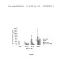 |  |
 | 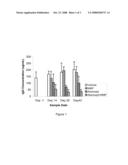 |
 |  |
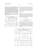 |  |
 |  |
 |  |
 |  |
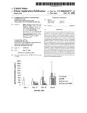 |  |
 |  |
 |  |
 | 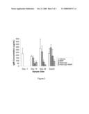 |
 |  |
 |  |
 |  |
 |  |
 |
| New patent applications in this class: | |
| Date | Title |
|---|---|
| 2022-05-05 | Dominant negative cd40l polypeptides |
| 2019-05-16 | Compositions and methods for antigen targeting to cd180 |
| 2019-05-16 | Synthetic membrane-receiver complexes |
| 2019-05-16 | Modular, controlled single chain variable fragment antibody switch |
| 2019-05-16 | Compositions comprising a p75 tumor necrosis factor receptor/ig fusion protein |
| New patent applications from these inventors: | |
| Date | Title |
|---|---|
| 2016-02-25 | Combination of blys inhibition and anti-cd 20 agents for treatment of autoimmune disease |
| 2015-01-15 | Combination of blys and/or april inhibition and immunosuppressants for treatment of autoimmune disease |
| Top Inventors for class "Drug, bio-affecting and body treating compositions" | |
| Rank | Inventor's name |
|---|---|
| 1 | David M. Goldenberg |
| 2 | Hy Si Bui |
| 3 | Lowell L. Wood, Jr. |
| 4 | Roderick A. Hyde |
| 5 | Yat Sun Or |