Patent application title: Reagents for the detection of protein phosphorylation in Leukemia signaling pathways
Inventors:
Roberto Polakiewicz (Lexington, MA, US)
Valerie Goss (Seabrook, NH, US)
Albrecht Moritz (Salem, MA, US)
Ting-Lei Gu (Woburn, MA, US)
Kimberly Lee (Seattle, WA, US)
IPC8 Class: AC07K1618FI
USPC Class:
435 71
Class name: Chemistry: molecular biology and microbiology measuring or testing process involving enzymes or micro-organisms; composition or test strip therefore; processes of forming such composition or test strip involving antigen-antibody binding, specific binding protein assay or specific ligand-receptor binding assay
Publication date: 2008-10-09
Patent application number: 20080248490
Inventors list |
Agents list |
Assignees list |
List by place |
Classification tree browser |
Top 100 Inventors |
Top 100 Agents |
Top 100 Assignees |
Usenet FAQ Index |
Documents |
Other FAQs |
Patent application title: Reagents for the detection of protein phosphorylation in Leukemia signaling pathways
Inventors:
Roberto Polakiewicz
Valerie Goss
Albrecht Moritz
Ting-Lei Gu
Kimberly Lee
Agents:
Simona Levi-Minzi, Ph.D.;General Counsel
Assignees:
Origin: DANVERS, MA US
IPC8 Class: AC07K1618FI
USPC Class:
435 71
Abstract:
The invention discloses nearly 288 novel phosphorylation sites identified
in signal transduction proteins and pathways underlying human Leukemia,
and provides phosphorylation-site specific antibodies and heavy-isotope
labeled peptides (AQUA peptides) for the selective detection and
quantification of these phosphorylated sites/proteins, as well as methods
of using the reagents for such purpose. Among the phosphorylation sites
identified are sites occurring in the following protein types:
Adaptor/Scaffold proteins, Cytoskeletal proteins, Cellular Metabolism
enzymes, G Protein/GTPase Activating/Guanine Nucleotide Exchange Factor
proteins, Immunoglobulin Superfamily proteins, Inhibitor proteins, Lipid
Kinases, Nuclear DNA Repair/RNA Binding/Transcription proteins,
Serine/Threonine Protein Kinases, Tyrosine Kinases, Protein Phosphatases,
and Translation/Transporter proteins.Claims:
1-14. (canceled)
15. An isolated phosphorylation site-specific antibody that specifically binds a human Leukemia-related signaling protein selected from Column A of Table 1 only when phosphorylated at the tyrosine listed in corresponding Column D of Table 1, comprised within the phosphorylatable peptide sequence listed in corresponding Column E of Table 1 (SEQ ID NOs: 1-288), wherein said antibody does not bind said signaling protein when not phosphorylated at said tyrosine.
16. An isolated phosphorylation site-specific antibody that specifically binds a human Leukemia-related signaling protein selected from Column A of Table 1 only when not phosphorylated at the tyrosine listed in corresponding Column D of Table 1, comprised within the phosphorylatable peptide sequence listed in corresponding Column E of Table 1 (SEQ ID NOs: 1-288), wherein said antibody does not bind said signaling protein when phosphorylated at said tyrosine.
17-51. (canceled)
52. An isolated phosphorylation site-specific antibody according to claim 15, that specifically binds a human Leukemia-related signaling protein selected from Column A, Rows 74, 87, 281, 117, 211, 90 and 64 of Table 1 only when phosphorylated at the tyrosine listed in corresponding Column D of Table 1, comprised within the phosphorylatable peptide sequence listed in corresponding Column E of Table 1 (SEQ ID NOs: 73, 86, 280, 116, 210, 89 and 63), wherein said antibody does not bind said signaling protein when not phosphorylated at said tyrosine.
53. An isolated phosphorylation site-specific antibody according to claim 17, that specifically binds a human Leukemia-related signaling protein selected from Column A, Rows 74, 87, 281, 117, 211, 90 and 64 of Table 1 only when not phosphorylated at the tyrosine listed in corresponding Column D of Table 1, comprised within the phosphorylatable peptide sequence listed in corresponding Column E of Table 1 (SEQ ID NOs: SEQ ID NOs: 73, 86, 280, 116, 210, 89 and 63), wherein said antibody does not bind said signaling protein when phosphorylated at said tyrosine.
54. A method selected from the group consisting of:(a) a method for detecting a human leukemia-related signaling protein selected from Column A of Table 1, wherein said human leukemia-related signaling protein is phosphorylated at the tyrosine listed in corresponding Column D of Table 1, comprised within the phosphorylatable peptide sequence listed in corresponding Column E of Table 1 (SEQ ID NOs: 1-288), comprising the step of adding an isolated phosphorylation-specific antibody according to claim 16, to a sample comprising said human leukemia-related signaling protein under conditions that permit the binding of said antibody to said human leukemia-related signaling protein, and detecting bound antibody;(b) a method for quantifying the amount of a human leukemia-related signaling protein listed in Column A of Table 1 that is phosphorylated at the corresponding tyrosine listed in Column D of Table 1, comprised within the phosphorylatable peptide sequence listed in corresponding Column E of Table 1 (SEQ ID NOs: 1-288), in a sample using a heavy-isotope labeled peptide (AQUA® peptide), said labeled peptide comprising a phosphorylated tyrosine at said corresponding tyrosine listed Column D of Table 1, comprised within the phosphorylatable peptide sequence listed in corresponding Column E of Table 1 as an internal standard; and(c) a method comprising step (a) followed by step (b).
55. The method of claim 54, wherein said isolated phosphorylation-specific antibody is capable of specifically binding ALDOA only when phosphorylated at Y3, comprised within the phosphorylatable peptide sequence listed in Column E, Row 74, of Table 1 (SEQ ID NO: 73), wherein said antibody does not bind said protein when not phosphorylated at said tyrosine.
56. The method of claim 54, wherein said isolated phosphorylation-specific antibody is capable of specifically binding ALDOA only when not phosphorylated at Y3, comprised within the phosphorylatable peptide sequence listed in Column E, Row 74, of Table 1 (SEQ ID NO: 73), wherein said antibody does not bind said protein when phosphorylated at said tyrosine.
57. The method of claim 54, wherein said isolated phosphorylation-specific antibody is capable of specifically binding TARS only when phosphorylated at Y298, comprised within the phosphorylatable peptide sequence listed in Column E, Row 87, of Table 1 (SEQ ID NO: 86), wherein said antibody does not bind said protein when not phosphorylated at said tyrosine.
58. The method of claim 54, wherein said isolated phosphorylation-specific antibody is capable of specifically binding TARS only when not phosphorylated at Y298, comprised within the phosphorylatable peptide sequence listed in Column E, Row 87, of Table 1 (SEQ ID NO: 86), wherein said antibody does not bind said protein when phosphorylated at said tyrosine.
59. The method of claim 54, wherein said isolated phosphorylation-specific antibody is capable of specifically binding SNAP23 only when phosphorylated at Y139, comprised within the phosphorylatable peptide sequence listed in Column E, Row 281, of Table 1 (SEQ ID NO: 280), wherein said antibody does not bind said protein when not phosphorylated at said tyrosine.
60. The method of claim 54, wherein said isolated phosphorylation-specific antibody is capable of specifically binding SNAP23 only when not phosphorylated at Y139, comprised within the phosphorylatable peptide sequence listed in Column E, Row 281, of Table 1 (SEQ ID NO: 280), wherein said antibody does not bind said protein when phosphorylated at said tyrosine.
61. The method of claim 54, wherein said isolated phosphorylation-specific antibody is capable of specifically binding PIN4 only when phosphorylated at Y147, comprised within the phosphorylatable peptide sequence listed in Column E, Row 117, of Table 1 (SEQ ID NO: 116), wherein said antibody does not bind said protein when not phosphorylated at said tyrosine.
62. The method of claim 54, wherein said isolated phosphorylation-specific antibody is capable of specifically binding PIN4 only when not phosphorylated at Y147, comprised within the phosphorylatable peptide sequence listed in Column E, Row 117, of Table 1 (SEQ ID NO: 116), wherein said antibody does not bind said protein when phosphorylated at said tyrosine.
63. The method of claim 54, wherein said isolated phosphorylation-specific antibody is capable of specifically binding ROD1 only when phosphorylated at Y127, comprised within the phosphorylatable peptide sequence listed in Column E, Row 211, of Table 1 (SEQ ID NO: 210), wherein said antibody does not bind said protein when not phosphorylated at said tyrosine.
64. The method of claim 54, wherein said isolated phosphorylation-specific antibody is capable of specifically binding ROD1 only when not phosphorylated at Y127, comprised within the phosphorylatable peptide sequence listed in Column E, Row 211, of Table 1 (SEQ ID NO: 210), wherein said antibody does not bind said protein when phosphorylated at said tyrosine.
65. The method of claim 54, wherein said isolated phosphorylation-specific antibody is capable of specifically binding WARS only when phosphorylated at Y316, comprised within the phosphorylatable peptide sequence listed in Column E, Row 90, of Table 1 (SEQ ID NO: 89), wherein said antibody does not bind said protein when not phosphorylated at said tyrosine.
66. The method of claim 54, wherein said isolated phosphorylation-specific antibody is capable of specifically binding WARS only when not phosphorylated at Y316, comprised within the phosphorylatable peptide sequence listed in Column E, Row 90, of Table 1 (SEQ ID NO: 89), wherein said antibody does not bind said protein when phosphorylated at said tyrosine.
67. The method of claim 54, wherein said isolated phosphorylation-specific antibody is capable of specifically binding hnRNPU only when phosphorylated at Y247, comprised within the phosphorylatable peptide sequence listed in Column E, Row 64, of Table 1 (SEQ ID NO: 63), wherein said antibody does not bind said protein when not phosphorylated at said tyrosine.
68. The method of claim 54, wherein said isolated phosphorylation-specific antibody is capable of specifically binding hnRNPU only when not phosphorylated at Y247, comprised within the phosphorylatable peptide sequence listed in Column E, Row 64, of Table 1 (SEQ ID NO: 63), wherein said antibody does not bind said protein when phosphorylated at said tyrosine.
Description:
RELATED APPLICATIONS
[0001]This application claims the benefit of, and priority to, PCT serial number PCT/US06/034050, filed Aug. 31, 2006, presently pending, the disclosure of which is incorporated herein, in its entirety, by reference.
FIELD OF THE INVENTION
[0002]The invention relates generally to antibodies and peptide reagents for the detection of protein phosphorylation, and to protein phosphorylation in cancer.
BACKGROUND OF THE INVENTION
[0003]The activation of proteins by post-translational modification is an important cellular mechanism for regulating most aspects of biological organization and control, including growth, development, homeostasis, and cellular communication. Protein phosphorylation, for example, plays a critical role in the etiology of many pathological conditions and diseases, including cancer, developmental disorders, autoimmune diseases, and diabetes. Yet, in spite of the importance of protein modification, it is not yet well understood at the molecular level, due to the extraordinary complexity of signaling pathways, and the slow development of technology necessary to unravel it.
[0004]Protein phosphorylation on a proteome-wide scale is extremely complex as a result of three factors: the large number of modifying proteins, e.g. kinases, encoded in the genome, the much larger number of sites on substrate proteins that are modified by these enzymes, and the dynamic nature of protein expression during growth, development, disease states, and aging. The human genome, for example, encodes over 520 different protein kinases, making them the most abundant class of enzymes known. See Hunter, Nature 411: 355-65 (2001). Most kinases phosphorylate many different substrate proteins, at distinct tyrosine, serine, and/or threonine residues. Indeed, it is estimated that one-third of all proteins encoded by the human genome are phosphorylated, and many are phosphorylated at multiple sites by different kinases. See Graves et al., Pharmacol. Ther. 82: 111-21 (1999).
[0005]Many of these phosphorylation sites regulate critical biological processes and may prove to be important diagnostic or therapeutic targets for molecular medicine. For example, of the more than 100 dominant oncogenes identified to date, 46 are protein kinases. See Hunter, supra. Understanding which proteins are modified by these kinases will greatly expand our understanding of the molecular mechanisms underlying oncogenic transformation. Therefore, the identification of, and ability to detect, phosphorylation sites on a wide variety of cellular proteins is crucially important to understanding the key signaling proteins and pathways implicated in the progression of diseases like cancer.
[0006]One form of cancer in which underlying signal transduction events are involved, but still poorly understood, is leukemia. Leukemia is a malignant disease of the bone marrow and blood, characterized by abnormal accumulation of blood cells, and is divided in four major categories. An estimated 33,500 new cases of leukemia will be diagnosed in the U.S. alone this year, affecting roughly 30,000 adults and 3,000 children, and close to 24,000 patients will die from the disease (Source: The Leukemia & Lymphoma Society (2004)). Depending of the cell type involved and the rate by which the disease progresses it can be defined as acute or chronic myelogenous leukemia (AML or CML), or acute and chronic lymphocytic leukemia (ALL or CLL). The acute forms of the disease rapidly progress, causing the accumulation of immature, functionless cells in the marrow and blood, which in turn results in anemia, immunodeficiency and coagulation deficiencies, respectively. Chronic forms of leukemia progress more slowly, allowing a greater number of mature, functional cells to be produced, which amass to high concentration in the blood over time.
[0007]More than half of adult leukemias occur in patients 67 years of age or older, and leukemia accounts for about 30% of all childhood cancers. The most common type of adult leukemia is acute myelogenous leukemia (AML), with an estimated 11,920 new cases annually. Without treatment patients rarely survive beyond 6-12 months, and despite continued development of new therapies, it remains fatal in 80% of treated patients (Source: The Leukemia & Lymphoma Society (2004)). The most common childhood leukemia is acute lymphocytic leukemia (ALL), but it can develop at any age. Chronic lymphocytic leukemia (CLL) is the second most prevalent adult leukemia, with approximately 8,200 new cases of CLL diagnosed annually in the U.S. The course of the disease is typically slower than acute forms, with a five-year relative survival of 74%. Chronic myelogenous leukemia (CML) is less prevalent, with about 4,600 new cases diagnosed each year in the U.S., and is rarely observed in children.
[0008]Most varieties of leukemia are generally characterized by genetic alterations associated with the etiology of the disease, and it has recently become apparent that, in many instances, such alterations (chromosomal translocations, deletions or point mutations) result in the constitutive activation of protein kinase genes, and their products, particularly tyrosine kinases. The most well known alteration is the oncogenic role of the chimeric BCR-Abl gene, which is generated by translocation of chromosome 9 to chromosome 22, creating the so-called Philadelphia chromosome characteristic of CML (see Nowell, Science 132:1497 (1960)). The resulting BCR-Abl kinase protein is constitutively active and elicits characteristic signaling pathways that have been shown to drive the proliferation and survival of CML cells (see Daley, Science 247: 824-830 (1990); Raitano et al., Biochim. Biophys. Acta. December 9; 1333(3): F201-16 (1997)). The recent success of Imanitib (also known as STI571 or Gleevec®), the first molecularly targeted compound designed to specifically inhibit the tyrosine kinase activity of BCR-Abl, provided critical confirmation of the central role of BCR-Abl signaling in the progression of CML (see Schindler et al., Science 289: 1938-1942 (2000); Nardi et al., Curr. Opin. Hematol. 11: 35-43 (2003)).
[0009]The success of Gleevec® now serves as a paradigm for the development of targeted drugs designed to block the activity of other tyrosine kinases known to be involved in leukemias and other malignancies (see, e.g., Sawyers, Curr. Opin. Genet. Dev. Feb; 12(1): 111-5 (2002); Druker, Adv. Cancer Res. 91:1-30 (2004)). For example, recent studies have demonstrated that mutations in the FLT3 gene occur in one third of adult patients with AML. FLT3 (Fms-like tyrosine kinase 3) is a member of the class III receptor tyrosine kinase (RTK) family including FMS, platelet-derived growth factor receptor (PDGFR) and c-KIT (see Rosnet et al., Crit. Rev. Oncog. 4: 595-613 (1993). In 20-27% of patients with AML, an internal tandem duplication in the juxta-membrane region of FLT3 can be detected (see Yokota et al., Leukemia 11: 1605-1609 (1997)). Another 7% of patients have mutations within the active loop of the second kinase domain, predominantly substitutions of aspartate residue 835 (D835), while additional mutations have been described (see Yamamoto et al., Blood 97: 2434-2439 (2001); Abu-Duhier et al., Br. J. Haematol. 113: 983-988 (2001)). Expression of mutated FLT3 receptors results in constitutive tyrosine phosphorylation of FLT3, and subsequent phosphorylation and activation of downstream molecules such as STAT5, Akt and MAPK, resulting in factor-independent growth of hematopoietic cell lines.
[0010]Altogether, FLT3 is the single most common activated gene in AML known to date. This evidence has triggered an intensive search for FLT3 inhibitors for clinical use leading to at least four compounds in advanced stages of clinical development, including: PKC412 (by Novartis), CEP-701 (by Cephalon), MLN518 (by Millenium Pharmaceuticals), and SU5614 (by Sugen/Pfizer) (see Stone et al., Blood (in press)(2004); Smith et al., Blood 103: 3669-3676 (2004); Clark et al., Blood 104: 2867-2872 (2004); and Spiekerman et al., Blood 101: 1494-1504 (2003)).
[0011]There is also evidence indicating that kinases such as FLT3, c-KIT and Abl are implicated in some cases of ALL (see Cools et al., Cancer Res. 64: 6385-6389 (2004); Hu, Nat. Genet. 36: 453-461 (2004); and Graux et al., Nat. Genet. 36: 1084-1089 (2004)). In contrast, very little is know regarding any causative role of protein kinases in CLL, except for a high correlation between high expression of the tyrosine kinase ZAP70 and the more aggressive form of the disease (see Rassenti et al., N. Eng. J. Med. 351: 893-901 (2004)).
[0012]Despite the identification of a few key molecules involved in progression of leukemia, the vast majority of signaling protein changes underlying this disease remains unknown. There is, therefore, relatively scarce information about kinase-driven signaling pathways and phosphorylation sites relevant to the different types of leukemia. This has hampered a complete and accurate understanding of how protein activation within signaling pathways is driving these complex cancers. Accordingly, there is a continuing and pressing need to unravel the molecular mechanisms of kinase-driven oncogenesis in leukemia by identifying the downstream signaling proteins mediating cellular transformation in this disease. Identifying particular phosphorylation sites on such signaling proteins and providing new reagents, such as phospho-specific antibodies and AQUA peptides, to detect and quantify them remains particularly important to advancing our understanding of the biology of this disease.
[0013]Presently, diagnosis of leukemia is made by tissue biopsy and detection of different cell surface markers. However, misdiagnosis can occur since some leukemia cases can be negative for certain markers, and because these markers may not indicate which genes or protein kinases may be deregulated. Although the genetic translocations and/or mutations characteristic of a particular form of leukemia can be sometimes detected, it is clear that other downstream effectors of constitutively active kinases having potential diagnostic, predictive, or therapeutic value, remain to be elucidated. Accordingly, identification of downstream signaling molecules and phosphorylation sites involved in different types of leukemia and development of new reagents to detect and quantify these sites and proteins may lead to improved diagnostic/prognostic markers, as well as novel drug targets, for the detection and treatment of this disease.
SUMMARY OF THE INVENTION
[0014]The invention discloses nearly 288 novel phosphorylation sites identified in signal transduction proteins and pathways underlying human Leukemias and provides new reagents, including phosphorylation-site specific antibodies and AQUA peptides, for the selective detection and quantification of these phosphorylated sites/proteins. Also provided are methods of using the reagents of the invention for the detection, quantification, and profiling of the disclosed phosphorylation sites.
BRIEF DESCRIPTION OF THE DRAWINGS
[0015]FIG. 1--Is a diagram broadly depicting the immunoaffinity isolation and mass-spectrometric characterization methodology (IAP) employed to identify the novel phosphorylation sites disclosed herein.
[0016]FIG. 2--Is a table (corresponding to Table 1) enumerating the Leukemia signaling protein phosphorylation sites disclosed herein: Column A=the name of the parent protein; Column B=the SwissProt accession number for the protein (human sequence); Column C=the protein type/classification; Column D=the tyrosine residue (in the parent protein amino acid sequence) at which phosphorylation occurs within the phosphorylation site; Column E=the phosphorylation site sequence encompassing the phosphorylatable residue (residue at which phosphorylation occurs (and corresponding to the respective entry in Column D) appears in lowercase; Column F=the type of leukemia in which the phosphorylation site was discovered; and Column G=the cell type(s), tissue(s) and/or patient(s) in which the phosphorylation site was discovered.
[0017]FIG. 3--is an exemplary mass spectrograph depicting the detection of the tyrosine 630 phosphorylation site in BANK1 (see Row 10 in FIG. 2/Table 1), as further described in Example 1 (red and blue indicate ions detected in MS/MS spectrum); Y* indicates the phosphorylated tyrosine (shown as lowercase "y" in FIG. 2).
[0018]FIG. 4--is an exemplary mass spectrograph depicting the detection of the tyrosine 289 phosphorylation site in FASN (see Row 10 in FIG. 2/Table 1), as further described in Example 1 (red and blue indicate ions detected in MS/MS spectrum); Y* indicates the phosphorylated tyrosine (shown as lowercase "y" in FIG. 2).
[0019]FIG. 5--is an exemplary mass spectrograph depicting the detection of the tyrosine 83 phosphorylation site in PUM1 (see Row 142 in FIG. 2/Table 1), as further described in Example 1 (red and blue indicate ions detected in MS/MS spectrum); Y* indicates the phosphorylated serine (shown as lowercase "y" in FIG. 2).
[0020]FIG. 6--is an exemplary mass spectrograph depicting the detection of the tyrosine 73 phosphorylation site in RAB11B (see Row 94 in FIG. 2/Table 1), as further described in Example 1 (red and blue indicate ions detected in MS/MS spectrum); Y* indicates the phosphorylated tyrosine (shown as lowercase "y" in FIG. 2).
[0021]FIG. 7--is an exemplary mass spectrograph depicting the detection of the tyrosine 189 phosphorylation site in RASGRP2 (see Row 105 in FIG. 2/Table 1), as further described in Example 1 (red and blue indicate ions detected in MS/MS spectrum); Y* indicates the phosphorylated tyrosine (shown as lowercase "y" in FIG. 2).
[0022]FIG. 8--is an exemplary mass spectrograph depicting the detection of the tyrosine 251 phosphorylation site in RBM15 (see Row 203 in FIG. 2/Table 1), as further described in Example 1 (red and blue indicate ions detected in MS/MS spectrum); Y* indicates the phosphorylated tyrosine (shown as lowercase "y" in FIG. 2).
DETAILED DESCRIPTION OF THE INVENTION
[0023]In accordance with the present invention, nearly 288 novel protein phosphorylation sites in signaling proteins and pathways underlying human Leukemia have now been discovered. These newly described phosphorylation sites were identified by employing the techniques described in "Immunoaffinity Isolation of Modified Peptides From Complex Mixtures," U.S. Patent Publication No. 20030044848, Rush et al., using cellular extracts from a variety of leukemia-derived cell lines, e.g. SEM, K562, etc., as further described below. The novel phosphorylation sites (tyrosine), and their corresponding parent proteins, disclosed herein are listed in Table 1. These phosphorylation sites correspond to numerous different parent proteins (the full sequences (human) of which are all publicly available in SwissProt database and their Accession numbers listed in Column B of Table 1/FIG. 2), each of which fall into discrete protein type groups, for example Acetyltransferases, oxyreductases, adaptor/scaffold proteins, cytoskeletal proteins, protein kinases, and adhesion proteins, etc. (see Column C of Table 1), the phosphorylation of which is relevant to signal transduction activity underlying Leukemias (AML, CML, CLL, and ALL), as disclosed herein.
[0024]The discovery of the nearly 288 novel protein phosphorylation sites described herein enables the production, by standard methods, of new reagents, such as phosphorylation site-specific antibodies and AQUA peptides (heavy-isotope labeled peptides), capable of specifically detecting and/or quantifying these phosphorylated sites/proteins. Such reagents are highly useful, inter alia, for studying signal transduction events underlying the progression of Leukemia. Accordingly, the invention provides novel reagents--phospho-specific antibodies and AQUA peptides--for the specific detection and/or quantification of a Leukemia-related signaling protein/polypeptide only when phosphorylated (or only when not phosphorylated) at a particular phosphorylation site disclosed herein. The invention also provides methods of detecting and/or quantifying one or more phosphorylated Leukemia-related signaling proteins using the phosphorylation-site specific antibodies and AQUA peptides of the invention, and methods of obtaining a phosphorylation profile of such proteins (e.g. Kinases).
[0025]In part, the invention provides an isolated phosphorylation site-specific antibody that specifically binds a given Leukemia-related signaling protein only when phosphorylated (or not phosphorylated, respectively) at a particular tyrosine enumerated in Column D of Table 1/FIG. 2 comprised within the phosphorylatable peptide site sequence enumerated in corresponding Column E. In further part, the invention provides a heavy-isotope labeled peptide (AQUA peptide) for the detection and quantification of a given Leukemia-related signaling protein, the labeled peptide comprising a particular phosphorylatable peptide site/sequence enumerated in Column E of Table 1/FIG. 2 herein. For example, among the reagents provided by the invention is an isolated phosphorylation site-specific antibody that specifically binds the PUM1 phosphatase only when phosphorylated (or only when not phosphorylated) at tyrosine 83 (see Row 142 (and Columns D and E) of Table 1/FIG. 2). By way of further example, among the group of reagents provided by the invention is an AQUA peptide for the quantification of phosphorylated TRPM3 channel protein, the AQUA peptide comprising the phosphorylatable peptide sequence listed in Column E, Row 48, of Table 1/FIG. 2 (which encompasses the phosphorylatable tyrosine at position 712).
[0026]In one embodiment, the invention provides an isolated phosphorylation site-specific antibody that specifically binds a human Leukemia-related signaling protein selected from Column A of Table 1 (Rows 2-289) only when phosphorylated at the tyrosine residue listed in corresponding Column D of Table 1, comprised within the phosphorylatable peptide sequence listed in corresponding Column E of Table 1 (SEQ ID NOs: 1-3, 6-28, 30-42, 44-93, 95-168, 170-183, 185-203, 205-278, and 280-288), wherein said antibody does not bind said signaling protein when not phosphorylated at said tyrosine. In another embodiment, the invention provides an isolated phosphorylation site-specific antibody that specifically binds a Leukemia-related signaling protein selected from Column A of Table 1 only when not phosphorylated at the tyrosine residue listed in corresponding Column D of Table 1, comprised within the peptide sequence listed in corresponding Column E of Table 1 (SEQ ID NOs: 1-3, 6-28, 30-42, 44-93, 95-168, 170-183, 185-203, 205-278, and 280-288), wherein said antibody does not bind said signaling protein when phosphorylated at said tyrosine. Such reagents enable the specific detection of phosphorylation (or non-phosphorylation) of a novel phosphorylatable site disclosed herein. The invention further provides immortalized cell lines producing such antibodies. In one preferred embodiment, the immortalized cell line is a rabbit or mouse hybridoma.
[0027]In another embodiment, the invention provides a heavy-isotope labeled peptide (AQUA peptide) for the quantification of a Leukemia-related signaling protein selected from Column A of Table 1, said labeled peptide comprising the phosphorylatable peptide sequence listed in corresponding Column E of Table 1 (SEQ ID NOs: 1-3, 6-28, 30-42, 44-93, 95-168, 170-183, 185-203, 205-278, and 280-288), which sequence comprises the phosphorylatable tyrosine listed in corresponding Column D of Table 1. In certain preferred embodiments, the phosphorylatable tyrosine within the labeled peptide is phosphorylated, while in other preferred embodiments, the phosphorylatable residue within the labeled peptide is not phosphorylated.
[0028]Reagents (antibodies and AQUA peptides) provided by the invention may conveniently be grouped by the type of Leukemia-related signaling protein in which a given phosphorylation site (for which reagents are provided) occurs. The protein types for each respective protein (in which a phosphorylation site has been discovered) are provided in Column C of Table 1/FIG. 2, and include: adaptor/scaffold proteins, acetyltransferases, actin binding proteins, adhesion proteins, apoptosis proteins, calcium channel proteins, cell cycle regulation proteins, cell surface proteins, channel proteins, chaperone proteins, contractile proteins, cytokine proteins, chaperone proteins, cytoskeletal proteins, DNA binding proteins, endoplasmic reticulum proteins, cellular metabolism enzymes, G protein regulators and GTPase activating proteins, guanine nucleotide exchange factors, helicase proteins, hydrolases, isomerases immunoglobulin superfamily proteins, inhibitor proteins, kinases, ligases, lyases, methyltransferases, motor proteins, mitochondrial proteins, myosin binding proteins, oxidoreductases, phosphotases, phosphodiesterases, proteases, receptor proteins, RNA binding proteins, transcription proteins, secreted proteins transferases, translation/transporter proteins, ubiquitin conjugating system proteins and vesicle proteins. Each of these distinct protein groups is considered a preferred subset of Leukemia-related signal transduction protein phosphorylation sites disclosed herein, and reagents for their detection/quantification may be considered a preferred subset of reagents provided by the invention.
[0029]Particularly preferred subsets of the phosphorylation sites (and their corresponding proteins) disclosed herein are those occurring on the following protein types/groups listed in Column C of Table 1/FIG. 2, are the protein kinases, adaptor/scaffold proteins, adhesion proteins, enzymes cell cycle regulation proteins, cell surface proteins, transcription proteins, phosphatases, proteases, receptor proteins, RNA binding proteins, G protein regulators/GTPase activators/Guanine nucleotide exchange factors, transporter proteins and ubiquitan conjugating system proteins. Accordingly, among preferred subsets of reagents provided by the invention are isolated antibodies and AQUA peptides useful for the detection and/or quantification of the foregoing preferred protein/phosphorylation site subsets.
[0030]In one subset of preferred embodiments, there is provided:
(i) An isolated phosphorylation site-specific antibody that specifically binds a protein kinase selected from Column A, Rows 118-120, of Table 1 only when phosphorylated at the tyrosine listed in corresponding Column D, Rows 118-120, of Table 1, comprised within the phosphorylatable peptide sequence listed in corresponding Column E, Rows 118-120, of Table 1 (SEQ ID NOs: 117-119), wherein said antibody does not bind said protein when not phosphorylated at said tyrosine.(ii) An equivalent antibody to (i) above that only binds the protein kinase when not phosphorylated at the disclosed site (and does not bind the protein when it is phosphorylated at the site).(iii) A heavy-isotope labeled peptide (AQUA peptide) for the quantification of a protein kinase selected from Column A, Rows 118-120, said labeled peptide comprising the phosphorylatable peptide sequence listed in corresponding Column E, Rows 118-120, of Table 1 (SEQ ID NOs: 117-119), which sequence comprises the phosphorylatable tyrosine listed in corresponding Column D, Rows 118-120, of Table 1.
[0031]In a second subset of preferred embodiments there is provided:
(i) An isolated phosphorylation site-specific antibody that specifically binds an adaptor/scaffold protein selected from Column A, Rows 8-22, of Table 1 only when phosphorylated at the tyrosine listed in corresponding Column D, Rows 8-22, of Table 1, comprised within the phosphorylatable peptide sequence listed in corresponding Column E, Rows 8-22, of Table 1 (SEQ ID NOs: 7-21), wherein said antibody does not bind said protein when not phosphorylated at said tyrosine.(ii) An equivalent antibody to (i) above that only binds the adaptor/scaffold protein when not phosphorylated at the disclosed site (and does not bind the protein when it is phosphorylated at the site).(iii) A heavy-isotope labeled peptide (AQUA peptide) for the quantification of a Leukemia-related signaling protein that is a adaptor/scaffold protein selected from Column A, Rows 8-22, said labeled peptide comprising the phosphorylatable peptide sequence listed in corresponding Column E, Rows 8-22, of Table 1 (SEQ ID NOs: 7-21), which sequence comprises the phosphorylatable tyrosine listed in corresponding Column D, Rows 8-22, of Table 1.
[0032]Among this preferred subset of reagents, antibodies and AQUA peptides for the detection/quantification of the following adaptor/scaffold protein phosphorylation sites are particularly preferred: BANK1 (Y630), LAX1 (Y373), and PIK3AP1 (Y163) (see SEQ ID NOs: 9, 14 and 18).
[0033]In another subset of preferred embodiments there is provided:
(i) An isolated phosphorylation site-specific antibody that specifically binds an adhesion protein selected from Column A, Rows 23-37, of Table 1 only when phosphorylated at the tyrosine listed in corresponding Column D, Rows 23-37, of Table 1, comprised within the phosphorylatable peptide sequence listed in corresponding Column E, Rows 23-37, of Table 1 (SEQ ID NOs: 22-28, and 30-36), wherein said antibody does not bind said protein when not phosphorylated at said tyrosine.(ii) An equivalent antibody to (i) above that only binds the adhesion protein when not phosphorylated at the disclosed site (and does not bind the protein when it is phosphorylated at the site).(iii) A heavy-isotope labeled peptide (AQUA peptide) for the quantification of a Leukemia-related signaling protein that is an adhesion protein selected from Column A, Rows 23-37, said labeled peptide comprising the phosphorylatable peptide sequence listed in corresponding Column E, Rows 23-37, of Table 1 (SEQ ID NOs: 22-28, and 30-36), which sequence comprises the phosphorylatable tyrosine listed in corresponding Column D, Rows 23-37, of Table 1.
[0034]Among this preferred subset of reagents, antibodies and AQUA peptides for the detection/quantification of the following adhesion protein phosphorylation sites are particularly preferred: FAT (Y400) (see SEQ ID NO: 22).
[0035]In still another subset of preferred embodiments there is provided:
(i) An isolated phosphorylation site-specific antibody that specifically binds an enzyme protein selected from Column A, Rows 68-90, of Table 1 only when phosphorylated at the tyrosine listed in corresponding Column D, Rows 68-90, of Table 1, comprised within the phosphorylatable peptide sequence listed in corresponding Column E, Rows 68-90, of Table 1 (SEQ ID NOs: 67-89), wherein said antibody does not bind said protein when not phosphorylated at said tyrosine.(ii) An equivalent antibody to (i) above that only binds the enzyme protein when not phosphorylated at the disclosed site (and does not bind the protein when it is phosphorylated at the site).(iii) A heavy-isotope labeled peptide (AQUA peptide) for the quantification of a Leukemia-related signaling protein that is a enzyme protein selected from Column A, Rows 68-90, said labeled peptide comprising the phosphorylatable peptide sequence listed in corresponding Column E, Rows 68-90, of Table 1 (SEQ ID NOs: 67-89), which sequence comprises the phosphorylatable tyrosine listed in corresponding Column D, Rows 68-90, of Table 1.
[0036]Among this preferred subset of reagents, antibodies and AQUA peptides for the detection/quantification of the following enzyme protein phosphorylation sites are particularly preferred: ACACA (Y306), FASN (Y289), GLA (Y329), MOGAT2 (Y154) (see SEQ ID NOs: 71, 78, 84 and 85).
[0037]In still another subset of preferred embodiments there is provided:
(i) An isolated phosphorylation site-specific antibody that specifically binds a G protein/GTPase activating protein/Guanine nucleotide exchange factor selected from Column A, Rows 92-105, of Table 1 only when phosphorylated at the tyrosine listed in corresponding Column D, Rows 92-105, of Table 1, comprised within the phosphorylatable peptide sequence listed in corresponding Column E, Rows 92-105, of Table 1 (SEQ ID NOs: 91-93, and 95-104), wherein said antibody does not bind said protein when not phosphorylated at said tyrosine.(ii) An equivalent antibody to (i) above that only binds the G protein/GTPase activating protein/Guanine nucleotide exchange factor when not phosphorylated at the disclosed site (and does not bind the protein when it is phosphorylated at the site).(iii) A heavy-isotope labeled peptide (AQUA peptide) for the quantification of a Leukemia-related signaling protein that is a G protein/GTPase activating protein/Guanine nucleotide exchange factor selected from Column A, Rows 92-105, said labeled peptide comprising the phosphorylatable peptide sequence listed in corresponding Column E, Rows 92-105, of Table 1 (SEQ ID NOs: 91-93, and 95-104), which sequence comprises the phosphorylatable tyrosine listed in corresponding Column D, Rows 92-105, of Table 1.
[0038]Among this preferred subset of reagents, antibodies and AQUA peptides for the detection/quantification of the following G protein/GTPase activating protein/Guanine nucleotide exchange factor phosphorylation sites are particularly preferred: RAB11B (Y73), RICS (Y1353), RASGRP2 (Y189) (see SEQ ID NOs: 93, 99 and 104).
[0039]In still another subset of preferred embodiments there is provided:
(i) An isolated phosphorylation site-specific antibody that specifically binds a phosphatase selected from Column A, Rows 136-142, of Table 1 only when phosphorylated at the tyrosine listed in corresponding Column D, Rows 136-142, of Table 1, comprised within the phosphorylatable peptide sequence listed in corresponding Column E, Rows 136-142 of Table 1 (SEQ ID NOs: 135-141), wherein said antibody does not bind said protein when not phosphorylated at said tyrosine.(ii) An equivalent antibody to (i) above that only binds phosphatase when not phosphorylated at the disclosed site (and does not bind the protein when it is phosphorylated at the site).(iii) A heavy-isotope labeled peptide (AQUA peptide) for the quantification of a Leukemia-related signaling protein that is a phosphatase selected from Column A, Rows 136-142, said labeled peptide comprising the phosphorylatable peptide sequence listed in corresponding Column E, Rows 136-142, of Table 1 (SEQ ID NOs: 135-141), which sequence comprises the phosphorylatable tyrosine listed in corresponding Column D, Rows 136-142, of Table 1.
[0040]Among this preferred subset of reagents, antibodies and AQUA peptides for the detection/quantification of the following phosphatase phosphorylation sites are particularly preferred: PP2R5B (Y244), PUM1 (Y83) (see SEQ ID NOs: 136-141).
[0041]In yet another subset of preferred embodiments, there is provided:
(i) An isolated phosphorylation site-specific antibody that specifically binds a protease selected from Column A, Rows 143-146, of Table 1 only when phosphorylated at the tyrosine listed in corresponding Column D, Rows 143-146, of Table 1, comprised within the phosphorylatable peptide sequence listed in corresponding Column E, Rows 143-146, of Table 1 (SEQ ID NOs: 142-145), wherein said antibody does not bind said protein when not phosphorylated at said tyrosine.(ii) An equivalent antibody to (i) above that only binds the protease when not phosphorylated at the disclosed site (and does not bind the protein when it is phosphorylated at the site).(iii) A heavy-isotope labeled peptide (AQUA peptide) for the quantification of a Leukemia-related signaling protein that is a protease selected from Column A, Rows 143-146, said labeled peptide comprising the phosphorylatable peptide sequence listed in corresponding Column E, Rows 143-146, of Table 1 (SEQ ID NOs: 142-145), which sequence comprises the phosphorylatable tyrosine listed in corresponding Column D, Rows 143-146, of Table 1.
[0042]Among this preferred subset of reagents, antibodies and AQUA peptides for the detection/quantification of the following protease phosphorylation sites are particularly preferred: ADAMTS14 (Y38) and SNEP2 (Y239) (see SEQ ID NOs: 142 and 143).
[0043]In yet another subset of preferred embodiments, there is provided:
(i) An isolated phosphorylation site-specific antibody specifically binds a receptor protein selected from Column A, Rows 149-170, of Table 1 only when phosphorylated at the tyrosine listed in corresponding Column D, Rows 149-170, of Table 1, comprised within the phosphorylatable peptide sequence listed in corresponding Column E, Rows 149-170, of Table 1 (SEQ ID NOs: 148-168), wherein said antibody does not bind said protein when not phosphorylated at said tyrosine.(ii) An equivalent antibody to (i) above that only binds the receptor protein when not phosphorylated at the disclosed site (and does not bind the protein when it is phosphorylated at the site).(iii) A heavy-isotope labeled peptide (AQUA peptide) for the quantification of a Leukemia-related signaling protein that is a receptor protein selected from Column A, Rows 149-170, said labeled peptide comprising the phosphorylatable peptide sequence listed in corresponding Column E, Rows 149-170, of Table 1 (SEQ ID NOs: 148-168), which sequence comprises the phosphorylatable tyrosine listed in corresponding Column D, Rows 149-170, of Table 1.
[0044]Among this preferred subset of reagents, antibodies and AQUA peptides for the detection/quantification of the following receptor protein phosphorylation sites are particularly preferred: ROBO1 (Y328) (see SEQ ID NOs: 166).
[0045]In yet another subset of preferred embodiments, there is provided:
(i) An isolated phosphorylation site-specific antibody that specifically binds a RNA binding protein selected from Column A, Rows 171-226, of Table 1 only when phosphorylated at the tyrosine listed in corresponding Column D, Rows 171-226, of Table 1, comprised within the phosphorylatable peptide sequence listed in corresponding Column E, Rows 171-226, of Table 1 (SEQ ID NOs: 170-183, 185-203, and 205-225), wherein said antibody does not bind said protein when not phosphorylated at said tyrosine.(ii) An equivalent antibody to (i) above that only binds the RNA binding protein when not phosphorylated at the disclosed site (and does not bind the protein when it is phosphorylated at the site).(iii) A heavy-isotope labeled peptide (AQUA peptide) for the quantification of a Leukemia-related signaling protein that is a RNA binding protein selected from Column A, Rows 171-226, said labeled peptide comprising the phosphorylatable peptide sequence listed in corresponding Column E, Rows 171-226, of Table 1 (SEQ ID NOs: 170-183, 185-203, and 205-225), which sequence comprises the phosphorylatable tyrosine listed in corresponding Column D, Rows 171-226, of Table 1.
[0046]Among this preferred subset of reagents, antibodies and AQUA peptides for the detection/quantification of the following RNA binding protein phosphorylation sites are particularly preferred: ARPP-19 (Y36), KHDRBS1 (Y435), MATR3 (Y243), and RBM15 (Y251) (see SEQ ID NO: 170, 188, 190 and 202).
[0047]In still another subset of preferred embodiments, there is provided:
(i) An isolated phosphorylation site-specific antibody that specifically binds a transcription protein selected from Column A, Rows 230-247, of Table 1 only when phosphorylated at the tyrosine listed in corresponding Column D, Rows 230-247, of Table 1, comprised within the phosphorylatable peptide sequence listed in corresponding Column E, Rows 230-247, of Table 1 (SEQ ID NOs: 229-246), wherein said antibody does not bind said protein when not phosphorylated at said tyrosine.(ii) An equivalent antibody to (i) above that only binds the transcription protein when not phosphorylated at the disclosed site (and does not bind the protein when it is phosphorylated at the site).(iii) A heavy-isotope labeled peptide (AQUA peptide) for the quantification of a Leukemia-related signaling protein that transcription protein selected from Column A, Rows 230-247, said labeled peptide comprising the phosphorylatable peptide sequence listed in corresponding Column E, Rows 230-247, of Table 1 (SEQ ID NOs: 229-246), which sequence comprises the phosphorylatable tyrosine listed in corresponding Column D, Rows 230-247, of Table 1.
[0048]Among this preferred subset of reagents, antibodies and AQUA peptides for the detection/quantification of the following transcription protein phosphorylation sites are particularly preferred: FOXJ1 (Y157) and IRFBP1 (Y268) (see SEQ ID NOs: 238 and 239).
[0049]In still another subset of preferred embodiments, there is provided:
(i) An isolated phosphorylation site-specific antibody that specifically binds a transporter protein selected from Column A, Rows 252-260, of Table 1 only when phosphorylated at the tyrosine listed in corresponding Column D, Rows 252-260, of Table 1, comprised within the phosphorylatable peptide sequence listed in corresponding Column E, Rows 252-260, of Table 1 (SEQ ID NOs: 251-259), wherein said antibody does not bind said protein when not phosphorylated at said tyrosine.(ii) An equivalent antibody to (i) above that only binds the transporter protein when not phosphorylated at the disclosed site (and does not bind the protein when it is phosphorylated at the site).(iii) A heavy-isotope labeled peptide (AQUA peptide) for the quantification of a Leukemia-related signaling protein that is an transporter protein selected from Column A, Rows 252-260, said labeled peptide comprising the phosphorylatable peptide sequence listed in corresponding Column E, Rows 252-260, of Table 1 (SEQ ID NOs: 251-259), which sequence comprises the phosphorylatable tyrosine listed in corresponding Column D, Rows 252-260, of Table 1.
[0050]In still another subset of preferred embodiments, there is provided:
(i) An isolated phosphorylation site-specific antibody that specifically binds an ubiquitin conjugating pathway protein selected from Column A, Rows 261-274, of Table 1 only when phosphorylated at the tyrosine listed in corresponding Column D, Rows 261-274, of Table 1, comprised within the phosphorylatable peptide sequence listed in corresponding Column E, Rows 261-274, of Table 1 (SEQ ID NOs: 260-273), wherein said antibody does not bind said protein when not phosphorylated at said tyrosine.(ii) An equivalent antibody to (i) above that only binds the ubiquitin conjugating pathway protein when not phosphorylated at the disclosed site (and does not bind the protein when it is phosphorylated at the site).(iii) A heavy-isotope labeled peptide (AQUA peptide) for the quantification of a Leukemia-related signaling protein that is an an ubiquitin conjugating pathway protein selected from Column A, Rows 261-274, said labeled peptide comprising the phosphorylatable peptide sequence listed in corresponding Column E, Rows 261-274, of Table 1 (SEQ ID NOs: 260-273), which sequence comprises the phosphorylatable tyrosine listed in corresponding Column D, Rows 261-274, of Table 1.
[0051]Among this preferred subset of reagents, antibodies and AQUA peptides for the detection/quantification of the following an ubiquitin conjugating pathway protein phosphorylation sites are particularly preferred: UBE3B (Y695) and CUL7 (Y786) (see SEQ ID NO: 261 and 270).
[0052]In yet a further subset of preferred embodiments, there is provided:
(i) An isolated phosphorylation site-specific antibody that specifically binds a protein selected from the group consisting of BIRC4BP (Y261), TRPM3 (Y712), C17orf31 (Y52), HIST1H2BO (Y43), TRAPPC1 (Y39), SUFU (Y60), AVO3 (Y1269), PARP3 (Y420), SCAMP3 (Y41) and SNAP23 (Y139) (Column A, Rows 38, 48, 58, 61, 67, 116, 147, 250, 280 and 281 of Table 1) only when phosphorylated at the tyrosine listed in corresponding Column D, Rows 38, 48, 58, 61, 67, 116, 147, 250, 280 and 281 of Table 1), said tyrosine comprised within the phosphorylatable peptide sequence listed in corresponding Column E, Rows 38, 48, 58, 61, 67, 116, 147, 250, 280 and 281 of Table 1 (SEQ ID NOs: 37, 47, 57, 60, 66, 115, 146, 249, 279 and 280), wherein said antibody does not bind said protein when not phosphorylated at said tyrosine.(ii) An equivalent antibody to (i) above that only binds the of BIRC4BP (Y261), TRPM3 (Y712), C17orf31 (Y52), HIST1H2BO (Y43), TRAPPC1 (Y39), SUFU (Y60), AVO3 (Y1269), PARP3 (Y420), SCAMP3 (Y41) and SNAP23 (Y139) protein when not phosphorylated at the disclosed site (and does not bind the protein when it is phosphorylated at the site).(iii) A heavy-isotope labeled peptide (AQUA peptide) for the quantification of a protein selected from the group consisting of BIRC4BP (Y261), TRPM3 (Y712), C17orf31 (Y52), HIST1H2BO (Y43), TRAPPC1 (Y39), SUFU (Y60), AVO3 (Y1269), PARP3 (Y420), SCAMP3 (Y41) and SNAP23 (Y139) (Column A, Rows 38, 48, 58, 61, 67, 116, 147, 250, 280 and 281 of Table 1), said labeled peptide comprising the phosphorylatable peptide sequence listed in corresponding Column E, Rows 38, 48, 58, 61, 67, 116, 147, 250, 280 and 281, of Table 1 (SEQ ID NOs: 37, 47, 57, 60, 66, 115, 146, 249, 279 and 280), which sequence comprises the phosphorylatable tyrosine listed in corresponding Column D, Rows 38, 48, 58, 61, 67, 116, 147, 250, 280 and 281 of Table 1.
[0053]The invention also provides, in part, an immortalized cell line producing an antibody of the invention, for example, a cell line producing an antibody within any of the foregoing preferred subsets of antibodies. In one preferred embodiment, the immortalized cell line is a rabbit hybridoma or a mouse hybridoma.
[0054]In certain other preferred embodiments, a heavy-isotope labeled peptide (AQUA peptide) of the invention (for example, an AQUA peptide within any of the foregoing preferred subsets of AQUA peptides) comprises a disclosed site sequence wherein the phosphorylatable tyrosine is phosphorylated. In certain other preferred embodiments, a heavy-isotope labeled peptide of the invention comprises a disclosed site sequence wherein the phosphorylatable tyrosine is not phosphorylated.
[0055]The foregoing subsets of preferred reagents of the invention should not be construed as limiting the scope of the invention, which, as noted above, includes reagents for the detection and/or quantification of disclosed phosphorylation sites on any of the other protein type/group subsets (each a preferred subset) listed in Column C of Table 1/FIG. 2.
[0056]Also provided by the invention are methods for detecting or quantifying a Leukemia-related signaling protein that is tyrosine phosphorylated, said method comprising the step of utilizing one or more of the above-described reagents of the invention to detect or quantify one or more Leukemia-related signaling protein(s) selected from Column A of Table 1 only when phosphorylated at the tyrosine listed in corresponding Column D of Table 1. In certain preferred embodiments of the methods of the invention, the reagents comprise a subset of preferred reagents as described above.
[0057]Also provided by the invention is a method for obtaining a phosphorylation profile of protein kinases that are phosphorylated in Leukemia signaling pathways, said method comprising the step of utilizing one or more isolated antibody that specifically binds a protein inase selected from Column A, Rows 210-291, of Table 1 only when phosphorylated at the tyrosine listed in corresponding Column D, Rows 210-291, of Table 1, comprised within the phosphorylation site sequence listed in corresponding Column E, Rows 210-291, of Table 1 (SEQ ID NOs: SEQ ID NOs: 210-221, 223-280, and 281-290), to detect the phosphorylation of one or more of said protein kinases, thereby obtaining a phosphorylation profile for said kinases.
[0058]The identification of the disclosed novel Leukemia-related signaling protein phosphorylation sites, and the standard production and use of the reagents provided by the invention are described in further detail below and in the Examples that follow.
[0059]All cited references are hereby incorporated herein, in their entirety, by reference. The Examples are provided to further illustrate the invention, and do not in any way limit its scope, except as provided in the claims appended hereto.
TABLE-US-00001 TABLE 1 Newly Discovered Leukemia-related Phosphorylation Sites. A B D Protein Accession C Phospho- E H 1 Name No. Protein Type Residue Phosphorylation Site Sequence SEQ ID NO 2 CAS1 NP_075051.3 Acetyltransferase Y184 IHNGSSEALSQyKMNITSIAPLLEK SEQ ID NO: 1 3 CPT1B NP_004368.1 Acetyltransferase Y644 NMyRLAMTGAGIDRHLFC SEQ ID NO: 2 4 FLJ10774 NP_078938.1 Acetyltransferase Y820 EELEALFLPyDLK SEQ ID NO: 3 5 Pstpip2 Actin binding Y323 RIPDDPDySVVEDYSLLYQ SEQ ID NO: 4 protein 6 Pstpip2 Actin binding Y333 RIPDDPDYSVVEDYSLLyQ SEQ ID NO: 5 protein 7 SHRM NP_065910.2 Actin binding Y1833 PNEFDKyRMFIGDLDK SEQ ID NO: 6 protein 8 AP4E1 NP_031373.2 Adaptor/scaffold Y830 DDyYSNTLHDTGDKE SEQ ID NO: 7 9 AP4E1 NP_031373.2 Adaptor/scaffold Y831 DDYySNTLHDTGDKE SEQ ID NO: 8 10 BANK1 NP_060405.2 Adaptor/scaffold Y630 PTSIPPKEETTPyIAQVFQQK SEQ ID NO: 9 11 FCRL2 NP_110391.2 Adaptor/scaffold Y502 TLLENKDSQVIySSVK SEQ ID NO: 10 12 FCRL3 NP_443171.2 Adaptor/scaffold Y722 GRAHEEDDEENyENVPR SEQ ID NO: 11 13 FGF14 NP_004106.1 Adaptor/scaffold Y81 QGyYLQMHPDGALDGTKDDSTNSTLFNLIPV SEQ ID NO: 12 GLR 14 FGF14 NP_004106.1 Adaptor/scaffold Y82 QGYyLQMHPDGALDGTKDDSTNSTLFNLIPV SEQ ID NO: 13 GLR 15 LAX1 NP_060243.2 Adaptor/scaffold Y373 HREEMSNEDSSDyENVLTAK SEQ ID NO: 14 16 LRRFIP2 NP_006300.1 Adaptor/scaffold Y304 SDKQYAENyTRPSSR SEQ ID NO: 15 17 LRRFIP2 NP_006300.1 Adaptor/scaffold Y348 DIyDLKDQIQDVEGR SEQ ID NO: 16 18 MAP3K7IP2 NP_055908.1 Adaptor/scaffold Y632 GPHFNPSAIHNFyDNIGFVGPVPPKPK SEQ ID NO: 17 19 PIK3AP1 NP_689522.2 Adaptor/scaffold Y163 AISEDSGCDSVTDTEPEDEKVVSySK SEQ ID NO: 18 20 PRKCABP NP_036539.1 Adaptor/scaffold Y275 EMDDEEySCIALGEPLYR SEQ ID NO: 19 21 PRKCABP NP_036539.1 Adaptor/scaffold Y285 EMDDEEYSCIALGEPLyR SEQ ID NO: 20 22 SPG20 NP_055902.1 Adaptor/scaffold Y45 GLNTDELGQKEEAKNyYK SEQ ID NO: 21 23 FAT NP_005236.2 Adhesion Y400 DVYRAEISEFAPPNTPVVMVKAIPAYSHLRyV SEQ ID NO: 22 FK 24 FAT2 NP_001438.1 Adhesion Y2139 yHLKVIARDGGTPSLQSEEEVLVTVR SEQ ID NO: 23 25 ITGBL1 NP_004782.1 Adhesion Y280 DCRAVyDRYSDDFCSGHGQCNCGR SEQ ID NO: 24 26 ITGBL1 NP_004782.1 Adhesion Y283 DCRAVYDRySDDFCSGHGQCNCGR SEQ ID NO: 25 27 NRXN1 NP_004792.1 Adhesion Y1027 ITTQITAGARNLDLKSDLyIGGVAKETYKSLPK SEQ ID NO: 26 28 NRXN1 NP_004792.1 Adhesion Y1036 ITTQITAGARNLDLKSDLYIGGVAKETyKSLPK SEQ ID NO: 27 29 PARVG NP_071424.1 Adhesion Y7 FLyDLLQLPKGVEPPAEEE SEQ ID NO: 28 30 PCDHB5 Adhesion Y191 DGRKyPELVLDK SEQ ID NO: 29 31 PCDHGB6 NP_061749.1 Adhesion Y418 EQTPEyNVTIVATDRGKPPLSSSR SEQ ID NO: 30 32 PVRL1 NP_976030.1 Adhesion Y370 LLAGTVAVFLILVAVLTVFFLyNR SEQ ID NO: 31 33 PVRL2 NP_002847.1 Adhesion Y408 KSPGGAGGGASGDGGFyDPK SEQ ID NO: 32 34 SIGLEC9 NP_055256.1 Adhesion Y456 GQEATDTEySEIK SEQ ID NO: 33 35 TES NP_056456.1 Adhesion Y251 EGDPAIyAER SEQ ID NO: 34 36 VEZATIN NP_060069.2 Adhesion Y514 KDDFyYLSQEDKERQKREHEESK SEQ ID NO: 35 37 VEZATIN NP_060069.2 Adhesion Y515 KDDFYyLSQEDKERQKREHEESK SEQ ID NO: 36 38 BIRC4BP NP_059993.2 Apoptosis Y261 GDKAAyDILR SEQ ID NO: 37 39 PAWR NP_002574.2 Apoptosis Y177 STGVVNIPAAECLDEyEDDEAGQKER SEQ ID NO: 38 40 PAWR NP_002574.2 Apoptosis Y241 YKSTTSVSEEDVSSRySR SEQ ID NO: 39 41 PDCD1 NP_005009.1 Apoptosis 121 NDSGTyLCGAISLAPKAQIK SEQ ID NO: 40 42 SCOTIN NP_057563.3 Apoptosis 232 PASQPPYNPAyMDAPKAAL SEQ ID NO: 41 43 POLS NP_008930.1 Cell cycle 339 IATCNGEQTQNREPESPyGQR SEQ ID NO: 42 regulation 44 CD300A Cell surface 267 EELHyASVVFDSNTNR SEQ ID NO: 43 45 CD300A NP_009192.2 Cell surface Y293 IAAQRPREEEPDSDySVIR SEQ ID NO: 44 46 MUC13 NP_149038.2 Cell surface Y500 DSQMQNPySR SEQ ID NO: 45 47 ITPR2 NP_002214.2 Channel, calcium Y2109 DVGHNIyILAHQLAR SEQ ID NO: 46 48 TRPM3 NP_001007472.1 Channel, calcium Y712 DFGQLAVELLDQSyKQDEQLAMK SEQ ID NO: 47 49 C21orf55 NP_060303.2 Chaperone Y31 SHLIKATVIPNRVKMLPyFGIIRNR SEQ ID NO: 48 50 TOMM34 NP_006800.2 Chaperone Y54 VLQAQGSSDPEEESVLySNR SEQ ID NO: 49 51 IL12A NP_000873.2 Cytokine Y162 KTSFMMALCLSSIyEDLK SEQ ID NO: 50 52 CKAP2 NP_060674.2 Cytoskeletal Y598 YNVSTTPyLQSVK SEQ ID NO: 51 protein 53 CKAP2 NP_060674.2 Cytoskeletal Y676 ETDAFVCRPNAALCRVyYEADTT SEQ ID NO: 52 protein 54 GAS2L2 NP_644814.1 Cytoskeletal Y801 RDHRPEKQPSRIPRPLAyVFLGPARQPPKDR SEQ ID NO: 53 protein 55 GAS2L3 NP_777602.1 Cytoskeletal Y683 KKEDDDHyFVMTGSK SEQ ID NO: 54 protein 56 HOOK3 NP_115786.1 Cytoskeletal Y347 NTMyMQNTVSLEEELRK SEQ ID NO: 55 protein 57 KA35 NP_998821.2 Cytoskeletal Y379 QNQEyEILLDVKSR SEQ ID NO: 56 protein 58 C17orf31 NP_060045.3 DNA binding Y52 RPDLEIyKPGLSR SEQ ID NO: 57 protein 59 C17orf31 NP_060045.3 DNA binding Y508 FQNSDNPyYYPR SEQ ID NO: 58 protein 60 HIST1H2BG NP_003509.1 DNA binding Y43 KESYSVYVyK SEQ ID NO: 59 protein 61 HIST1H2BO NP_003518.2 DNA binding Y43 KESYSIYVyK SEQ ID NO: 60 protein 62 PCM1 NP_006188.2 DNA binding Y1176 TEyMAFPKPFESSSSIGAEKPR SEQ ID NO: 61 protein 63 SMARCE1 NP_003070.3 DNA binding Y170 GEPyMSIQPAEDPDDYDDGFSMK SEQ ID NO: 62 protein 64 HNRPU NP_004492.2 DNA binding Y247 GYFEYIEENKySR SEQ ID NO: 63 protein; RNA binding protein 65 RTN4 NP_065393.1 Endoplasmic Y659 SIKHEPENPPPyEE SEQ ID NO: 64 reticulum 66 RTN4 NP_065393.1 Endoplasmic Y718 TKLSAEPAPDFSDySE SEQ ID NO: 65 reticulum 67 TRAPPC1 NP_067033.1 Endoplasmic Y39 LMyGMLFSIRSFVSKMSPLDMK SEQ ID NO: 66 reticulum 68 RARS NP_002878.2 Enzym, misc. Y384 SDGGYTyDTSDLAAIK SEQ ID NO: 67 69 ALDH2 NP_000681.2 Enzyme, cellular Y396 GyFIQPTVFGDVQDGMTIAK SEQ ID NO: 68 metabolism 70 GLUD1 NP_005262.1 Enzyme, cellular Y464 DSNyHLLMSVQESLERK SEQ ID NO: 69 metabolism 71 LDHA NP_005557.1 Enzyme, cellular Y127 NVNIFKFIIPNVVKySPNCK SEQ ID NO: 70 metabolism 72 ACACA AAC50139.1 Enzyme, cellular Y306 GYVKDVDDGLKAAEKVGyPVMIK SEQ ID NO: 71 metabolism; Transferase 73 AARS NP_001596.2 Enzyme, misc. Y543 TCFYAEQGGQIyDEGYLVK SEQ ID NO: 72 74 ALDOA NP_000025.1 Enzyme, misc. Y3 PyQYPALTPEQK SEQ ID NO: 73 75 BG1 NP_055977.3 Enzyme, misc. Y719 LTVLEKYKGIIDSFyQEQK SEQ ID NO: 74 76 CAD NP_004332.2 Enzyme, misc. Y1890 KVAEPELMGTPDGTCyPPPPVPR SEQ ID NO: 75 77 CSAD NP_057073.3 Enzyme, misc. Y158 LRALVGWSSGDGIFCPGGSISNMyAVNLAR SEQ ID NO: 76 78 FA2H NP_077282.2 Enzyme, misc. Y311 CMQLILPEAVGGTVFAGGLLGYVLyDMTH SEQ ID NO: 77 79 FASN NP_004095.4 Enzyme, misc. Y289 SLYQSAGVAPESFEyIEAHGTGTK SEQ ID NO: 78 80 FASN AAC50259.1 Enzyme, misc. Y2433 AKySGNVMLLR SEQ ID NO: 79 81 FUCA1 NP_000138.1 Enzyme, misc. Y301 FKPQSLPDHKWEMCTSIDKFSWGyRR SEQ ID NO: 80 82 GALE NP_000394.2 Enzyme, misc. Y267 IyNLGTGTGYSVLQMVQAMEKASGKKIPYK SEQ ID NO: 81 83 GALE NP_000394.2 Enzyme, misc. Y275 IYNLGTGTGySVLQMVQAMEKASGKKIPYK SEQ ID NO: 82 84 GLA NP_000160.1 Enzyme, misc. Y134 LGIyADVGNK SEQ ID NO: 83
85 GLA NP_000160.1 Enzyme, misc. Y329 ALLQDKDVIAINQDPLGKQGyQLRQGDNFEV SEQ ID NO: 84 WER 86 MOGAT2 NP_079374.2 Enzyme, misc. Y154 DyIMSAGLVTSEKESAAHILNRK SEQ ID NO: 85 87 TARS NP_689508.3 Enzyme, misc. Y298 IyGISFPDPK SEQ ID NO: 86 88 UROC1 NP_653240.1 Enzyme, misc. Y185 LVITNGMVIPNySSRTEYEK SEQ ID NO: 87 89 VARS2 NP_006286.1 Enzyme, misc. Y280 DPGVITyDLPTPPGEK SEQ ID NO: 88 90 WARS NP_004175.2 Enzyme, misc. Y316 DRTDIQCLIPCAIDQDPyFR SEQ ID NO: 89 91 USH2A NP_996816.1 Extracelluar Y3701 HIIINSTTVELyWSLPEK SEQ ID NO: 90 matrix 92 SYTL4 NP_542775.1 G protein regula- Y554 EAKNLTAAKAGGTSDSFVKGyLLPMRNK SEQ ID NO: 91 tor, misc. 93 SPG3A NP_056999.2 G protein, mono- Y538 HLyHQAFPTPKSESTEQSEKKK SEQ ID NO: 92 meric (non-Rab) 94 RAB11B NP_004209.1 G protein, Rab Y73 AQIWDTAGQERyR SEQ ID NO: 93 95 ARFGAP3 GTPase activating Y349 KKYNDDSDDSyFTSSSR SEQ ID NO: 94 protein, ARF 96 GPSM1 NP_056412.2 GTPase activating Y376 LTSPAASEKPDLAGyEAQGARPK SEQ ID NO: 95 protein, misc. 97 TBC1D15 NP_073608.2 GTPase activating Y215 NCQNKSLSQSFENLLDEPAyGLIQAG SEQ ID NO: 96 protein, misc. 98 DLC1 NP_006085.2 GTPase activating Y919 EKFKGWVSYSTSEQAELSyK SEQ ID NO: 97 protein, Rac/Rho 99 RICS NP_055530.2 GTPase activating Y1283 SDyHVTQLQPYFENGR SEQ ID NO: 98 protein, Rac/Rho 100 RICS NP_055530.2 GTPase activating Y1353 SLySYAGLAPRPR SEQ ID NO: 99 protein, Rac/Rho 101 RICS NP_055530.2 GTPase activating Y1369 ANVTGyFSPNDHNVVSMPPMDVK SEQ ID NO: 100 protein, Rac/Rho 102 DOCK8 NP_982272.1 Guanine nucleotide Y869 MSyYCSGSSDAPSSPMPRPASK SEQ ID NO: 101 exchange factor, misc. 103 ARHGEF18 NP_056133.2 Guanine nucleotide Y845 VSMLPSGVGPEyAERPEVAR SEQ ID NO: 102 exchange factor, Rac/Rho 104 MCF2L2 NP_055893.2 Guanine nucleotide Y751 yLKGPSQRLIK SEQ ID NO: 103 exchange factor, Rac/Rho 105 RASGRP2 NP_005816.2 Guanine nucleotide Y189 HSSLIDIDSVPTyK SEQ ID NO: 104 exchange factor, Ras 106 DDX17 NP_006377.2 Helicase Y580 TTSSANNPNLMyQDECDRR SEQ ID NO: 105 107 DDX23 NP_004809.2 Helicase Y599 MLANFESGKHKyR SEQ ID NO: 106 108 ASPA NP_000040.1 Hydrolase Y64 yIDCDLNRIFDLENLGKK SEQ ID NO: 107 109 HAGH NP_005317.2 Hydrolase, Y145 FyEGTADEMCKALLEVLGR SEQ ID NO: 108 esterase 110 HINT1 NP_005331.1 Hydrolase, Y109 MVVNEGSDGGQSVyHVHLHVLGGR SEQ ID NO: 109 esterase 111 MPG NP_001015052.1 Hydrolase, non- Y66 CLGPPTTPGPyR SEQ ID NO: 110 esterase 112 RENT1 NP_002902.2 Hydrolase, non- Y114 TSQLLAELNFEEDEEDTYyTK SEQ ID NO: 111 esterase 113 UNG NP_550433.1 Hydrolase, non- Y8 MIGQKTLySFFSPSPAR SEQ ID NO: 112 esterase 114 NCDN NP_001014839.1 Inhibitor protein Y378 EAIGAVIHyLLQVGSEKQK SEQ ID NO: 113 115 SPRED1 NP_689807.1 Inhibitor protein Y187 RVyMQSQANQITFGQPGLDIQSRSMEYVQR SEQ ID NO: 114 116 SUFU NP_057253.2 Inhibitor protein Y60 yWLGGPDPLDYVSMYR SEQ ID NO: 115 117 PIN4 NP_006214.2 Isomerase Y147 FGyHIIMVEGR SEQ ID NO: 116 118 IPMK NP_689416.1 Kinase (non- Y127 YLPKYYGIWSPPTAPNDLyLKLEDVTHK SEQ ID NO: 117 protein) 119 TAOK3 NP_057365.2 KINASE; Protein Y429 PTQSVQSQALHyR SEQ ID NO: 118 kinase, Ser/Thr non-receptor) 120 TLK1 NP_036422.3 KINASE; Protein Y669 EPPKISNKVDVWSVGVIFFQCLyGR SEQ ID NO: 119 kinase, Ser/Thr (non-receptor) 121 ACAS2L NP_115890.2 Ligase Y623 IAKyAVPDEILVVKRLPKTR SEQ ID NO: 120 122 SCLY NP_057594.2 Lyase Y33 VyMDYNATTPLEPEVIQAMTK SEQ ID NO: 121 123 SCLY NP_057594.2 Lyase Y36 VYMDyNATTPLEPEVIQAMTK SEQ ID NO: 122 124 NSD1 NP_071900.2 Methyltransferase Y1400 TPGNyESKRQRKPTKKLLESNDLDPGFMPK SEQ ID NO: 123 125 MRPL38 NP_115867.1 Mitochondrial Y154 MPVyCGNEVTPTEAAQAPEV SEQ ID NO: 124 126 RTN4IP1 NP_116119.2 Mitochondrial Y94 MRSGyGATALNMK SEQ ID NO: 125 127 DNAH11 NP_003768.2 Motor protein Y437 VQVAVNILKTFKNSFFNyRK SEQ ID NO: 126 128 DNAH11 NP_003768.2 Motor protein Y759 yIGNLDLLVQGYNKLK SEQ ID NO: 127 129 DNAH3 NP_060009.1 Motor protein Y1559 FRTVAMMVPDyALIGEISL SEQ ID NO: 128 130 DNAH8 NP_001362.1 Motor protein Y1010 DISKLVLLLSSSVNSLRKAAHEALQDFQKyK SEQ ID NO: 129 131 MYH14 NP_079005.2 Motor protein Y1045 RRRRSRASISyGSNMRPQSQTWRDRLR SEQ ID NO: 130 132 MYH15 XP_036988.9 Motor protein Y362 YGCyKLTGAIMHFGNMK SEQ ID NO: 131 133 MYO1G NP_149043.1 Motor protein Y548 LLyNSTDPTLR SEQ ID NO: 132 134 MYBPC3 NP_000247.1 Myosin binding Y1119 KTMEWFTVLEHyRR SEQ ID NO: 133 protein 135 COX11 NP_004366.1 Oxidoreductase Y117 QNKTTLTYVAAVAVGMLGASYAAVPLyR SEQ ID NO: 134 136 NUDT11 NP_060629.2 Phosphatase (non- Y11 MKCKPNQTRTyDPEGFKK SEQ ID NO: 135 protein) 137 PPP2R5B NP_006235.1 Phosphatase, Y244 FIyEFEHFNGVAELLEILGSIINGFALPLK SEQ ID NO: 136 regulatory subunit 138 PTPN22 NP_036543.2 Phosphatase; Y526 HHDSSALGVySYIPLVENPYFSSWPPSGTSSK SEQ ID NO: 137 Protein phospha- tase, tyrosine (non-receptor) 139 PTPN22 NP_036543.2 Phosphatase; Y528 HHDSSALGVYSyIPLVENPYFSSWPPSGTSSK SEQ ID NO: 138 Protein phospha- tase, tyrosine (non-receptor) 140 PTPN22 NP_036543.2 Phosphatase; Y536 HHDSSALGVYSYIPLVENPyFSSWPPSGTSSK SEQ ID NO: 139 Protein phospha- tase, tyrosine (non-receptor) 141 PTPRCAP NP_005599.1 Phosphatase; Y64 DSGGyYHPAR SEQ ID NO: 140 Receptor protein phosphatase, tyrosine 142 PUM1 NP_055491.1 Phosphatase; Y83 SQDDAMVDyFFQR SEQ ID NO: 141 Receptor protein phosphatase, tyrosine 143 ADAMTS14 NP_542453.2 Protease (non- Y38 LSDyGVTVPCSTDFR SEQ ID NO: 142 proteasomal) 144 SENP2 NP_067640.2 Protease (non- Y239 LKESGHGNSVCPVTSNyHSSQR SEQ ID NO: 143 proteasomal) 145 TRHDE NP_037513.1 Protease (non- Y179 NATRyVVLHASR SEQ ID NO: 144 proteasomal) 146 TRHDE NP_037513.1 Protease (non- Y672 ITyLDKGSWLLGNINQTGYFR SEQ ID NO: 145 proteasomal) 147 AVO3 NP_689969.2 Protein kinase, Y1269 TSHyLTPQSNHLSLSK SEQ ID NO: 146 regulatory subunit 148 BCCIP NP_057651.1 Protein kinase, Y257 AALMFANAEEEFFyEEQGKPEVLGGPDTR SEQ ID NO: 147 regulatory subunit 149 CELSR2 NP_001399.1 Receptor, GPCR Y1459 yYNKPLLGQTGLPQGPSEQK SEQ ID NO: 148 150 CELSR2 NP_001399.1 Receptor, GPCR Y1460 YyNKPLLGQTGLPQGPSEQK SEQ ID NO: 149 151 GPR172A NP_078807.1 Receptor, GPCR Y430 PALLAAGVAIQVGSLLGAVAMFPPTSIYHVFHSR SEQ ID NO: 150 152 OR10A6 NP_001004461.1 Receptor, GPCR Y259 AFSTCAAHLTSVTLFYGTASMTyLQPK SEQ ID NO: 151 153 OR2A7 NP_001005328.1 Receptor, GPCR Y258 AFCTCFSHLCVIGLFYGTAIIMyVGPR SEQ ID NO: 152 154 OR2B2 NP_149046.1 Receptor, GPCR Y290 GKMVSLFCGIIAPMLNPLIyTLR SEQ ID NO: 153 155 OR2G3 NP_001001914.1 Receptor, GPCR Y102 TITYGGCVAQLyISLALGSTECILLADMALDR SEQ ID NO: 154 156 OR2T27 NP_001001824.1 Receptor, GPCR Y290 AVSAFYTILTPMLNPLIySLR SEQ ID NO: 155 157 OR2T29 NP_001004694.1 Receptor, GPCR Y276 DMMVSVFyTILTPVLNPLIYSLRNKDVMGALK SEQ ID NO: 156 158 OR2T29 NP_001004694.1 Receptor, GPCR Y288 DMMVSVFYTILTPVLNPLIySLRNKDVMGALK SEQ ID NO: 157 159 OR5P3 NP_703146.1 Receptor, GPCR Y290 SSYSTDQNKVVSVFYTVVIPMLNPLIySLR SEQ ID NO: 158
160 OR7G1 NP_001005192.1 Receptor, GPCR Y235 MPSARGKyK SEQ ID NO: 159 161 OR9A4 NP_001001656.1 Receptor, GPCR Y34 yLVTLMGNTVIIMIVCVDKRL SEQ ID NO: 160 162 JMJD1C NP_004232.2 Receptor, misc. Y377 yVSYISPLSAVSVMEDK SEQ ID NO: 161 163 JMJD1C NP_004232.2 Receptor, misc. Y380 YVSyISPLSAVSVMEDK SEQ ID NO: 162 164 LILRB4 NP_006838.2 Receptor, misc. Y360 QSPHDEDPQAVTyAK SEQ ID NO: 163 165 LILRB4 NP_006838.2 Receptor, misc. Y442 QKATEPPPSQEGASPAEPSVyATLAIH SEQ ID NO: 164 166 NRBF2 NP_910386.1 Receptor, misc. Y143 CLPEIQGIFDRDPDTLLyLLQQK SEQ ID NO: 165 167 ROBO1 NP_002932.1 Receptor, misc. Y328 VTAGDMGSyTCVAENMVGK SEQ ID NO: 166 168 ROBO1 NP_002932.1 Receptor, misc. Y932 NGLTSTyAGIR SEQ ID NO: 167 169 SCARB1 NP_005496.3 Receptor, misc. Y490 DKEAIQAySESLMTSAPK SEQ ID NO: 168 170 TREM1 Receptor, misc. Y116 MVNLQVEDSGLYQCVIyQPPK SEQ ID NO: 169 171 ARPP-19 NP_006619.1 RNA binding Y36 ARyPHLGQKPGGSDFLR SEQ ID NO: 170 protein 172 CASC3 NP_031385.2 RNA binding Y181 HLDDDEDRKNPAyIPR SEQ ID NO: 171 protein 173 CPSF6 NP_008938.1 RNA binding Y76 GAAPNVVYTyTGK SEQ ID NO: 172 protein 174 CPSF6 NP_008938.1 RNA binding Y390 GPPPTDPYGRPPPyDRGDYGPPGR SEQ ID NO: 173 protein 175 CPSF6 NP_008938.1 RNA binding Y395 GPPPTDPYGRPPPYDRGDyGPPGR SEQ ID NO: 174 protein 176 ELAVL1 NP_001410.2 RNA binding Y200 NVALLSQLyHSPAR SEQ ID NO: 175 protein 177 GEMIN4 NP_056536.1 RNA binding Y343 EWGEELQAVLRSSQGTSyDSYR SEQ ID NO: 176 protein 178 GRSF1 NP_002083.2 RNA binding Y79 SQESKTTYLEDLPPPPEyELAPSKLEEEVDDVF SEQ ID NO: 177 protein 179 HNRPA0 NP_006796.1 RNA binding Y145 GFGFVyFQNHDAADKAAVVK SEQ ID NO: 178 protein 180 HNRPA1 NP_002127.1 RNA binding Y167 yHTVNGHNCEVR SEQ ID NO: 179 protein 181 HNRPA2B1 NP_002128.1 RNA binding Y162 yHTINGHNAEVR SEQ ID NO: 180 protein 182 HNRPC NP_004491.1 RNA binding Y124 DYYDRMySYPAR SEQ ID NO: 181 protein 183 HNRPH2 NP_062543.1 RNA binding Y240 GAYGGGyGGYDDYGGYNDGYGFGSDR SEQ ID NO: 182 protein 184 HNRPH2 NP_062543.1 RNA binding Y249 GAYGGGYGGYDDYGGyNDGYGFGSDR SEQ ID NO: 183 protein 185 HNRPK RNA binding Y380 GSyGDLGGPIITTQVTIPK SEQ ID NO: 184 protein 186 HNRPUL1 NP_008971.2 RNA binding Y124 QNQFYDTQVIKQENESGyER SEQ ID NO: 185 protein 187 KHDRBS1 NP_006550.1 RNA binding Y396 SQSQGDSEyYDYGHGEVQDSY SEQ ID NO: 186 protein 188 KHDRBS1 NP_006550.1 RNA binding Y397 GYYSQSQGDSEYyDYGHGE SEQ ID NO: 187 protein 189 KHDRBS1 NP_006550.1 RNA binding Y435 GAyREHPYGRY SEQ ID NO: 188 protein 190 MATR3 NP_061322.2 RNA binding Y171 SATREPPyRVPR SEQ ID NO: 189 protein 191 MATR3 NP_061322.2 RNA binding Y243 CRDDSFFGETSHNyHKFDSEYER SEQ ID NO: 190 protein 192 MATR3 NP_061322.2 RNA binding Y250 CRDDSFFGETSHNYHKFDSEyER SEQ ID NO: 191 protein 193 NOB1P NP_054781.1 RNA binding Y366 QKTNVFAPDyIAGVSPFVENDISSR SEQ ID NO: 192 protein 194 NOLA1 NP_061856.1 RNA binding Y149 FYIDPyKLLPLQR SEQ ID NO: 193 protein 195 NXF1 NP_006353.2 RNA binding Y75 YNPyTTRPNR SEQ ID NO: 194 protein 196 PABPC3 NP_112241.2 RNA binding Y54 ICRDLITSGSSNyAYVNFQHTK SEQ ID NO: 195 protein 197 PABPC3 NP_112241.2 RNA binding Y56 ICRDLITSGSSNYAyVNFQHTK SEQ ID NO: 196 protein 198 PAI-RBP1 NP_001018077.1 RNA binding Y231 GGSGSHNWGTVKDELTESPKyIQK SEQ ID NO: 197 protein 199 PCBP2 NP_005007.2 RNA binding Y230 GPPLEAyTIQGQYAIPQPD SEQ ID NO: 198 protein 200 PRPF31 NP_056444.2 RNA binding Y207 HRIYEyVESR SEQ ID NO: 199 protein 201 PTBP2 NP_067013.1 RNA binding Y127 NQPIyIQYSNHK SEQ ID NO: 200 protein 202 RBM14 NP_006319.1 RNA binding Y614 LAELSDyR SEQ ID NO: 201 protein 203 RBM15 NP_073605.4 RNA binding Y251 IEAVyVSR SEQ ID NO: 202 protein 204 RBM22 NP_060517.1 RNA binding Y116 SDVNKEyYTQNMER SEQ ID NO: 203 protein 205 RBM3 RNA binding Y143 NQGGyDRYSGGNYRDNYDN SEQ ID NO: 204 protein 206 RBM3 NP_006734.1 RNA binding Y151 DYNGRNQGGYDRYSGGNyR SEQ ID NO: 205 protein 207 RBMX NP_002130.2 RNA binding Y134 GGHMDDGGySMNFNMSSSR SEQ ID NO: 206 protein 208 RBMX NP_002130.2 RNA binding Y220 DSySSRDYPSSR SEQ ID NO: 207 protein 209 RBMX NP_002130.2 RNA binding Y255 DYGHSSSRDDyPSR SEQ ID NO: 208 protein 210 RNASEH1 NP_002927.2 RNA binding Y114 EPLDGDGHESAEPyAKHMKPSVEPAPPVSR SEQ ID NO: 209 protein 211 ROD1 NP_005147.3 RNA binding Y127 SQPVyIQYSNHR SEQ ID NO: 210 protein 212 RPL23A NP_000975.2 RNA binding Y74 LDHyAIIKFPLTTESAMK SEQ ID NO: 211 protein 213 RPL4 NP_000959.2 RNA binding Y264 KLDELyGTWR SEQ ID NO: 212 protein 214 SF1 NP_004621.2 RNA binding Y52 EQERAyIVQLQIEDLTR SEQ ID NO: 213 protein 215 SF3A2 NP_009096.2 RNA binding Y45 QLALETIDINKDPyFMK SEQ ID NO: 214 protein 216 SFPQ NP_005057.1 RNA binding Y527 DAKDKLESEMEDAyHEHQANLLR SEQ ID NO: 215 protein 217 SFPQ NP_005057.1 RNA binding Y698 GREEyEGPNKKPR SEQ ID NO: 216 protein 218 SFRS10 NP_004584.1 RNA binding Y128 HVGNRANPDPNCCLGVFGLSLyTTER SEQ ID NO: 217 protein 219 SFRS2 NP_003007.2 RNA binding Y44 VGDVyIPR SEQ ID NO: 218 protein 220 SFRS3 NP_003008.1 RNA binding Y32 AFGyYGPLR SEQ ID NO: 219 protein 221 SFRS6 NP_006266.2 RNA binding Y191 PRTSHRRSySGSRSR SEQ ID NO: 220 protein 222 SFRS9 NP_003760.1 RNA binding Y214 GSPHyFSPFRPY SEQ ID NO: 221 protein 223 SR140 XP_031553.8 RNA binding Y173 AAAEIyEEFLAAFEGSDGNK SEQ ID NO: 222 protein 224 XRN1 NP_061874.3 RNA binding Y1248 MQyFQPTIQEK SEQ ID NO: 223 protein 225 HNRPM NP_005959.2 RNA binding Y64 GGNRFEPyANPTK SEQ ID NO: 224 proteins 226 HNRPM NP_005959.2 RNA binding Y681 DKFNECGHVLyADIK SEQ ID NO: 225 proteins 227 AZGP1 NP_001176.1 Secreted protein Y107 DIVEyYNDSNGSHVLQGR SEQ ID NO: 226 228 FGF10 NP_004456.1 Secreted protein Y70 GQDMVSPEATNSSSSSFSSPSSAGRHVRSy SEQ ID NO: 227 229 FRZB NP_001454.2 Secreted protein Y197 CKPIRATQKTYFRNNYNyVIR SEQ ID NO: 228 230 MAML2 NP_115803.1 Transcription, Y513 IPSPSFGQQTFSPQSSPMPGVAGGSGQSKV SEQ ID NO: 229 coactivator/ MANyMYK corepressor 231 MAML2 NP_115803.1 Transcription Y515 IPSPSFGQQTFSPQSSPMPGVAGGSGQSKV SEQ ID NO: 230 coactivator/ MANYMyK corepressor 232 SLB NP_056477.1 Transcription, Y222 KIVAyGKEGHMLQTFDYSRDPQER SEQ ID NO: 231 coactivator/ corepressor 233 SUPT16H NP_009123.1 Transcription, Y1006 KADRESRyEEEEEQSR SEQ ID NO: 232 coactivator/ corepressor 234 UNC5CL NP_775832.1 Transcription, Y194 PCTLTFKHCAEQPSHARTySSNTTLLDAKVWR SEQ ID NO: 233 coactivator/
corepressor 235 CNOT2 NP_055330.1 Transcription Y37 FVEGVDSDyHDENMYYSQSSMFPHR SEQ ID NO: 234 factor 236 CNOT2 NP_055330.1 Transcription Y43 FVEGVDSDYHDENMyYSQSSMFPHR SEQ ID NO: 235 factor 237 CNOT2 NP_055330.1 Transcription Y44 FVEGVDSDYHDENMYySQSSMFPHR SEQ ID NO: 236 factor 238 FOXJ1 NP_001445.2 Transcription Y148 ITLSAIyKWITDNFCYFR SEQ ID NO: 237 factor 239 FOXJ1 NP_001445.2 Transcription Y157 ITLSAIYKWITDNFCyFR SEQ ID NO: 238 factor 240 IRF2BP1 NP_056464.1 Transcription Y268 VFAFDATARPPGyEFELK SEQ ID NO: 239 factor 241 LITAF NP_004853.2 Transcription Y32 NSyYPTPPAPMPGPT SEQ ID NO: 240 factor 242 LITAF NP_004853.2 Transcription Y62 TGLVTGPDGKGMNPPSYyTQPAPIPNNNPIT SEQ ID NO: 241 factor 243 SNAPC3 NP_001034786.1 Transcription Y157 QETFVyEMESHAIGKK SEQ ID NO: 242 factor 244 SPDEF NP_036523.1 Transcription Y312 LSRSIRQyYKKGIIRKPDISQRLVYQFVHPI SEQ ID NO: 243 factor 245 SPDEF NP_036523.1 Transcription Y313 LSRSIRQYyKKGIIRKPDISQRLVYQFVHPI SEQ ID NO: 244 factor 246 ZHX2 NP_055758.1 Transcription Y731 KATKPMAESPKNGGDWPQYyKDPK SEQ ID NO: 245 factor 247 POLR3B NP_060552.3 Transcription Y714 IDTLMYLLAyPQKPMVK SEQ ID NO: 246 initiation complex 248 CSS3 NP_787052.3 Transferase Y677 GyQNKYPKAEMTLIPMKGEFSR SEQ ID NO: 247 249 GALNT4 NP_003765.2 Transferase Y181 TIHSVLETSPAVLLKEIILVDDLSDRVyLK SEQ ID NO: 248 250 PARP3 NP_001003931.1 Transferase Y420 VGKGIyFASENSKSAGYVIGMK SEQ ID NO: 249 251 PARP3 NP_001003931.1 Transferase Y431 VGKGIYFASENSKSAGyVIGMK SEQ ID NO: 250 252 SLC27A1 NP_940982.1 Transporter, Y488 GDSAyLSGDVLVMDELGYMYFR SEQ ID NO: 251 active 253 SLC27A1 NP_940982.1 Transporter, Y501 GDSAYLSGDVLVMDELGyMYFR SEQ ID NO: 252 active 254 SLC27A1 NP_940982.1 Transporter, Y503 GDSAYLSGDVLVMDELGYMyFR SEQ ID NO: 253 active 255 SLC29A4 NP_694979.2 Transporter, Y198 RyTQGVMTGESTAGVMISLSRILTK SEQ ID NO: 254 active 256 SLC7A6 AAH28216.1 Transporter, Y13 EPGRPTPTyHLVPNTSQSQVEEDVSSPPQR SEQ ID NO: 255 active 257 SLC12A7 NP_006589.1 Transporter, Y991 LIAEKyR SEQ ID NO: 256 facilitator 258 SLC26A1 NP_602297.1 Transporter, Y191 VATALTLMTGLyQTSWGR SEQ ID NO: 257 facilitator 259 SLC3582 NP_835361.1 Transporter, Y54 MVPGyLLVQYF SEQ ID NO: 258 facilitator 260 SLC6A5 NP_004202.2 Transporter, Y710 yPNWSMVLGWLMLACSVIWIPIMFVIKMHLAPGR SEQ ID NO: 259 facilitator 261 RNF139 NP_009149.2 Ubiquitin Y450 VIVSLTVYTLFMIDGYyNVLWEKLDDYVYYVR SEQ ID NO: 260 conjugating pathway 262 UBE3B NP_569733.2 Ubiquitin Y695 MLEDGyEQLRQLSQHAMK SEQ ID NO: 261 conjugating pathway 263 USP15 NP_006304.1 Ubiquitin Y234 NSNyCLPSYTAYKNYDYSEPGR SEQ ID NO: 262 conjugating pathway 264 USP15 NP_006304.1 Ubiquitin Y245 NSNYCLPSYTAYKNyDYSEPGR SEQ ID NO: 263 conjugating pathway 265 USP15 NP_006304.1 Ubiquitin Y247 NSNYCLPSYTAYKNYDySEPGR SEQ ID NO: 264 conjugating pathway 266 USP2S NP_.037528.3 Ubiquitin Y740 ESETSVTTAQAAGDPEyLEQPSRSDFSK SEQ ID NO: 265 conjugating pathway 267 USP3 NP_006528.2 Ubiquitin Y383 SFTDLEELDETELyMCHKCKK SEQ ID NO: 266 conjugating pathway 268 U5P38 NPJ 15946.2 Ubiquitin Y987 LyLQEQELNARAR SEQ ID NO: 267 conjugating pathway 269 U5P48 NP_115612.4 Ubiquitin Y575 ILRLKNQLNEDyKTVNNLLK SEQ ID NO: 268 conjugating pathway 270 CACYBP NP_001007215.1 Ubiquitin Y28 KAELLDNEKPAAVVAPITTGyTVK SEQ ID NO: 269 conjugating system 271 CUL7 NP_055595.2 Ubiquitin Y786 CEKHAHLyRKLITNILGGCIQMVLGQIEDHR SEQ ID NO: 270 conjugating system 272 HACEl NP_065822.1 Ubiquitin Y677 HILGIPVNyQDVASIDPEYAK SEQ ID NO: 271 conjugating system 273 HACEl NP_065822.1 Ubiquitin Y687 HILGIPVNYQDVASIDPEyAK SEQ ID NO: 272 conjugating system 274 RNF25 NP_071898.2 Ubiquitin Y432 TPGSSyPR SEQ ID NO: 273 conjugating system 275 CLTA NP_001824.1 Vesicle protein Y83 DGGAPGPQPHGEPPGGPDAVDGVMNGEyY SEQ ID NO: 274 QESNGPTDSY 276 CLTA NP_001824.1 Vesicle protein Y84 GGAPGPQPHGEPPGGPDAVDGVMNGEYyQE SEQ ID NO: 275 277 COPB2 NP_004757.1 Vesicle protein Y354 DMGSCEIyPQTIQHNPNGR SEQ ID NO: 276 278 HPS3 NP_115759.2 Vesicle protein Y922 CPEAVIPyANHELKEENR SEQ ID NO: 277 279 NSF NP_006169.1 Vesicle protein Y45 yTFTLKTHPSVVPGSIAFSLPQRK SEQ ID NO: 278 280 SCAMP3 Vesicle protein Y41 QYATLDVyNPFETR SEQ ID NO: 279 281 SNAP23 NP_003816.2 Vesicle protein Y139 QPGPVTNGQLQQPTTGAASGGyIK SEQ ID NO: 280 282 SNAP29 NP_004773.1 Vesicle protein Y122 SVFGGLVNyFK SEQ ID NO: 281 283 SNAP29 NP_004773.1 Vesicle protein Y189 GAGSAMSTDAyPKNPHLR SEQ ID NO: 282 284 STX1A NP_004594.1 Vesicle protein Y141 FVEVMSEYNATQSDyRER SEQ ID NO: 283 285 SV2A NP_055664.2 Vesicle protein Y41 GLDRVQDEySR SEQ ID NO: 284 286 VPS13B NP_060360.3 Vesicle protein Y1453 LLDGTHQQHGFLSLTyTK SEQ ID NO: 285 287 VPS41 NP_055211.1 Vesicle protein Y518 KDSQNKTLLKTLAELyTYDK SEQ ID NO: 286 288 VPS41 NP_055211.1 Vesicle protein Y520 KDSQNKTLLKTLAELYTyDK SEQ ID NO: 287 289 VTI1B NP_006361.1 Vesicle protein Y115 YGIyAVENEHMNR SEQ ID NO: 288
[0060]The short name for each protein in which a phosphorylation site has presently been identified is provided in Column A, and its SwissProt accession number (human) is provided Column B. The protein type/group into which each protein falls is provided in Column C. The identified tyrosine residue at which phosphorylation occurs in a given protein is identified in Column D, and the amino acid sequence of the phosphorylation site encompassing the tyrosine residue is provided in Column E (lower case y=the tyrosine (identified in Column D)) at which phosphorylation occurs. Table 1 above is identical to FIG. 2, except that the latter includes the disease and cell type(s) in which the particular phosphorylation site was identified (Columns F and G).
[0061]The identification of these 288 phosphorylation sites is described in more detail in Part A below and in Example 1.
DEFINITIONS
[0062]As used herein, the following terms have the meanings indicated:
[0063]Antibody" or "antibodies" refers to all types of immunoglobulins, including IgG, IgM, IgA, IgD, and IgE, including Fab or antigen-recognition fragments thereof, including chimeric, polyclonal, and monoclonal antibodies. The term "does not bind" with respect to an antibody's binding to one phospho-form of a sequence means does not substantially react with as compared to the antibody's binding to the other phospho-form of the sequence for which the antibody is specific.
[0064]Leukemia-related signaling protein" means any protein (or poly-peptide derived therefrom) enumerated in Column A of Table 1/FIG. 2, which is disclosed herein as being phosphorylated in one or more leukemia cell line(s). Leukemia-related signaling proteins may be tyrosine kinases, such as Flt-3 or BCR-Abl, or serine/threonine kinases, or direct substrates of such kinases, or may be indirect substrates downstream of such kinases in signaling pathways. A Leukemia-related signaling protein may also be phosphorylated in other cell lines (non-leukemic) harboring activated kinase activity.
[0065]Heavy-isotope labeled peptide" (used interchangeably with AQUA peptide) means a peptide comprising at least one heavy-isotope label, which is suitable for absolute quantification or detection of a protein as described in WO/03016861, "Absolute Quantification of Proteins and Modified Forms Thereof by Multistage Mass Spectrometry" (Gygi et al.), further discussed below.
[0066]Protein" is used interchangeably with polypeptide, and includes protein fragments and domains as well as whole protein.
[0067]Phosphorylatable amino acid" means any amino acid that is capable of being modified by addition of a phosphate group, and includes both forms of such amino acid.
[0068]Phosphorylatable peptide sequence" means a peptide sequence comprising a phosphorylatable amino acid.
[0069]Phosphorylation site-specific antibody" means an antibody that specifically binds a phosphorylatable peptide sequence/epitope only when phosphorylated, or only when not phosphorylated, respectively. The term is used interchangeably with "phospho-specific" antibody.
A. Identification of Novel Leukemia-related Protein Phosphorylation Sites.
[0070]The nearly 288 novel Leukemia-related signaling protein phosphorylation sites disclosed herein and listed in Table 1/FIG. 2 were discovered by employing the modified peptide isolation and characterization techniques described in "Immunoaffinity Isolation of Modified Peptides From Complex Mixtures," U.S. Patent Publication No. 20030044848, Rush et al. (the teaching of which is hereby incorporated herein by reference, in its entirety) using cellular extracts from the following human Leukemia (AML, ALL, CML and CLL) derived cell lines and patient samples: Jurkat, K562, CMK, MV4-11, SEM, HT-93, CTV-1, MOLT15, CLL-9, H1993, OCI-ly3, KBM-3, UT-7, SUPT-13, MKPL-1, HU-3, M-07e, HU-3, EHEB, SU-DHL1, OCI-ly1, DU-528, CMK, OCI-ly8, ELF-153, OCI-ly18, MEC-1, Karpas 299, CLL23LB4, OCI-ly12, M01043, CLL-10, HL60, Molm 14, MV4-11, CLL-1202, EOL-1, CLL-19, CV-1, PL21; or from the following cell lines expressing activated BCR-Abl wild type and mutant kinases such as: Baf3-p210 BCR-Abl, Baf3-M351T-BCR-ABL, Baf3-E255K-BCR-Abl, Baf3-Y253F-BCR-Abl, Baf3-T315I-BCR-ABI, 3T3-v-Abl; or activated Flt3 kinase such as Baf3-FLT3 or FLT3-ITD; or JAK2 such as Baf3/Jak2; or mutant JAK2 V617F such as Baf3-V617F-JAK2, or Tyk2 such as Baf3/Tyk2; or TEL-FGFR3 such as Baf3-Tel/FGFR3; or TpoR such as Baf3/TpoR and Baf3/cc-TpoR-IV; or FGFR1 such as 293T-FGFR. The isolation and identification of phosphopeptides from these cell lines, using an immobilized general phosphotyrosine-specific antibody, or an antibody recognizing the phosphorylated motif PXpSP is described in detail in Example 1 below. In addition to the nearly 288 previously unknown protein phosphorylation sites (tyrosine) discovered, many known phosphorylation sites were also identified (not described herein). The immunoaffinity/mass spectrometric technique described in the '848 patent Publication (the "IAP" method)--and employed as described in detail in the Examples--is briefly summarized below.
[0071]The IAP method employed generally comprises the following steps: (a) a proteinaceous preparation (e.g. a digested cell extract) comprising phosphopeptides from two or more different proteins is obtained from an organism; (b) the preparation is contacted with at least one immobilized general phosphotyrosine-specific antibody; (c) at least one phosphopeptide specifically bound by the immobilized antibody in step (b) is isolated; and (d) the modified peptide isolated in step (c) is characterized by mass spectrometry (MS) and/or tandem mass spectrometry (MS-MS). Subsequently, (e) a search program (e.g. Sequest) may be utilized to substantially match the spectra obtained for the isolated, modified peptide during the characterization of step (d) with the spectra for a known peptide sequence. A quantification step employing, e.g. SILAC or AQUA, may also be employed to quantify isolated peptides in order to compare peptide levels in a sample to a baseline.
[0072]In the IAP method as employed herein, a general phosphotyrosine-specific monoclonal antibody (commercially available from Cell Signaling Technology, Inc., Beverly, Mass., Cat #9411 (p-Tyr-100)) was used in the immunoaffinity step to isolate the widest possible number of phospho-tyrosine and phospho-serine containing peptides from the cell extracts.
[0073]Extracts from the following human Leukemia cell lines (ALL, AML, CLL, CML, respectively) were employed: Jurkat, K562, SEM, HT-93, CTV-1, MOLT15, CLL-9, H1993, OCI-ly3, KBM-3, UT-7, SUPT-13, MKPL-1, HU-3, M-07e, HU-3, EHEB, SU-DHL1, OCI-ly1, DU-528, CMK, OCI-ly8, ELF-153, OCI-ly18, MEC-1, Karpas 299, CLL23LB4, OCI-ly12, M01043, CLL-10, HL60, Molm 14, MV4-11, CLL-1202, EOL-1, CLL-19, CV-1, PL21; or from the following cell lines expressing activated BCR-Abl wild type and mutant kinases such as: Baf3-p210 BCR-Abl, Baf3-M351T-BCR-ABL, Baf3-E255K-BCR-Abl, Baf3-Y253F-BCR-Abl, Baf3-T3151-BCR-ABI, 3T3-v-Abl; or activated Flt3 kinase such as Baf3-FLT3 or FLT3-ITD; or JAK2 such as Baf3/Jak2; or mutant JAK2 V617F such as Baf3-V617F-JAK2, or Tyk2 such as Baf3/Tyk2; or TEL-FGFR3 such as Baf3-Tel/FGFR3; or TpoR such as Baf3/TpoR and Baf3/cc-TpoR-IV; or FGFR1 such as 293T-FGFR.
[0074]As described in more detail in the Examples, lysates were prepared from these cells line and digested with trypsin after treatment with DTT and iodoacetamide to alkylate cysteine residues. Before the immunoaffinity step, peptides were pre-fractionated by reversed-phase solid phase extraction using Sep-Pak C18 columns to separate peptides from other cellular components. The solid phase extraction cartridges were eluted with varying steps of acetonitrile. Each lyophilized peptide fraction was redissolved in PBS and treated with phosphotyrosine (P-Tyr-100, CST #9411) immobilized on protein G-Sepharose or Protein A-Sepharose. Immunoaffinity-purified peptides were eluted with 0.1% TFA and a portion of this fraction was concentrated with Stage or Zip tips and analyzed by LC-MS/MS, using a ThermoFinnigan LTQ ion trap mass spectrometer. Peptides were eluted from a 10 cm×75 μm reversed-phase column with a 45-min linear gradient of acetonitrile. MS/MS spectra were evaluated using the program Sequest with the NCBI human protein database.
[0075]This revealed a total of nearly 288 novel tyrosine phosphorylation sites in signaling pathways affected by kinase activation or active in leukemia cells. The identified phosphorylation sites and their parent proteins are enumerated in Table 1/FIG. 2. The tyrosine (human sequence) at which phosphorylation occurs is provided in Column D, and the peptide sequence encompassing the phosphorylatable tyrosine residue at the site is provided in Column E. FIG. 2 also shows the particular type of leukemic disease (see Column G) and cell line(s) (see Column F) in which a particular phosphorylation site was discovered.
[0076]As a result of the discovery of these phosphorylation sites, phospho-specific antibodies and AQUA peptides for the detection of and quantification of these sites and their parent proteins may now be produced by standard methods, described below. These new reagents will prove highly useful in, e.g., studying the signaling pathways and events underlying the progression of leukemias and the identification of new biomarkers and targets for diagnosis and treatment of such diseases.
B. Antibodies and Cell Lines
[0077]Isolated phosphorylation site-specific antibodies that specifically bind a Leukemia-related signaling protein disclosed in Column A of Table 1 only when phosphorylated (or only when not phosphorylated) at the corresponding amino acid and phosphorylation site listed in Columns D and E of Table 1/FIG. 2 may now be produced by standard antibody production methods, such as anti-peptide antibody methods, using the phosphorylation site sequence information provided in Column E of Table 1. For example, two previously unknown AP4E1 adaptor/scaffold phosphorylation sites (tyrosines 830 and 831) (see Rows 8 and 9 of Table 1/FIG. 2) are presently disclosed. Thus, antibodies that specifically bind either of these novel AP4E1 adaptor/scaffold sites can now be produced, e.g. by immunizing an animal with a peptide antigen comprising all or part of the amino acid sequence encompassing the respective phosphorylated residue (e.g. a peptide antigen comprising the sequence set forth in Row 357, Column E, of Table 1 (SEQ ID NO: 7) (which encompasses the phosphorylated tyrosine at position 830 in AP4E1), to produce an antibody that only binds AP4E1 adaptor/scaffold when phosphorylated at that site.
[0078]Polyclonal antibodies of the invention may be produced according to standard techniques by immunizing a suitable animal (e.g., rabbit, goat, etc.) with a peptide antigen corresponding to the Leukemia-related phosphorylation site of interest (i.e. a phosphorylation site enumerated in Column E of Table 1, which comprises the corresponding phosphorylatable amino acid listed in Column D of Table 1), collecting immune serum from the animal, and separating the polyclonal antibodies from the immune serum, in accordance with known procedures. For example, a peptide antigen corresponding to all or part of the novel RASGRP2 guanine nucleotide exchange factor phosphorylation site disclosed herein (SEQ ID NO: 104=HSSLIDIDSVPTyK, encompassing phosphorylated tyrosine 189 (see Row 104 of Table 1)) may be used to produce antibodies that only bind RASGRP2 when phosphorylated at Tyr 104. Similarly, a peptide comprising all or part of any one of the phosphorylation site sequences provided in Column E of Table 1 may employed as an antigen to produce an antibody that only binds the corresponding protein listed in Column A of Table 1 when phosphorylated (or when not phosphorylated) at the corresponding residue listed in Column D. If an antibody that only binds the protein when phosphorylated at the disclosed site is desired, the peptide antigen includes the phosphorylated form of the amino acid. Conversely, if an antibody that only binds the protein when not phosphorylated at the disclosed site is desired, the peptide antigen includes the non-phosphorylated form of the amino acid.
[0079]Peptide antigens suitable for producing antibodies of the invention may be designed, constructed and employed in accordance with well-known techniques. See, e.g., ANTIBODIES: A LABORATORY MANUAL, Chapter 5, p. 75-76, Harlow & Lane Eds., Cold Spring Harbor Laboratory (1988); Czernik, Methods In Enzymology, 201: 264-283 (1991); Merrifield, J. Am. Chem. Soc. 85:21-49 (1962)).
[0080]It will be appreciated by those of skill in the art that longer or shorter phosphopeptide antigens may be employed. See Id. For example, a peptide antigen may comprise the full sequence disclosed in Column E of Table 1/FIG. 2, or it may comprise additional amino acids flanking such disclosed sequence, or may comprise of only a portion of the disclosed sequence immediately flanking the phosphorylatable amino acid (indicated in Column E by lowercase "y"). Typically, a desirable peptide antigen will comprise four or more amino acids flanking each side of the phosphorylatable amino acid and encompassing it. Polyclonal antibodies produced as described herein may be screened as further described below.
[0081]Monoclonal antibodies of the invention may be produced in a hybridoma cell line according to the well-known technique of Kohler and Milstein. See Nature 265:495-97 (1975); Kohler and Milstein, Eur. J. Immunol. 6: 511 (1976); see also, CURRENT PROTOCOLS IN MOLECULAR BIOLOGY, Ausubel et al. Eds. (1989). Monoclonal antibodies so produced are highly specific, and improve the selectivity and specificity of diagnostic assay methods provided by the invention. For example, a solution containing the appropriate antigen may be injected into a mouse or other species and, after a sufficient time (in keeping with conventional techniques), the animal is sacrificed and spleen cells obtained. The spleen cells are then immortalized by fusing them with myeloma cells, typically in the presence of polyethylene glycol, to produce hybridoma cells. Rabbit fusion hybridomas, for example, may be produced as described in U.S. Pat. No. 5,675,063, C. Knight, Issued Oct. 7, 1997. The hybridoma cells are then grown in a suitable selection media, such as hypoxanthine-aminopterin-thymidine (HAT), and the supernatant screened for monoclonal antibodies having the desired specificity, as described below. The secreted antibody may be recovered from tissue culture supernatant by conventional methods such as precipitation, ion exchange or affinity chromatography, or the like.
[0082]Monoclonal Fab fragments may also be produced in Escherichia coli by recombinant techniques known to those skilled in the art. See, e.g., W. Huse, Science 246: 1275-81 (1989); Mullinax et al., Proc. Nat'l Acad. Sci. 87: 8095 (1990). If monoclonal antibodies of one isotype are preferred for a particular application, particular isotypes can be prepared directly, by selecting from the initial fusion, or prepared secondarily, from a parental hybridoma secreting a monoclonal antibody of different isotype by using the sib selection technique to isolate class-switch variants (Steplewski, et al., Proc. Nat'l. Acad. Sci., 82: 8653 (1985); Spira et al., J. Immunol. Methods, 74: 307 (1984)).
[0083]The preferred epitope of a phosphorylation-site specific antibody of the invention is a peptide fragment consisting essentially of about 8 to 17 amino acids including the phosphorylatable tyrosine, wherein about 3 to 8 amino acids are positioned on each side of the phosphorylatable tyrosine (for example, the BIRC4BP tyrosine 261 phosphorylation site sequence disclosed in Row 8, Column E of Table 1), and antibodies of the invention thus specifically bind a target Leukemia-related signaling polypeptide comprising such epitopic sequence. Particularly preferred epitopes bound by the antibodies of the invention comprise all or part of a phosphorylatable site sequence listed in Column E of Table 1, including the phosphorylatable amino acid.
[0084]Included in the scope of the invention are equivalent non-antibody molecules, such as protein binding domains or nucleic acid aptamers, which bind, in a phospho-specific manner, to essentially the same phosphorylatable epitope to which the phospho-specific antibodies of the invention bind. See, e.g., Neuberger et al., Nature 312: 604 (1984). Such equivalent non-antibody reagents may be suitably employed in the methods of the invention further described below.
[0085]Antibodies provided by the invention may be any type of immunoglobulins, including IgG, IgM, IgA, IgD, and IgE, including Fab or antigen-recognition fragments thereof. The antibodies may be monoclonal or polyclonal and may be of any species of origin, including (for example) mouse, rat, rabbit, horse, or human, or may be chimeric antibodies. See, e.g., M. Walker et al., Molec. Immunol. 26: 403-11 (1989); Morrision et al., Proc. Nat'l. Acad. Sci. 81: 6851 (1984); Neuberger et al., Nature 312: 604 (1984)). The antibodies may be recombinant monoclonal antibodies produced according to the methods disclosed in U.S. Pat. No. 4,474,893 (Reading) or U.S. Pat. No. 4,816,567 (Cabilly et al.) The antibodies may also be chemically constructed by specific antibodies made according to the method disclosed in U.S. Pat. No. 4,676,980 (Segel et al.)
[0086]The invention also provides immortalized cell lines that produce an antibody of the invention. For example, hybridoma clones, constructed as described above, that produce monoclonal antibodies to the Leukemia-related signaling protein phosphorylation sites disclosed herein are also provided. Similarly, the invention includes recombinant cells producing an antibody of the invention, which cells may be constructed by well known techniques; for example the antigen combining site of the monoclonal antibody can be cloned by PCR and single-chain antibodies produced as phage-displayed recombinant antibodies or soluble antibodies in E. coli (see, e.g., ANTIBODY ENGINEERING PROTOCOLS, 1995, Humana Press, Sudhir Paul editor.)
[0087]Phosphorylation site-specific antibodies of the invention, whether polyclonal or monoclonal, may be screened for epitope and phospho-specificity according to standard techniques. See, e.g. Czemik et al., Methods in Enzymology, 201: 264-283 (1991). For example, the antibodies may be screened against the phospho and non-phospho peptide library by ELISA to ensure specificity for both the desired antigen (i.e. that epitope including a phosphorylation site sequence enumerated in Column E of Table 1) and for reactivity only with the phosphorylated (or non-phosphorylated) form of the antigen. Peptide competition assays may be carried out to confirm lack of reactivity with other phospho-epitopes on the given Leukemia-related signaling protein. The antibodies may also be tested by Western blotting against cell preparations containing the signaling protein, e.g. cell lines over-expressing the target protein, to confirm reactivity with the desired phosphorylated epitope/target.
[0088]Specificity against the desired phosphorylated epitope may also be examined by constructing mutants lacking phosphorylatable residues at positions outside the desired epitope that are known to be phosphorylated, or by mutating the desired phospho-epitope and confirming lack of reactivity. Phosphorylation-site specific antibodies of the invention may exhibit some limited cross-reactivity to related epitopes in non-target proteins. This is not unexpected as most antibodies exhibit some degree of cross-reactivity, and anti-peptide antibodies will often cross-react with epitopes having high homology to the immunizing peptide. See, e.g., Czernik, supra. Cross-reactivity with non-target proteins is readily characterized by Western blotting alongside markers of known molecular weight. Amino acid sequences of cross-reacting proteins may be examined to identify sites highly homologous to the Leukemia-related signaling protein epitope for which the antibody of the invention is specific.
[0089]In certain cases, polyclonal antisera may exhibit some undesirable general cross-reactivity to phosphotyrosine or phosphoserine itself, which may be removed by further purification of antisera, e.g. over a phosphotyramine column. Antibodies of the invention specifically bind their target protein (i.e. a protein listed in Column A of Table 1) only when phosphorylated (or only when not phosphorylated, as the case may be) at the site disclosed in corresponding Columns D/E, and do not (substantially) bind to the other form (as compared to the form for which the antibody is specific).
[0090]Antibodies may be further characterized via immunohistochemical (IHC) staining using normal and diseased tissues to examine Leukemia-related phosphorylation and activation status in diseased tissue. IHC may be carried out according to well-known techniques. See, e.g., ANTIBODIES: A LABORATORY MANUAL, Chapter 10, Harlow & Lane Eds., Cold Spring Harbor Laboratory (1988). Briefly, paraffin-embedded tissue (e.g. tumor tissue) is prepared for immunohistochemical staining by deparaffinizing tissue sections with xylene followed by ethanol; hydrating in water then PBS; unmasking antigen by heating slide in sodium citrate buffer; incubating sections in hydrogen peroxide; blocking in blocking solution; incubating slide in primary antibody and secondary antibody; and finally detecting using ABC avidin/biotin method according to manufacturer's instructions.
[0091]Antibodies may be further characterized by flow cytometry carried out according to standard methods. See Chow et al., Cytometry (Communications in Clinical Cytometry) 46: 72-78 (2001). Briefly and by way of example, the following protocol for cytometric analysis may be employed: samples may be centrifuged on Ficoll gradients to remove erythrocytes, and cells may then be fixed with 2% paraformaldehyde for 10 minutes at 37° C. followed by permeabilization in 90% methanol for 30 minutes on ice. Cells may then be stained with the primary phosphorylation-site specific antibody of the invention (which detects a Leukemia-related signal transduction protein enumerated in Table 1), washed and labeled with a fluorescent-labeled secondary antibody. Additional fluorochrome-conjugated marker antibodies (e.g. CD45, CD34) may also be added at this time to aid in the subsequent identification of specific hematopoietic cell types. The cells would then be analyzed on a flow cytometer (e.g. a Beckman Coulter FC500) according to the specific protocols of the instrument used.
[0092]Antibodies of the invention may also be advantageously conjugated to fluorescent dyes (e.g. Alexa488, PE) for use in multi-parametric analyses along with other signal transduction (phospho-CrkL, phospho-Erk 1/2) and/or cell marker (CD34) antibodies.
[0093]Phosphorylation-site specific antibodies of the invention specifically bind to a human Leukemia-related signal transduction protein or polypeptide only when phosphorylated at a disclosed site, but are not limited only to binding the human species, per se. The invention includes antibodies that also bind conserved and highly homologous or identical phosphorylation sites in respective Leukemia-related proteins from other species (e.g. mouse, rat, monkey, yeast), in addition to binding the human phosphorylation site. Highly homologous or identical sites conserved in other species can readily be identified by standard sequence comparisons, such as using BLAST, with the human Leukemia-related signal transduction protein phosphorylation sites disclosed herein.
C. Heavy-isotope Labeled Peptides (AQUA Peptides).
[0094]The novel Leukemia-related signaling protein phosphorylation sites disclosed herein now enable the production of corresponding heavy-isotope labeled peptides for the absolute quantification of such signaling proteins (both phosphorylated and not phosphorylated at a disclosed site) in biological samples. The production and use of AQUA peptides for the absolute quantification of proteins (AQUA) in complex mixtures has been described. See WO/03016861, "Absolute Quantification of Proteins and Modified Forms Thereof by Multistage Mass Spectrometry," Gygi et al. and also Gerber et al. Proc. Natl. Acad. Sci. U.S.A. 100: 6940-5 (2003) (the teachings of which are hereby incorporated herein by reference, in their entirety).
[0095]The AQUA methodology employs the introduction of a known quantity of at least one heavy-isotope labeled peptide standard (which has a unique signature detectable by LC-SRM chromatography) into a digested biological sample in order to determine, by comparison to the peptide standard, the absolute quantity of a peptide with the same sequence and protein modification in the biological sample. Briefly, the AQUA methodology has two stages: peptide internal standard selection and validation and method development; and implementation using validated peptide internal standards to detect and quantify a target protein in sample. The method is a powerful technique for detecting and quantifying a given peptide/protein within a complex biological mixture, such as a cell lysate, and may be employed, e.g., to quantify change in protein phosphorylation as a result of drug treatment, or to quantify differences in the level of a protein in different biological states.
[0096]Generally, to develop a suitable internal standard, a particular peptide (or modified peptide) within a target protein sequence is chosen based on its amino acid sequence and the particular protease to be used to digest. The peptide is then generated by solid-phase peptide synthesis such that one residue is replaced with that same residue containing stable isotopes (13C, 15N). The result is a peptide that is chemically identical to its native counterpart formed by proteolysis, but is easily distinguishable by MS via a 7-Da mass shift. A newly synthesized AQUA internal standard peptide is then evaluated by LC-MS/MS. This process provides qualitative information about peptide retention by reverse-phase chromatography, ionization efficiency, and fragmentation via collision-induced dissociation. Informative and abundant fragment ions for sets of native and internal standard peptides are chosen and then specifically monitored in rapid succession as a function of chromatographic retention to form a selected reaction monitoring (LC-SRM) method based on the unique profile of the peptide standard.
[0097]The second stage of the AQUA strategy is its implementation to measure the amount of a protein or modified protein from complex mixtures. Whole cell lysates are typically fractionated by SDS-PAGE gel electrophoresis, and regions of the gel consistent with protein migration are excised. This process is followed by in-gel proteolysis in the presence of the AQUA peptides and LC-SRM analysis. (See Gerber et al. supra.) AQUA peptides are spiked in to the complex peptide mixture obtained by digestion of the whole cell lysate with a proteolytic enzyme and subjected to immunoaffinity purification as described above. The retention time and fragmentation pattern of the native peptide formed by digestion (e.g. trypsinization) is identical to that of the AQUA internal standard peptide determined previously; thus, LC-MS/MS analysis using an SRM experiment results in the highly specific and sensitive measurement of both internal standard and analyte directly from extremely complex peptide mixtures. Because an absolute amount of the AQUA peptide is added (e.g. 250 fmol), the ratio of the areas under the curve can be used to determine the precise expression levels of a protein or phosphorylated form of a protein in the original cell lysate. In addition, the internal standard is present during in-gel digestion as native peptides are formed, such that peptide extraction efficiency from gel pieces, absolute losses during sample handling (including vacuum centrifugation), and variability during introduction into the LC-MS system do not affect the determined ratio of native and AQUA peptide abundances.
[0098]An AQUA peptide standard is developed for a known phosphorylation site sequence previously identified by the IAP-LC-MS/MS method within a target protein. One AQUA peptide incorporating the phosphorylated form of the particular residue within the site may be developed, and a second AQUA peptide incorporating the non-phosphorylated form of the residue developed. In this way, the two standards may be used to detect and quantify both the phosphorylated and non-phosphorylated forms of the site in a biological sample.
[0099]Peptide internal standards may also be generated by examining the primary amino acid sequence of a protein and determining the boundaries of peptides produced by protease cleavage. Alternatively, a protein may actually be digested with a protease and a particular peptide fragment produced can then sequenced. Suitable proteases include, but are not limited to, serine proteases (e.g. trypsin, hepsin), metallo proteases (e.g. PUMP1), chymotrypsin, cathepsin, pepsin, thermolysin, carboxypeptidases, etc.
[0100]A peptide sequence within a target protein is selected according to one or more criteria to optimize the use of the peptide as an internal standard. Preferably, the size of the peptide is selected to minimize the chances that the peptide sequence will be repeated elsewhere in other non-target proteins. Thus, a peptide is preferably at least about 6 amino acids. The size of the peptide is also optimized to maximize ionization frequency. Thus, peptides longer than about 20 amino acids are not preferred. The preferred ranged is about 7 to 15 amino acids. A peptide sequence is also selected that is not likely to be chemically reactive during mass spectrometry, thus sequences comprising cysteine, tryptophan, or methionine are avoided.
[0101]A peptide sequence that does not include a modified region of the target region may be selected so that the peptide internal standard can be used to determine the quantity of all forms of the protein. Alternatively, a peptide internal standard encompassing a modified amino acid may be desirable to detect and quantify only the modified form of the target protein. Peptide standards for both modified and unmodified regions can be used together, to determine the extent of a modification in a particular sample (i.e. to determine what fraction of the total amount of protein is represented by the modified form). For example, peptide standards for both the phosphorylated and unphosphorylated form of a protein known to be phosphorylated at a particular site can be used to quantify the amount of phosphorylated form in a sample.
[0102]The peptide is labeled using one or more labeled amino acids (i.e. the label is an actual part of the peptide) or less preferably, labels may be attached after synthesis according to standard methods. Preferably, the label is a mass-altering label selected based on the following considerations: The mass should be unique to shift fragment masses produced by MS analysis to regions of the spectrum with low background; the ion mass signature component is the portion of the labeling moiety that preferably exhibits a unique ion mass signature in MS analysis; the sum of the masses of the constituent atoms of the label is preferably uniquely different than the fragments of all the possible amino acids. As a result, the labeled amino acids and peptides are readily distinguished from unlabeled ones by the ion/mass pattern in the resulting mass spectrum. Preferably, the ion mass signature component imparts a mass to a protein fragment that does not match the residue mass for any of the natural amino acids.
[0103]The label should be robust under the fragmentation conditions of MS and not undergo unfavorable fragmentation. Labeling chemistry should be efficient under a range of conditions, particularly denaturing conditions, and the labeled tag preferably remains soluble in the MS buffer system of choice. The label preferably does not suppress the ionization efficiency of the protein and is not chemically reactive. The label may contain a mixture of two or more isotopically distinct species to generate a unique mass spectrometric pattern at each labeled fragment position. Stable isotopes, such as 2H, 13C, 15N, 17O, 18O, or 34S, are among preferred labels. Pairs of peptide internal standards that incorporate a different isotope label may also be prepared. Preferred amino acid residues into which a heavy isotope label may be incorporated include leucine, proline, valine, and phenylalanine.
[0104]Peptide internal standards are characterized according to their mass-to-charge (m/z) ratio, and preferably, also according to their retention time on a chromatographic column (e.g. an HPLC column). Internal standards that co-elute with unlabeled peptides of identical sequence are selected as optimal internal standards. The internal standard is then analyzed by fragmenting the peptide by any suitable means, for example by collision-induced dissociation (CID) using, e.g., argon or helium as a collision gas. The fragments are then analyzed, for example by multi-stage mass spectrometry (MSn) to obtain a fragment ion spectrum, to obtain a peptide fragmentation signature. Preferably, peptide fragments have significant differences in m/z ratios to enable peaks corresponding to each fragment to be well separated, and a signature that is unique for the target peptide is obtained. If a suitable fragment signature is not obtained at the first stage, additional stages of MS are performed until a unique signature is obtained.
[0105]Fragment ions in the MS/MS and MS3 spectra are typically highly specific for the peptide of interest, and, in conjunction with LC methods, allow a highly selective means of detecting and quantifying a target peptide/protein in a complex protein mixture, such as a cell lysate, containing many thousands or tens of thousands of proteins. Any biological sample potentially containing a target protein/peptide of interest may be assayed. Crude or partially purified cell extracts are preferably employed. Generally, the sample has at least 0.01 mg of protein, typically a concentration of 0.1-10 mg/mL, and may be adjusted to a desired buffer concentration and pH.
[0106]A known amount of a labeled peptide internal standard, preferably about 10 femtomoles, corresponding to a target protein to be detected/quantified is then added to a biological sample, such as a cell lysate. The spiked sample is then digested with one or more protease(s) for a suitable time period to allow digestion. A separation is then performed (e.g. by HPLC, reverse-phase HPLC, capillary electrophoresis, ion exchange chromatography, etc.) to isolate the labeled internal standard and its corresponding target peptide from other peptides in the sample. Microcapillary LC is a preferred method.
[0107]Each isolated peptide is then examined by monitoring of a selected reaction in the MS. This involves using the prior knowledge gained by the characterization of the peptide internal standard and then requiring the MS to continuously monitor a specific ion in the MS/MS or MSn spectrum for both the peptide of interest and the internal standard. After elution, the area under the curve (AUC) for both peptide standard and target peptide peaks are calculated. The ratio of the two areas provides the absolute quantification that can be normalized for the number of cells used in the analysis and the protein's molecular weight, to provide the precise number of copies of the protein per cell. Further details of the AQUA methodology are described in Gygi et al., and Gerber et al. supra.
[0108]In accordance with the present invention, AQUA internal peptide standards (heavy-isotope labeled peptides) may now be produced, as described above, for any of the nearly 288 novel Leukemia-related signaling protein phosphorylation sites disclosed herein (see Table 1/FIG. 2). Peptide standards for a given phosphorylation site (e.g. the tyrosine 712 in TRPM3--see Row 48 of Table 1) may be produced for both the phosphorylated and non-phosphorylated forms of the site (e.g. see TRPM3 site sequence in Column E, Row 48 of Table 1 (SEQ ID NO: 47) and such standards employed in the AQUA methodology to detect and quantify both forms of such phosphorylation site in a biological sample.
[0109]AQUA peptides of the invention may comprise all, or part of, a phosphorylation site peptide sequence disclosed herein (see Column E of Table 1/FIG. 2). In a preferred embodiment, an AQUA peptide of the invention comprises a phosphorylation site sequence disclosed herein in Table 1/FIG. 2. For example, an AQUA peptide of the invention for detection/quantification of C17orf31 DNA binding protein when phosphorylated at tyrosine Y52 may comprise the sequence RPDLElyKPGLSR (y=phosphotyrosine), which comprises phosphorylatable tyrosine 52 (see Row 329, Column E; (SEQ ID NO: 57)). Heavy-isotope labeled equivalents of the peptides enumerated in Table 1/FIG. 2 (both in phosphorylated and unphosphorylated form) can be readily synthesized and their unique MS and LC-SRM signature determined, so that the peptides are validated as AQUA peptides and ready for use in quantification experiments.
[0110]The phosphorylation site peptide sequences disclosed herein (see Column E of Table 1/FIG. 2) are particularly well suited for development of corresponding AQUA peptides, since the IAP method by which they were identified (see Part A above and Example 1) inherently confirmed that such peptides are in fact produced by enzymatic digestion (trypsinization) and are in fact suitably fractionated/ionized in MS/MS. Thus, heavy-isotope labeled equivalents of these peptides (both in phosphorylated and unphosphorylated form) can be readily synthesized and their unique MS and LC-SRM signature determined, so that the peptides are validated as AQUA peptides and ready for use in quantification experiments.
[0111]Accordingly, the invention provides heavy-isotope labeled peptides (AQUA peptides) for the detection and/or quantification of any of the Leukemia-related phosphorylation sites disclosed in Table 1/FIG. 2 (see Column E) and/or their corresponding parent proteins/polypeptides (see Column A). A phosphopeptide sequence comprising any of the phosphorylation sequences listed in Table 1 may be considered a preferred AQUA peptide of the invention. For example, an AQUA peptide comprising the sequence KESYSIYVyK (SEQ ID NO: 60) (where y may be either phosphotyrosine or tyrosine, and where V=labeled valine (e.g. 14C)) is provided for the quantification of phosphorylated (or non-phosphorylated) HIST1H2BO (Tyr43) in a biological sample (see Row 61 of Table 1, tyrosine 43 being the phosphorylatable residue within the site). However, it will be appreciated that a larger AQUA peptide comprising a disclosed phosphorylation site sequence (and additional residues downstream or upstream of it) may also be constructed. Similarly, a smaller AQUA peptide comprising less than all of the residues of a disclosed phosphorylation site sequence (but still comprising the phosphorylatable residue enumerated in Column D of Table 1/FIG. 2) may alternatively be constructed. Such larger or shorter AQUA peptides are within the scope of the present invention, and the selection and production of preferred AQUA peptides may be carried out as described above (see Gygi et al., Gerber et al. supra.).
[0112]Certain particularly preferred subsets of AQUA peptides provided by the invention are described above (corresponding to particular protein types/groups in Table 1, for example, Tyrosine Protein Kinases or Protein Phosphatases). Example 4 is provided to further illustrate the construction and use, by standard methods described above, of exemplary AQUA peptides provided by the invention. For example, the above-described AQUA peptides corresponding to both the phosphorylated and non-phosphorylated forms of the disclosed RAB11B G protein tyrosine 73 phosphorylation site (see Row 94 of Table 1/FIG. 2) may be used to quantify the amount of phosphorylated RAB11B (Tyr73) in a biological sample, e.g. a tumor cell sample (or a sample before or after treatment with a test drug).
[0113]AQUA peptides of the invention may also be employed within a kit that comprises one or multiple AQUA peptide(s) provided herein (for the quantification of a Leukemia-related signal transduction protein disclosed in Table 1/FIG. 2), and, optionally, a second detecting reagent conjugated to a detectable group. For example, a kit may include AQUA peptides for both the phosphorylated and non-phosphorylated form of a phosphorylation site disclosed herein. The reagents may also include ancillary agents such as buffering agents and protein stabilizing agents, e.g., polysaccharides and the like. The kit may further include, where necessary, other members of the signal-producing system of which system the detectable group is a member (e.g., enzyme substrates), agents for reducing background interference in a test, control reagents, apparatus for conducting a test, and the like. The test kit may be packaged in any suitable manner, typically with all elements in a single container along with a sheet of printed instructions for carrying out the test.
[0114]AQUA peptides provided by the invention will be highly useful in the further study of signal transduction anomalies underlying cancer, including leukemias, and in identifying diagnostic/bio-markers of these diseases, new potential drug targets, and/or in monitoring the effects of test compounds on Leukemia-related signal transduction proteins and pathways.
D. Immunoassay Formats
[0115]Antibodies provided by the invention may be advantageously employed in a variety of standard immunological assays (the use of AQUA peptides provided by the invention is described separately above). Assays may be homogeneous assays or heterogeneous assays. In a homogeneous assay the immunological reaction usually involves a phosphorylation-site specific antibody of the invention), a labeled analyte, and the sample of interest. The signal arising from the label is modified, directly or indirectly, upon the binding of the antibody to the labeled analyte. Both the immunological reaction and detection of the extent thereof are carried out in a homogeneous solution. Immunochemical labels that may be employed include free radicals, radioisotopes, fluorescent dyes, enzymes, bacteriophages, coenzymes, and so forth.
[0116]In a heterogeneous assay approach, the reagents are usually the specimen, a phosphorylation-site specific antibody of the invention, and suitable means for producing a detectable signal. Similar specimens as described above may be used. The antibody is generally immobilized on a support, such as a bead, plate or slide, and contacted with the specimen suspected of containing the antigen in a liquid phase. The support is then separated from the liquid phase and either the support phase or the liquid phase is examined for a detectable signal employing means for producing such signal. The signal is related to the presence of the analyte in the specimen. Means for producing a detectable signal include the use of radioactive labels, fluorescent labels, enzyme labels, and so forth. For example, if the antigen to be detected contains a second binding site, an antibody which binds to that site can be conjugated to a detectable group and added to the liquid phase reaction solution before the separation step. The presence of the detectable group on the solid support indicates the presence of the antigen in the test sample. Examples of suitable immunoassays are the radioimmunoassay, immunofluorescence methods, enzyme-linked immunoassays, and the like.
[0117]Immunoassay formats and variations thereof that may be useful for carrying out the methods disclosed herein are well known in the art. See generally E. Maggio, Enzyme-Immunoassay, (1980) (CRC Press, Inc., Boca Raton, Fla.); see also, e.g., U.S. Pat. No. 4,727,022 (Skold et al., "Methods for Modulating Ligand-Receptor Interactions and their Application"); U.S. Pat. No. 4,659,678 (Forrest et al., "Immunoassay of Antigens"); U.S. Pat. No. 4,376,110 (David et al., "Immunometric Assays Using Monoclonal Antibodies"). Conditions suitable for the formation of reagent-antibody complexes are well described. See id. Monoclonal antibodies of the invention may be used in a "two-site" or "sandwich" assay, with a single cell line serving as a source for both the labeled monoclonal antibody and the bound monoclonal antibody. Such assays are described in U.S. Pat. No. 4,376,110. The concentration of detectable reagent should be sufficient such that the binding of a target Leukemia-related signal transduction protein is detectable compared to background.
[0118]Phosphorylation site-specific antibodies disclosed herein may be conjugated to a solid support suitable for a diagnostic assay (e.g., beads, plates, slides or wells formed from materials such as latex or polystyrene) in accordance with known techniques, such as precipitation. Antibodies, or other target protein or target site-binding reagents, may likewise be conjugated to detectable groups such as radiolabels (e.g., 35S, 125I, 131I), enzyme labels (e.g., horseradish peroxidase, alkaline phosphatase), and fluorescent labels (e.g., fluorescein) in accordance with known techniques.
[0119]Antibodies of the invention may also be optimized for use in a flow cytometry (FC) assay to determine the activation/phosphorylation status of a target Leukemia-related signal transduction protein in patients before, during, and after treatment with a drug targeted at inhibiting phosphorylation at such a protein at the phosphorylation site disclosed herein. For example, bone marrow cells or peripheral blood cells from patients may be analyzed by flow cytometry for target Leukemia-related signal transduction protein phosphorylation, as well as for markers identifying various hematopoietic cell types. In this manner, activation status of the malignant cells may be specifically characterized. Flow cytometry may be carried out according to standard methods. See, e.g. Chow et al., Cytometry (Communications in Clinical Cytometry) 46: 72-78 (2001). Briefly and by way of example, the following protocol for cytometric analysis may be employed: fixation of the cells with 1% para-formaldehyde for 10 minutes at 37° C. followed by permeabilization in 90% methanol for 30 minutes on ice. Cells may then be stained with the primary antibody (a phospho-specific antibody of the invention), washed and labeled with a fluorescent-labeled secondary antibody. Alternatively, the cells may be stained with a fluorescent-labeled primary antibody. The cells would then be analyzed on a flow cytometer (e.g. a Beckman Coulter EPICS-XL) according to the specific protocols of the instrument used. Such an analysis would identify the presence of activated Leukemia-related signal transduction protein(s) in the malignant cells and reveal the drug response on the targeted protein.
[0120]Alternatively, antibodies of the invention may be employed in immunohistochemical (IHC) staining to detect differences in signal transduction or protein activity using normal and diseased tissues. IHC may be carried out according to well-known techniques. See, e.g., ANTIBODIES: A LABORATORY MANUAL, supra. Briefly, paraffin-embedded tissue (e.g. tumor tissue) is prepared for immunohistochemical staining by deparaffinizing tissue sections with xylene followed by ethanol; hydrating in water then PBS; unmasking antigen by heating slide in sodium citrate buffer; incubating sections in hydrogen peroxide; blocking in blocking solution; incubating slide in primary antibody and secondary antibody; and finally detecting using ABC avidin/biotin method according to manufacturer's instructions.
[0121]Antibodies of the invention may be also be optimized for use in other clinically-suitable applications, for example bead-based multiplex-type assays, such as IGEN, Luminex® and/or Bioplex® assay formats, or otherwise optimized for antibody arrays formats, such as reversed-phase array applications (see, e.g. Paweletz et al., Oncogene 20(16): 1981-89 (2001)). Accordingly, in another embodiment, the invention provides a method for the multiplex detection of Leukemia-related protein phosphorylation in a biological sample, the method comprising utilizing two or more antibodies or AQUA peptides of the invention to detect the presence of two or more phosphorylated Leukemia-related signaling proteins enumerated in Column A of Table 1/FIG. 2. In one preferred embodiment, two to five antibodies or AQUA peptides of the invention are employed in the method. In another preferred embodiment, six to ten antibodies or AQUA peptides of the invention are employed, while in another preferred embodiment eleven to twenty such reagents are employed.
[0122]Antibodies and/or AQUA peptides of the invention may also be employed within a kit that comprises at least one phosphorylation site-specific antibody or AQUA peptide of the invention (which binds to or detects a Leukemia-related signal transduction protein disclosed in Table 1/FIG. 2), and, optionally, a second antibody conjugated to a detectable group. In some embodies, the kit is suitable for multiplex assays and comprises two or more antibodies or AQUA peptides of the invention, and in some embodiments, comprises two to five, six to ten, or eleven to twenty reagents of the invention. The kit may also include ancillary agents such as buffering agents and protein stabilizing agents, e.g., polysaccharides and the like. The kit may further include, where necessary, other members of the signal-producing system of which system the detectable group is a member (e.g., enzyme substrates), agents for reducing background interference in a test, control reagents, apparatus for conducting a test, and the like. The test kit may be packaged in any suitable manner, typically with all elements in a single container along with a sheet of printed instructions for carrying out the test.
[0123]The following Examples are provided only to further illustrate the invention, and are not intended to limit its scope, except as provided in the claims appended hereto. The present invention encompasses modifications and variations of the methods taught herein which would be obvious to one of ordinary skill in the art.
EXAMPLE 1
Isolation of Phosphotyrosine-Containing Peptides from Extracts of Leukemia Cell Lines and Identification of Novel Phosphorylation Sites
[0124]In order to discover previously unknown Leukemia-related signal transduction protein phosphorylation sites, IAP isolation techniques were employed to identify phosphotyrosine- and/or phosphoserine-containing peptides in cell extracts from the following human Leukemia cell lines and patient cell lines: Jurkat, K562, SEM, HT-93, CTV-1, MOLT15, CLL-9, H1993, OCI-ly3, KBM-3, UT-7, SUPT-13, MKPL-1, HU-3, M-07e, HU-3, EHEB, SU-DHL1, OCI-ly1, DU-528, CMK, OCI-ly8, ELF-153, OCI-ly18, MEC-1, Karpas 299, CLL23LB4, OCI-ly12, M01043, CLL-10, HL60, Molm 14, MV4-11, CLL-1202, EOL-1, CLL-19, CV-1, PL21; or from the following cell lines expressing activated BCR-Abl wild type and mutant kinases such as: Baf3-p210 BCR-Abl, Baf3-M351T-BCR-ABL, Baf3-E255K-BCR-Abl, Baf3-Y253F-BCR-Abl, Baf3-T315I-BCR-ABI, 3T3-v-Abl; or activated Flt3 kinase such as Baf3-FLT3 or FLT3-ITD; or JAK2 such as Baf3/Jak2; or mutant JAK2 V617F such as Baf3-V617F-JAK2, or Tyk2 such as Baf3/Tyk2; or TEL-FGFR3 such as Baf3-Tel/FGFR3; or TpoR such as Baf3/TpoR and Baf3/cc-TpoR-IV; or FGFR1 such as 293T-FGFR.
[0125]Tryptic phosphotyrosine- and phosphoserine-containing peptides were purified and analyzed from extracts of each of the 29 cell lines mentioned above, as follows. Cells were cultured in DMEM medium or RPMI 1640 medium supplemented with 10% fetal bovine serum and penicillin/streptomycin.
[0126]Suspension cells were harvested by low speed centrifugation. After complete aspiration of medium, cells were resuspended in 1 mL lysis buffer per 1.25×10⁸ cells (20 mM HEPES pH 8.0, 9 M urea, 1 mM sodium vanadate, supplemented or not with 2.5 mM sodium pyro-phosphate, 1 mM β-glycerol-phosphate) and sonicated.
[0127]Sonicated cell lysates were cleared by centrifugation at 20,000×g, and proteins were reduced with DTT at a final concentration of 4.1 mM and alkylated with iodoacetamide at 8.3 mM. For digestion with trypsin, protein extracts were diluted in 20 mM HEPES pH 8.0 to a final concentration of 2 M urea and soluble TLCK-trypsin (Worthington) was added at 10-20 μg/mL. Digestion was performed for 1-2 days at room temperature.
[0128]Trifluoroacetic acid (TFA) was added to protein digests to a final concentration of 1%, precipitate was removed by centrifugation, and digests were loaded onto Sep-Pak C18 columns (Waters) equilibrated with 0.1% TFA. A column volume of 0.7-1.0 ml was used per 2×10⁸ cells. Columns were washed with 15 volumes of 0.1% TFA, followed by 4 volumes of 5% acetonitrile (MeCN) in 0.1% TFA. Peptide fraction I was obtained by eluting columns with 2 volumes each of 8, 12, and 15% MeCN in 0.1% TFA and combining the eluates. Fractions II and III were a combination of eluates after eluting columns with 18, 22, 25% MeCN in 0.1% TFA and with 30, 35, 40% MeCN in 0.1% TFA, respectively. All peptide fractions were lyophilized.
[0129]Peptides from each fraction corresponding to 2×10⁸ cells were dissolved in 1 ml of IAP buffer (20 mM Tris/HCl or 50 mM MOPS pH 7.2, 10 mM sodium phosphate, 50 mM NaCl) and insoluble matter (mainly in peptide fractions III) was removed by centrifugation. IAP was performed on each peptide fraction separately. The phosphotyrosine monoclonal antibody P-Tyr-100 (Cell Signaling Technology, Inc., catalog number 9411) was coupled at 4 mg/ml beads to protein G or protein A agarose (Roche), respectively. Immobilized antibody (15 μl, 60 μg) was added as 1:1 slurry in IAP buffer to 1 ml of each peptide fraction, and the mixture was incubated overnight at 4° C. with gentle rotation. The immobilized antibody beads were washed three times with 1 ml IAP buffer and twice with 1 ml water, all at 4° C. Peptides were eluted from beads by incubation with 75 μl of 0.1% TFA at room temperature for 10 minutes.
[0130]Alternatively, one single peptide fraction was obtained from Sep-Pak C18 columns by elution with 2 volumes each of 10%, 15%, 20%, 25%, 30%, 35% and 40% acetonitirile in 0.1% TFA and combination of all eluates. IAP on this peptide fraction was performed as follows: After lyophilization, peptide was dissolved in 1.4 ml IAP buffer (MOPS pH 7.2, 10 mM sodium phosphate, 50 mM NaCl) and insoluble matter was removed by centrifugation. Immobilized antibody (40 μl, 160 μg) was added as 1:1 slurry in IAP buffer, and the mixture was incubated overnight at 4° C. with gentle shaking. The immobilized antibody beads were washed three times with 1 ml IAP buffer and twice with 1 ml water, all at 4° C. Peptides were eluted from beads by incubation with 55 μl of 0.15% TFA at room temperature for 10 min (eluate 1), followed by a wash of the beads (eluate 2) with 45 μl of 0.15% TFA. Both eluates were combined.
Analysis by LC-MS/MS Mass Spectrometry.
[0131]40 μl or more of IAP eluate were purified by 0.2 μl StageTips or ZipTips. Peptides were eluted from the microcolumns with 1 μl of 40% MeCN, 0.1% TFA (fractions I and II) or 1 μl of 60% MeCN, 0.1% TFA (fraction III) into 7.6 μl of 0.4% acetic acid/0.005% heptafluorobutyric acid. This sample was loaded onto a 10 cm×75 μm PicoFrit capillary column (New Objective) packed with Magic C18 AQ reversed-phase resin (Michrom Bioresources) using a Famos autosampler with an inert sample injection valve (Dionex). The column was then developed with a 45-min linear gradient of acetonitrile delivered at 200 nl/min (Ultimate, Dionex), and tandem mass spectra were collected in a data-dependent manner with an LTQ ion trap mass spectrometer essentially as described by Gygi et al., supra.
Database Analysis & Assignments.
[0132]MS/MS spectra were evaluated using TurboSequest in the Sequest Browser package (v. 27, rev. 12) supplied as part of BioWorks 3.0 (ThermoFinnigan). Individual MS/MS spectra were extracted from the raw data file using the Sequest Browser program CreateDta, with the following settings: bottom MW, 700; top MW, 4,500; minimum number of ions, 20; minimum TIC, 4×10⁵; and precursor charge state, unspecified. Spectra were extracted from the beginning of the raw data file before sample injection to the end of the eluting gradient. The IonQuest and VuDta programs were not used to further select MS/MS spectra for Sequest analysis. MS/MS spectra were evaluated with the following TurboSequest parameters: peptide mass tolerance, 2.5; fragment ion tolerance, 0.0; maximum number of differential amino acids per modification, 4; mass type parent, average; mass type fragment, average; maximum number of internal cleavage sites, 10; neutral losses of water and ammonia from b and y ions were considered in the correlation analysis. Proteolytic enzyme was specified except for spectra collected from elastase digests.
[0133]Searches were performed against the NCBI human protein database (as released on Feb. 23, 2004 and containing 27,175 protein sequences). Cysteine carboxamidomethylation was specified as a static modification, and phosphorylation was allowed as a variable modification on serine, threonine, and tyrosine residues or on tyrosine residues alone. It was determined that restricting phosphorylation to tyrosine residues had little effect on the number of phosphorylation sites assigned. Furthermore, it should be noted that certain peptides were originally isolated in mouse and later normalized to human sequences as shown by Table 1/FIG. 2.
[0134]In proteomics research, it is desirable to validate protein identifications based solely on the observation of a single peptide in one experimental result, in order to indicate that the protein is, in fact, present in a sample. This has led to the development of statistical methods for validating peptide assignments, which are not yet universally accepted, and guidelines for the publication of protein and peptide identification results (see Carr et al., Mol. Cell. Proteomics 3: 531-533 (2004)), which were followed in this Example. However, because the immunoaffinity strategy separates phosphorylated peptides from unphosphorylated peptides, observing just one phosphopeptide from a protein is a common result, since many phosphorylated proteins have only one tyrosine-phosphorylated site. For this reason, it is appropriate to use additional criteria to validate phosphopeptide assignments. Assignments are likely to be correct if any of these additional criteria are met: (i) the same sequence is assigned to co-eluting ions with different charge states, since the MS/MS spectrum changes markedly with charge state; (ii) the site is found in more than one peptide sequence context due to sequence overlaps from incomplete proteolysis or use of proteases other than trypsin; (iii) the site is found in more than one peptide sequence context due to homologous but not identical protein isoforms; (iv) the site is found in more than one peptide sequence context due to homologous but not identical proteins among species; and (v) sites validated by MS/MS analysis of synthetic phosphopeptides corresponding to assigned sequences, since the ion trap mass spectrometer produces highly reproducible MS/MS spectra. The last criterion is routinely employed to confirm novel site assignments of particular interest.
[0135]All spectra and all sequence assignments made by Sequest were imported into a relational database. The following Sequest scoring thresholds were used to select phosphopeptide assignments that are likely to be correct: RSp<6, XCorr≧2.2, and DeltaCN>0.099. Further, the assigned sequences could be accepted or rejected with respect to accuracy by using the following conservative, two-step process.
[0136]In the first step, a subset of high-scoring sequence assignments should be selected by filtering for XCorr values of at least 1.5 for a charge state of +1, 2.2 for +2, and 3.3 for +3, allowing a maximum RSp value of 10. Assignments in this subset should be rejected if any of the following criteria were satisfied: (i) the spectrum contains at least one major peak (at least 10% as intense as the most intense ion in the spectrum) that can not be mapped to the assigned sequence as an a, b, or y ion, as an ion arising from neutral-loss of water or ammonia from a b or y ion, or as a multiply protonated ion; (ii) the spectrum does not contain a series of b or y ions equivalent to at least six uninterrupted residues; or (iii) the sequence is not observed at least five times in all the studies conducted (except for overlapping sequences due to incomplete proteolysis or use of proteases other than trypsin).
[0137]In the second step, assignments with below-threshold scores should be accepted if the low-scoring spectrum shows a high degree of similarity to a high-scoring spectrum collected in another study, which simulates a true reference library-searching strategy.
EXAMPLE 2
Production of Phospho-Specific Polyclonal Antibodies for the Detection of Leukemia-related Signaling Protein Phosphorylation
[0138]Polyclonal antibodies that specifically bind a Leukemia-related signal transduction protein only when phosphorylated at the respective phosphorylation site disclosed herein (see Table 1/FIG. 2) are produced according to standard methods by first constructing a synthetic peptide antigen comprising the phosphorylation site sequence and then immunizing an animal to raise antibodies against the antigen, as further described below. Production of exemplary polyclonal antibodies is provided below.
A. SCAMP3 (Tyrosine 41).
[0139]A 14 amino acid phospho-peptide antigen, QYATLDVy*NPFETR (where y*=phosphotyrosine) that corresponds to the sequence encompassing the tyrosine 41 phosphorylation site in human SCAMP3 vesicle protein (see Row 280 of Table 1; SEQ ID NO: 279), plus cysteine on the C-terminal for coupling, is constructed according to standard synthesis techniques using, e.g., a Rainin/Protein Technologies, Inc., Symphony peptide synthesizer. See ANTIBODIES: A LABORATORY MANUAL, supra.; Merrifield, supra. This peptide is then coupled to KLH and used to immunize animals to produce (and subsequently screen) phospho-specific SCAMP3 (tyr41) polyclonal antibodies as described in Immunization/Screening below.
B. BIRC4BP (Tyrosine 261).
[0140]A 10 amino acid phospho-peptide antigen, GDKAAy*DILR (where s*=phosphotyrosine) that corresponds to the sequence encompassing the tyrosine 261 phosphorylation site in human BIRC4BP apoptosis protein (see Row 38 of Table 1 (SEQ ID NO: 37)), plus cysteine on the C-terminal for coupling, is constructed according to standard synthesis techniques using, e.g., a Rainin/Protein Technologies, Inc., Symphony peptide synthesizer. See ANTIBODIES: A LABORATORY MANUAL, supra.; Merrifield, supra. This peptide is then coupled to KLH and used to immunize animals to produce (and subsequently screen) phospho-specific BIRC4BP (tyr261) polyclonal antibodies as described in Immunization/Screening below.
C. PUM1 (Tyrosine 83).
[0141]A 13 amino acid phospho-peptide antigen, SQDDAMVDYy*FFQR (where y*=phosphotyrosine) that corresponds to the sequence encompassing the tyrosine 83 phosphorylation site in human PUM1 phosphatase protein (see Row 142 of Table 1 (SEQ ID NO: 141), plus cysteine on the C-terminal for coupling, is constructed according to standard synthesis techniques using, e.g., a Rainin/Protein Technologies, Inc., Symphony peptide synthesizer. See ANTIBODIES: A LABORATORY MANUAL, supra.; Merrifield, supra. This peptide is then coupled to KLH and used to immunize animals to produce (and subsequently screen) phospho-specific PUM1 (tyr83) antibodies as described in
Immunization/Screening Below.
[0142]Immunization/Screening.
[0143]A synthetic phospho-peptide antigen as described in A-C above is coupled to KLH, and rabbits are injected intradermally (ID) on the back with antigen in complete Freunds adjuvant (500 μg antigen per rabbit). The rabbits are boosted with same antigen in incomplete Freund adjuvant (250 μg antigen per rabbit) every three weeks. After the fifth boost, bleeds are collected. The sera are purified by Protein A-affinity chromatography by standard methods (see ANTIBODIES: A LABORATORY MANUAL, Cold Spring Harbor, supra.). The eluted immunoglobulins are further loaded onto a non-phosphorylated synthetic peptide antigen-resin Knotes column to pull out antibodies that bind the non-phosphorylated form of the phosphorylation site. The flow through fraction is collected and applied onto a phospho-synthetic peptide antigen-resin column to isolate antibodies that bind the phosphorylated form of the site. After washing the column extensively, the bound antibodies (i.e. antibodies that bind a phosphorylated peptide described in A-C above, but do not bind the non-phosphorylated form of the peptide) are eluted and kept in antibody storage buffer.
[0144]The isolated antibody is then tested for phospho-specificity using Western blot assay using an appropriate cell line that expresses (or overexpresses) target phospho-protein (i.e. phosphorylated SCAMP3, PUM1 or BIRC4BP), for example, SEM and Jurkat cells, respectively. Cells are cultured in DMEM or RPMI supplemented with 10% FCS. Cell are collected, washed with PBS and directly lysed in cell lysis buffer. The protein concentration of cell lysates is then measured. The loading buffer is added into cell lysate and the mixture is boiled at 100° C. for 5 minutes. 20 μl (10 μg protein) of sample is then added onto 7.5% SDS-PAGE gel.
[0145]A standard Western blot may be performed according to the Immunoblotting Protocol set out in the CELL SIGNALING TECHNOLOGY, INC. 2003-04 Catalogue, p. 390. The isolated phospho-specific antibody is used at dilution 1:1000. Phosphorylation-site specificity of the antibody will be shown by binding of only the phosphorylated form of the target protein. Isolated phospho-specific polyclonal antibody does not (substantially) recognize the target protein when not phosphorylated at the appropriate phosphorylation site in the non-stimulated cells (e.g. SCAMP3 is not bound when not phosphorylated at tyrosine 41).
[0146]In order to confirm the specificity of the isolated antibody, different cell lysates containing various phosphorylated signal transduction proteins other than the target protein are prepared. The Western blot assay is performed again using these cell lysates. The phospho-specific polyclonal antibody isolated as described above is used (1:1000 dilution) to test reactivity with the different phosphorylated non-target proteins on Western blot membrane. The phospho-specific antibody does not significantly cross-react with other phosphorylated signal transduction proteins, although occasionally slight binding with a highly homologous phosphorylation-site on another protein may be observed. In such case the antibody may be further purified using affinity chromatography, or the specific immunoreactivity cloned by rabbit hybridoma technology.
EXAMPLE 3
Production of Phospho-Specific Monoclonal Antibodies for the Detection of Leukemia-Related Signaling Protein Phosphorylation
[0147]Monoclonal antibodies that specifically bind a Leukemia-related signal transduction protein only when phosphorylated at the respective phosphorylation site disclosed herein (see Table 1/FIG. 2) are produced according to standard methods by first constructing a synthetic peptide antigen comprising the phosphorylation site sequence and then immunizing an animal to raise antibodies against the antigen, and harvesting spleen cells from such animals to produce fusion hybridomas, as further described below. Production of exemplary monoclonal antibodies is provided below.
A. ADAMTS14 (Tyrosine 38).
[0148]A 15 amino acid phospho-peptide antigen, LSDy*GVTVPCSTDFR (where y*=phosphotyrosine) that corresponds to the sequence encompassing the tyrosine 38 phosphorylation site in human ADAMTS14 protease (see Row 368 of Table 1 (SEQ ID NO: 142)), plus cysteine on the C-terminal for coupling, is constructed according to standard synthesis techniques using, e.g., a Rainin/Protein Technologies, Inc., Symphony peptide synthesizer. See ANTIBODIES: A LABORATORY MANUAL, supra.; Merrifield, supra. This peptide is then coupled to KLH and used to immunize animals and harvest spleen cells for generation (and subsequent screening) of phospho-specific monoclonal ADAMTS14 (tyr38) antibodies as described in Immunization/Fusion/Screening below.
B. AVO3 (Tyrosine 1269).
[0149]A 16 amino acid phospho-peptide antigen, TSHy*LTPQSNHLSLSK (where y*=phosphotyrosine) that corresponds to the sequence encompassing the tyrosine 1269 phosphorylation site in human LRRK1 kinase (see Row 147 of Table 1 (SEQ ID NO: 146)), plus cysteine on the C-terminal for coupling, is constructed according to standard synthesis techniques using, e.g., a Rainin/Protein Technologies, Inc., Symphony peptide synthesizer. See ANTIBODIES: A LABORATORY MANUAL, supra.; Merrifield, supra. This peptide is then coupled to KLH and used to immunize animals and harvest spleen cells for generation (and subsequent screening) of phospho-specific monoclonal AVO3 (tyr1269) antibodies as described in Immunization/Fusion/Screening below.
C. RBM15 (Tyrosine 251).
[0150]An 8 amino acid phospho-peptide antigen, IEAVy*VSR (where y*=phosphotyrosine) that corresponds to the sequence encompassing the tyrosine 251 phosphorylation site in human RBM15 RNA binding protein (see Row 203 of Table 1 (SEQ ID NO: 202), plus cysteine on the C-terminal for coupling, is constructed according to standard synthesis techniques using, e.g., a Rainin/Protein Technologies, Inc., Symphony peptide synthesizer. See ANTIBODIES: A LABORATORY MANUAL, supra.; Merrifield, supra. This peptide is then coupled to KLH and used to immunize animals and harvest spleen cells for generation (and subsequent screening) of phospho-specific monoclonal RBM15 (tyr251) antibodies as described in Immunization/Fusion/Screening below.
Immunization/Fusion/Screening.
[0151]A synthetic phospho-peptide antigen as described in A-C above is coupled to KLH, and BALB/C mice are injected intradermally (ID) on the back with antigen in complete Freunds adjuvant (e.g. 50 μg antigen per mouse). The mice are boosted with same antigen in incomplete Freund adjuvant (e.g. 25 μg antigen per mouse) every three weeks. After the fifth boost, the animals are sacrificed and spleens are harvested.
[0152]Harvested spleen cells are fused to SP2/0 mouse myeloma fusion partner cells according to the standard protocol of Kohler and Milstein (1975). Colonies originating from the fusion are screened by ELISA for reactivity to the phospho-peptide and non-phospho-peptide forms of the antigen and by Western blot analysis (as described in Example 1 above). Colonies found to be positive by ELISA to the phospho-peptide while negative to the non-phospho-peptide are further characterized by Western blot analysis. Colonies found to be positive by Western blot analysis are subcloned by limited dilution. Mouse ascites are produced from a single clone obtained from subcloning, and tested for phospho-specificity (against the ADAMTS14, AVO3 or RBM15 phospho-peptide antigen, as the case may be) on ELISA. Clones identified as positive on Western blot analysis using cell culture supernatant as having phospho-specificity, as indicated by a strong band in the induced lane and a weak band in the uninduced lane of the blot, are isolated and subcloned as clones producing monoclonal antibodies with the desired specificity.
[0153]Ascites fluid from isolated clones may be further tested by Western blot analysis. The ascites fluid should produce similar results on Western blot analysis as observed previously with the cell culture supernatant, indicating phospho-specificity against the phosphorylated target (e.g. RBM phosphorylated at tyrosine 251).
EXAMPLE 4
Production and Use of AQUA Peptides for the Quantification of Leukemia-related Signaling Protein Phosphorylation
[0154]Heavy-isotope labeled peptides (AQUA peptides (internal standards)) for the detection and quantification of a Leukemia-related signal transduction protein only when phosphorylated at the respective phosphorylation site disclosed herein (see Table 1/FIG. 2) are produced according to the standard AQUA methodology (see Gygi et al., Gerber et al., supra.) methods by first constructing a synthetic peptide standard corresponding to the phosphorylation site sequence and incorporating a heavy-isotope label. Subsequently, the MSn and LC-SRM signature of the peptide standard is validated, and the AQUA peptide is used to quantify native peptide in a biological sample, such as a digested cell extract. Production and use of exemplary AQUA peptides is provided below.
A. RICS (Tyrosine 1353).
[0155]An AQUA peptide comprising the sequence, SLy*SYAGLAPRPR (y*=phosphotyrosine; sequence incorporating 14C/15N-labeled leucine (indicated by bold L), which corresponds to the tyrosine 1353 phosphorylation site in human RICS GTPase activating protein (see Row 100 in Table 1 (SEQ ID NO: 99)), is constructed according to standard synthesis techniques using, e.g., a Rainin/Protein Technologies, Inc., Symphony peptide synthesizer (see Merrifield, supra.) as further described below in Synthesis & MS/MS Signature. The RICS (tyr1353) AQUA peptide is then spiked into a biological sample to quantify the amount of phosphorylated RICS (tyr1353) in the sample, as further described below in Analysis & Quantification.
B. LAX1 (Tyrosine 373).
[0156]An AQUA peptide comprising the sequence HREEMSNEDSSDy*ENVLTAK (y*=phosphotyrosine; sequence incorporating 14C/15N-labeled leucine (indicated by bold L), which corresponds to the tyrosine 373 phosphorylation site in human LAX1 adaptor/scaffold protein (see Row 15 in Table 1 (SEQ ID NO: 14)), is constructed according to standard synthesis techniques using, e.g., a Rainin/Protein Technologies, Inc., Symphony peptide synthesizer (see Merrifield, supra.) as further described below in Synthesis & MS/MS Signature. The LAX1 (tyr373) AQUA peptide is then spiked into a biological sample to quantify the amount of phosphorylated LAX1 (tyr373) in the sample, as further described below in Analysis & Quantification.
C. FAT (Tyrosine 400)
[0157]An AQUA peptide comprising the sequence, DVYRAEISEFAPPNTPWMVKAIPAYSHLRy*VFK (y*=phosphotyrosine; sequence incorporating 14C/15N-labeled phenylalanine (indicated by bold F), which corresponds to the tyrosine 400 phosphorylation site in human FAT adhesion protein (see Row 23 in Table 1 (SEQ ID NO: 22)), is constructed according to standard synthesis techniques using, e.g., a Rainin/Protein Technologies, Inc., Symphony peptide synthesizer (see Merrifield, supra.) as further described below in Synthesis & MS/MS Signature. The FAT (tyr400) AQUA peptide is then spiked into a biological sample to quantify the amount of phosphorylated FAT (tyr400) in the sample, as further described below in Analysis & Quantification.
D. SNAP23 (Tyrosine 139).
[0158]An AQUA peptide comprising the sequence, QPGPVTNGQLQQPTTGASGGy*IK (y*=phosphotyrosine; sequence incorporating 14C/15N-labeled proline (indicated by bold P), which corresponds to the tyrosine 139 phosphorylation site in human SNAP23 vesicle protein (see Row 281 in Table 1 (SEQ ID NO: 280)), is constructed according to standard synthesis techniques using, e.g., a Rainin/Protein Technologies, Inc., Symphony peptide synthesizer (see Merrifield, supra.) as further described below in Synthesis & MS/MS Signature. The SNAP23 (tyr139) AQUA peptide is then spiked into a biological sample to quantify the amount of phosphorylated SNAP23 (tyr139) in the sample, as further described below in Analysis & Quantification.
Synthesis & MS/MS Spectra.
[0159]Fluorenylmethoxycarbonyl (Fmoc)-derivatized amino acid monomers may be obtained from AnaSpec (San Jose, Calif.). Fmoc-derivatized stable-isotope monomers containing one 15N and five to nine 13C atoms may be obtained from Cambridge Isotope Laboratories (Andover, Mass.). Preloaded Wang resins may be obtained from Applied Biosystems. Synthesis scales may vary from 5 to 25 μmol. Amino acids are activated in situ with 1-H-benzotriazolium, 1-bis(dimethylamino) methylene]-hexafluorophosphate (1-),3-oxide:1-hydroxybenzotriazole hydrate and coupled at a 5-fold molar excess over peptide. Each coupling cycle is followed by capping with acetic anhydride to avoid accumulation of one-residue deletion peptide by-products. After synthesis peptide-resins are treated with a standard scavenger-containing trifluoroacetic acid (TFA)-water cleavage solution, and the peptides are precipitated by addition to cold ether. Peptides (i.e. a desired AQUA peptide described in A-D above) are purified by reversed-phase C18 HPLC using standard TFA/acetonitrile gradients and characterized by matrix-assisted laser desorption ionization-time of flight (Biflex III, Bruker Daltonics, Billerica, Mass.) and ion-trap (ThermoFinnigan, LCQ DecaXP) MS.
[0160]MS/MS spectra for each AQUA peptide should exhibit a strong y-type ion peak as the most intense fragment ion that is suitable for use in an SRM monitoring/analysis. Reverse-phase microcapillary columns (0.1 Ř150-220 mm) are prepared according to standard methods. An Agilent 1100 liquid chromatograph may be used to develop and deliver a solvent gradient [0.4% acetic acid/0.005% heptafluorobutyric acid (HFBA)/7% methanol and 0.4% acetic acid/0.005% HFBA/65% methanol/35% acetonitrile] to the microcapillary column by means of a flow splitter. Samples are then directly loaded onto the microcapillary column by using a FAMOS inert capillary autosampler (LC Packings, San Francisco) after the flow split. Peptides are reconstituted in 6% acetic acid/0.01% TFA before injection.
Analysis & Quantification.
[0161]Target protein (e.g. a phosphorylated protein of A-D above) in a biological sample is quantified using a validated AQUA peptide (as described above). The IAP method is then applied to the complex mixture of peptides derived from proteolytic cleavage of crude cell extracts to which the AQUA peptides have been spiked in.
[0162]LC-SRM of the entire sample is then carried out. MS/MS may be performed by using a ThermoFinnigan (San Jose, Calif.) mass spectrometer (LTQ ion trap or TSQ Quantum triple quadrupole). On the LTQ, parent ions are isolated at 1.6 m/z width, the ion injection time being limited to 100 ms per microscan, with one microscans per peptide, and with an AGC setting of 1×10⁵; on the Quantum, Q1 is kept at 0.4 and Q3 at 0.8 m/z with a scan time of 200 ms per peptide. On both instruments, analyte and internal standard are analyzed in alternation within a previously known reverse-phase retention window; well-resolved pairs of internal standard and analyte are analyzed in separate retention segments to improve duty cycle. Data are processed by integrating the appropriate peaks in an extracted ion chromatogram (60.15 m/z from the fragment monitored) for the native and internal standard, followed by calculation of the ratio of peak areas multiplied by the absolute amount of internal standard (e.g., 500 fmol).
Sequence CWU
1
288125PRTHomo sapiensMOD_RES(12)..(12)PHOSPHORYLATION tyrosine at
position 12 is phosphorylated 1Ile His Asn Gly Ser Ser Glu Ala Leu
Ser Gln Tyr Lys Met Asn Ile1 5 10
15Thr Ser Ile Ala Pro Leu Leu Glu Lys20
25218PRTHomo sapiensMOD_RES(3)..(3)PHOSPHORYLATION tyrosine at position
3 is phosphorylated 2Asn Met Tyr Arg Leu Ala Met Thr Gly Ala Gly Ile
Asp Arg His Leu1 5 10
15Phe Cys313PRTHomo sapiensMOD_RES(10)..(10)PHOSPHORYLATION; tyrosine at
position 10 is phosphorylated 3Glu Glu Leu Glu Ala Leu Phe Leu Pro
Tyr Asp Leu Lys1 5 10419PRTHomo
sapiensMOD_RES(8)..(8)PHOSPHORYLATION; tyrosine at position 8 is
phosphorylated 4Arg Ile Pro Asp Asp Pro Asp Tyr Ser Val Val Glu Asp Tyr
Ser Leu1 5 10 15Leu Tyr
Gln519PRTHomo sapiensMOD_RES(18)..(18)PHOSPHORYLATION; tyrosine at
position 18 is phosphorylated 5Arg Ile Pro Asp Asp Pro Asp Tyr Ser
Val Val Glu Asp Tyr Ser Leu1 5 10
15Leu Tyr Gln616PRTHomo sapiensMOD_RES(7)..(7)PHOSPHORYLATION;
tyrosine at position 7 is phosphorylated 6Pro Asn Glu Phe Asp Lys
Tyr Arg Met Phe Ile Gly Asp Leu Asp Lys1 5
10 15715PRTHomo sapiensMOD_RES(3)..(3)PHOSPHORYLATION;
tyrosine at position 3 is phosphorylated 7Asp Asp Tyr Tyr Ser Asn
Thr Leu His Asp Thr Gly Asp Lys Glu1 5 10
15815PRTHomo sapiensMOD_RES(4)..(4)PHOSPHORYLATION;
tyrosinie at position 4 is phosphorylated 8Asp Asp Tyr Tyr Ser Asn
Thr Leu His Asp Thr Gly Asp Lys Glu1 5 10
15921PRTHomo sapiensMOD_RES(13)..(13)PHOSPHORYLATION;
tyrosine at position 13 is phosphorylated 9Pro Thr Ser Ile Pro Pro
Lys Glu Glu Thr Thr Pro Tyr Ile Ala Gln1 5
10 15Val Phe Gln Gln Lys201016PRTHomo
sapiensMOD_RES(12)..(12)PHOSPHORYLATION; tyrosine at position 12 is
phosphorylated 10Thr Leu Leu Glu Asn Lys Asp Ser Gln Val Ile Tyr Ser Ser
Val Lys1 5 10
151117PRTHomo sapiensMOD_RES(12)..(12)PHOSPHORYLATION; tyrosine at
position 12 is phosphorylated 11Gly Arg Ala His Glu Glu Asp Asp Glu
Glu Asn Tyr Glu Asn Val Pro1 5 10
15Arg1234PRTHomo sapiensMOD_RES(3)..(3)PHOSPHORYLATION; tyrosine
at position 3 is phosphorylated 12Gln Gly Tyr Tyr Leu Gln Met His
Pro Asp Gly Ala Leu Asp Gly Thr1 5 10
15Lys Asp Asp Ser Thr Asn Ser Thr Leu Phe Asn Leu Ile Pro
Val Gly 20 25 30Leu
Arg1334PRTHomo sapiensMOD_RES(4)..(4)PHOSPHORYLATION; tyrosine at
position 4 is phosphorylated 13Gln Gly Tyr Tyr Leu Gln Met His Pro
Asp Gly Ala Leu Asp Gly Thr1 5 10
15Lys Asp Asp Ser Thr Asn Ser Thr Leu Phe Asn Leu Ile Pro Val
Gly 20 25 30Leu
Arg1420PRTHomo sapiensMOD_RES(13)..(13)PHOSPHORYLATION; tyrosine at
position 13 is phosphorylated 14His Arg Glu Glu Met Ser Asn Glu Asp
Ser Ser Asp Tyr Glu Asn Val1 5 10
15Leu Thr Ala Lys 201515PRTHomo
sapiensMOD_RES(9)..(9)PHOSPHORYLATION; tyrosine at position 9 is
phosphorylated 15Ser Asp Lys Gln Tyr Ala Glu Asn Tyr Thr Arg Pro Ser Ser
Arg1 5 10 151615PRTHomo
sapiensMOD_RES(3)..(3)PHOSPHORYLATION; tyrosine at position 3 is
phosphorylated 16Asp Ile Tyr Asp Leu Lys Asp Gln Ile Gln Asp Val Glu Gly
Arg1 5 10 151727PRTHomo
sapiensMOD_RES(13)..(13)PHOSPHORYLATION; tyrosine at position 13 is
phosphorylated 17Gly Pro His Phe Asn Pro Ser Ala Ile His Asn Phe Tyr Asp
Asn Ile1 5 10 15Gly Phe
Val Gly Pro Val Pro Pro Lys Pro Lys 20
251826PRTHomo sapiensMOD_RES(24)..(24)PHOSPHORYLATION; tyrsoine at
position 24 is phosphorylated 18Ala Ile Ser Glu Asp Ser Gly Cys Asp
Ser Val Thr Asp Thr Glu Pro1 5 10
15Glu Asp Glu Lys Val Val Ser Tyr Ser Lys 20
251918PRTHomo sapiensMOD_RES(7)..(7)PHOSPHORYLATION; tyrosine at
position 7 is phosphorylated 19Glu Met Asp Asp Glu Glu Tyr Ser Cys
Ile Ala Leu Gly Glu Pro Leu1 5 10
15Tyr Arg2018PRTHomo sapiensMOD_RES(17)..(17)PHOSPHORYLATION;
tyrosine at position 17 is phosphorylated 20Glu Met Asp Asp Glu Glu
Tyr Ser Cys Ile Ala Leu Gly Glu Pro Leu1 5
10 15Tyr Arg2118PRTHomo
sapiensMOD_RES(16)..(16)PHOSPHORYLATION; tyrosiine at position 16 is
phosphorylated 21Gly Leu Asn Thr Asp Glu Leu Gly Gln Lys Glu Glu Ala Lys
Asn Tyr1 5 10 15Tyr
Lys2234PRTHomo sapiensMOD_RES(31)..(31)PHOSPHORYLATION; tyrosine at
position 31 is phosphorylated 22Asp Val Tyr Arg Ala Glu Ile Ser Glu
Phe Ala Pro Pro Asn Thr Pro1 5 10
15Val Val Met Val Lys Ala Ile Pro Ala Tyr Ser His Leu Arg Tyr
Val 20 25 30Phe
Lys2326PRTHomo sapiensMOD_RES(1)..(1)PHOSPHORYLATION; tyrosine at
position 1 is phosphorylated 23Tyr His Leu Lys Val Ile Ala Arg Asp
Gly Gly Thr Pro Ser Leu Gln1 5 10
15Ser Glu Glu Glu Val Leu Val Thr Val Arg 20
252424PRTHomo sapiensMOD_RES(6)..(6)PHOSPHORYLATION; tyrosine at
position 6 is phosphorylated 24Asp Cys Arg Ala Val Tyr Asp Arg Tyr
Ser Asp Asp Phe Cys Ser Gly1 5 10
15His Gly Gln Cys Asn Cys Gly Arg 202524PRTHomo
sapiensMOD_RES(9)..(9)PHOSPHORYLATION; tyrosine at position 9 is
phosphorylated 25Asp Cys Arg Ala Val Tyr Asp Arg Tyr Ser Asp Asp Phe Cys
Ser Gly1 5 10 15His Gly
Gln Cys Asn Cys Gly Arg 202633PRTHomo
sapiensMOD_RES(19)..(19)PHOSPHORYLATION; tyrosine at position 19 is
phosphorylated 26Ile Thr Thr Gln Ile Thr Ala Gly Ala Arg Asn Leu Asp Leu
Lys Ser1 5 10 15Asp Leu
Tyr Ile Gly Gly Val Ala Lys Glu Thr Tyr Lys Ser Leu Pro 20
25 30Lys2733PRTHomo
sapiensMOD_RES(28)..(28)PHOSPHORYLATION; tyrosine at position 28 is
phosphorylated 27Ile Thr Thr Gln Ile Thr Ala Gly Ala Arg Asn Leu Asp Leu
Lys Ser1 5 10 15Asp Leu
Tyr Ile Gly Gly Val Ala Lys Glu Thr Tyr Lys Ser Leu Pro20
25 30Lys2819PRTHomo
sapiensMOD_RES(3)..(3)PHOSPHORYLATION; tyrosine at position 3 is
phosphorylated 28Phe Leu Tyr Asp Leu Leu Gln Leu Pro Lys Gly Val Glu Pro
Pro Ala1 5 10 15Glu Glu
Glu2912PRTHomo sapiensMOD_RES(5)..(5)PHOSPHORYLATION; tyrosine at
position 5 is phosphorylated 29Asp Gly Arg Lys Tyr Pro Glu Leu Val
Leu Asp Lys1 5 103024PRTHomo
sapiensMOD_RES(6)..(6)PHOSPHORYLATION; tyrosine at position 6 is
phosphorylated 30Glu Gln Thr Pro Glu Tyr Asn Val Thr Ile Val Ala Thr Asp
Arg Gly1 5 10 15Lys Pro
Pro Leu Ser Ser Ser Arg 203124PRTHomo
sapiensMOD_RES(22)..(22)PHOSPHORYLATION; tyrosine at position 22 is
phosphorylated 31Leu Leu Ala Gly Thr Val Ala Val Phe Leu Ile Leu Val Ala
Val Leu1 5 10 15Thr Val
Phe Phe Leu Tyr Asn Arg 203220PRTHomo
sapiensMOD_RES(17)..(17)PHOSPHORYLATION; tyrosine at position 17 is
phosphorylated 32Lys Ser Pro Gly Gly Ala Gly Gly Gly Ala Ser Gly Asp Gly
Gly Phe1 5 10 15Tyr Asp
Pro Lys 203313PRTHomo sapiensMOD_RES(9)..(9)PHOSPHORYLATION;
tyrosine at position 9 is phosphorylated 33Gly Gln Glu Ala Thr Asp
Thr Glu Tyr Ser Glu Ile Lys1 5
103410PRTHomo sapiensMOD_RES(7)..(7)PHOSPHORYLATION; tyrosine at position
7 is phosphorylated 34Glu Gly Asp Pro Ala Ile Tyr Ala Glu Arg1
5 103523PRTHomo
sapiensMOD_RES(5)..(5)PHOSPHORYLATION; tyrosine at position 5 is
phosphorylated 35Lys Asp Asp Phe Tyr Tyr Leu Ser Gln Glu Asp Lys Glu Arg
Gln Lys1 5 10 15Arg Glu
His Glu Glu Ser Lys 203623PRTHomo
sapiensMOD_RES(6)..(6)PHOSPHORYLATION; tyrosine at position 6 is
phosphorylated 36Lys Asp Asp Phe Tyr Tyr Leu Ser Gln Glu Asp Lys Glu Arg
Gln Lys1 5 10 15Arg Glu
His Glu Glu Ser Lys 203710PRTHomo
sapiensMOD_RES(6)..(6)PHOSPHORYLATION; tyrosine at position 6 is
phosphorylated 37Gly Asp Lys Ala Ala Tyr Asp Ile Leu Arg1 5
103826PRTHomo sapiensMOD_RES(16)..(16)PHOSPHORYLATION;
tyrosine at position 16 is phosphorylated 38Ser Thr Gly Val Val Asn
Ile Pro Ala Ala Glu Cys Leu Asp Glu Tyr1 5
10 15Glu Asp Asp Glu Ala Gly Gln Lys Glu Arg
20 253918PRTHomo
sapiensMOD_RES(16)..(16)PHOSPHORYLATION; tyrosine at position 16 is
phosphorylated 39Tyr Lys Ser Thr Thr Ser Val Ser Glu Glu Asp Val Ser Ser
Arg Tyr1 5 10 15Ser
Arg4020PRTHomo sapiensMOD_RES(6)..(6)PHOSPHORYLATION tyrosine at position
6 is phosphorylated 40Asn Asp Ser Gly Thr Tyr Leu Cys Gly Ala Ile
Ser Leu Ala Pro Lys1 5 10
15Ala Gln Ile Lys 204119PRTHomo
sapiensMOD_RES(11)..(11)PHOSPHORYLATION; tyrosine at position 11 is
phosphorylated 41Pro Ala Ser Gln Pro Pro Tyr Asn Pro Ala Tyr Met Asp Ala
Pro Lys1 5 10 15Ala Ala
Leu4221PRTHomo sapiensMOD_RES(18)..(18)PHOSPHORYLATION; tyrsoine at
position 18 is phosphorylated 42Ile Ala Thr Cys Asn Gly Glu Gln Thr
Gln Asn Arg Glu Pro Glu Ser1 5 10
15Pro Tyr Gly Gln Arg 204316PRTHomo
sapiensMOD_RES(5)..(5)PHOSPHORYLATION tyrosine at position 5 is
phosphorylated 43Glu Glu Leu His Tyr Ala Ser Val Val Phe Asp Ser Asn Thr
Asn Arg1 5 10
154419PRTHomo sapiensMOD_RES(15)..(15)PHOSPHORYLATION; tyrosine at
position 15 is phosphorylated 44Ile Ala Ala Gln Arg Pro Arg Glu Glu
Glu Pro Asp Ser Asp Tyr Ser1 5 10
15Val Ile Arg4510PRTHomo sapiensMOD_RES(8)..(8)PHOSPHORYLATION;
tyrosine at position 8 is phosphorylated 45Asp Ser Gln Met Gln Asn
Pro Tyr Ser Arg1 5 104615PRTHomo
sapiensMOD_RES(7)..(7)PHOSPHORYLATION; tyrosine at position 7 is
phosphorylated 46Asp Val Gly His Asn Ile Tyr Ile Leu Ala His Gln Leu Ala
Arg1 5 10 154723PRTHomo
sapiensMOD_RES(14)..(14)PHOSPHORYLATION; tyrosine at position 14 is
phosphorylated 47Asp Phe Gly Gln Leu Ala Val Glu Leu Leu Asp Gln Ser Tyr
Lys Gln1 5 10 15Asp Glu
Gln Leu Ala Met Lys 204825PRTHomo
sapiensMOD_RES(18)..(18)PHOSPHORYLATION; tyrosine at position 18 is
phosphorylated 48Ser His Leu Ile Lys Ala Thr Val Ile Pro Asn Arg Val Lys
Met Leu1 5 10 15Pro Tyr
Phe Gly Ile Ile Arg Asn Arg 20 254920PRTHomo
sapiensMOD_RES(17)..(17)PHOSPHORYLATION; tyrosine at position 17 is
phosphorylated 49Val Leu Gln Ala Gln Gly Ser Ser Asp Pro Glu Glu Glu Ser
Val Leu1 5 10 15Tyr Ser
Asn Arg 205018PRTHomo sapiensMOD_RES(14)..(14)PHOSPHORYLATION;
tyrosine at position 14 is phosphorylated 50Lys Thr Ser Phe Met Met
Ala Leu Cys Leu Ser Ser Ile Tyr Glu Asp1 5
10 15Leu Lys5113PRTHomo
sapiensMOD_RES(8)..(8)PHOSPHORYLATION; tyrosine at position 8 is
phosphorylated 51Tyr Asn Val Ser Thr Thr Pro Tyr Leu Gln Ser Val Lys1
5 105223PRTHomo
sapiensMOD_RES(17)..(17)PHOSPHORYLATION; tyrosine at position 17 is
phosphorylated 52Glu Thr Asp Ala Phe Val Cys Arg Pro Asn Ala Ala Leu Cys
Arg Val1 5 10 15Tyr Tyr
Glu Ala Asp Thr Thr 205331PRTHomo
sapiensMOD_RES(18)..(18)PHOSPHORYLATION; tyrosine at position 18 is
phosphorylated 53Arg Asp His Arg Pro Glu Lys Gln Pro Ser Arg Ile Pro Arg
Pro Leu1 5 10 15Ala Tyr
Val Phe Leu Gly Pro Ala Arg Gln Pro Pro Lys Asp Arg 20
25 305415PRTHomo
sapiensMOD_RES(8)..(8)PHOSPHORYLATION; tyrosine at position 8 is
phosphorylated 54Lys Lys Glu Asp Asp Asp His Tyr Phe Val Met Thr Gly Ser
Lys1 5 10 155517PRTHomo
sapiensMOD_RES(4)..(4)PHOSPHORYLATION; tyrosine at position 4 is
phosphorylated 55Asn Thr Met Tyr Met Gln Asn Thr Val Ser Leu Glu Glu Glu
Leu Arg1 5 10
15Lys5614PRTHomo sapiensMOD_RES(5)..(5)PHOSPHORYLATION; tyrosine at
position 5 is phosphorylated 56Gln Asn Gln Glu Tyr Glu Ile Leu Leu
Asp Val Lys Ser Arg1 5 105713PRTHomo
sapiensMOD_RES(7)..(7)PHOSPHORYLATION; tyrosine at position 7 is
phosphorylated 57Arg Pro Asp Leu Glu Ile Tyr Lys Pro Gly Leu Ser Arg1
5 105812PRTHomo
sapiensMOD_RES(8)..(8)PHOSPHORYLATION; tyrosine at position 8 is
phosphorylated 58Phe Gln Asn Ser Asp Asn Pro Tyr Tyr Tyr Pro Arg1
5 105910PRTHomo
sapiensMOD_RES(9)..(9)PHOSPHORYLATION; tyrosine at position 9 is
phosphorylated 59Lys Glu Ser Tyr Ser Val Tyr Val Tyr Lys1 5
106010PRTHomo sapiensMOD_RES(9)..(9)PHOSPHORYLATION;
tyrsoine at position 9 is phosphorylated 60Lys Glu Ser Tyr Ser Ile
Tyr Val Tyr Lys1 5 106122PRTHomo
sapiensMOD_RES(3)..(3)PHOSPHORYLATION; tyrsosine at position 3 is
phosphorylated 61Thr Glu Tyr Met Ala Phe Pro Lys Pro Phe Glu Ser Ser Ser
Ser Ile1 5 10 15Gly Ala
Glu Lys Pro Arg 206223PRTHomo
sapiensMOD_RES(4)..(4)PHOSPHORYLATION; tyrosine at position 4 is
phosphorylated 62Gly Glu Pro Tyr Met Ser Ile Gln Pro Ala Glu Asp Pro Asp
Asp Tyr1 5 10 15Asp Asp
Gly Phe Ser Met Lys 206313PRTHomo
sapiensMOD_RES(11)..(11)PHOSPHORYLATION; tyrosine at position 11 is
phosphorylated 63Gly Tyr Phe Glu Tyr Ile Glu Glu Asn Lys Tyr Ser Arg1
5 106414PRTHomo
sapiensMOD_RES(12)..(12)PHOSPHORYLATION; tyrosine at position 12 is
phosphorylated 64Ser Ile Lys His Glu Pro Glu Asn Pro Pro Pro Tyr Glu Glu1
5 106516PRTHomo
sapiensMOD_RES(14)..(14)PHOSPHORYLATION; tyrosine at position 14 is
phosphorylated 65Thr Lys Leu Ser Ala Glu Pro Ala Pro Asp Phe Ser Asp Tyr
Ser Glu1 5 10
156622PRTHomo sapiensMOD_RES(3)..(3)PHOSPHORYLATION; tyrosine at position
3 is phosphorylated 66Leu Met Tyr Gly Met Leu Phe Ser Ile Arg Ser
Phe Val Ser Lys Met1 5 10
15Ser Pro Leu Asp Met Lys 206716PRTHomo
sapiensMOD_RES(7)..(7)PHOSPHORYLATION; tyrosine at position 7 is
phosphorylated 67Ser Asp Gly Gly Tyr Thr Tyr Asp Thr Ser Asp Leu Ala Ala
Ile Lys1 5 10
156820PRTHomo sapiensMOD_RES(2)..(2)PHOSPHORYLATION; tyrosine at position
2 is phosphorylated 68Gly Tyr Phe Ile Gln Pro Thr Val Phe Gly Asp
Val Gln Asp Gly Met1 5 10
15Thr Ile Ala Lys 206917PRTHomo
sapiensMOD_RES(4)..(4)PHOSPHORYLATION; tyrosine at position 4 is
phosphorylated 69Asp Ser Asn Tyr His Leu Leu Met Ser Val Gln Glu Ser Leu
Glu Arg1 5 10
15Lys7020PRTHomo sapiensMOD_RES(15)..(15)PHOSPHORYLATION; tyrosine at
position 15 is phosphorylated 70Asn Val Asn Ile Phe Lys Phe Ile Ile
Pro Asn Val Val Lys Tyr Ser1 5 10
15Pro Asn Cys Lys 207123PRTHomo
sapiensMOD_RES(18)..(18)PHOSPHORYLATION; tyrosine at position 18 is
phosphorylated 71Gly Tyr Val Lys Asp Val Asp Asp Gly Leu Lys Ala Ala Glu
Lys Val1 5 10 15Gly Tyr
Pro Val Met Ile Lys 207219PRTHomo
sapiensMOD_RES(12)..(12)PHOSPHORYLATION; tyrosine at position 12 is
phosphorylated 72Thr Cys Phe Tyr Ala Glu Gln Gly Gly Gln Ile Tyr Asp Glu
Gly Tyr1 5 10 15Leu Val
Lys7312PRTHomo sapiensMOD_RES(2)..(2)PHOSPHORYLATION; tyrosine at
position 2 is phosphorylated 73Pro Tyr Gln Tyr Pro Ala Leu Thr Pro
Glu Gln Lys1 5 107419PRTHomo
sapiensMOD_RES(15)..(15)PHOSPHORYLATION; tyrosine at position 15 is
phosphorylated 74Leu Thr Val Leu Glu Lys Tyr Lys Gly Ile Ile Asp Ser Phe
Tyr Gln1 5 10 15Glu Gln
Lys7523PRTHomo sapiensMOD_RES(16)..(16)PHOSPHORYLATION; tyrosine at
position 16 is phosphorylated 75Lys Val Ala Glu Pro Glu Leu Met Gly
Thr Pro Asp Gly Thr Cys Tyr1 5 10
15Pro Pro Pro Pro Val Pro Arg 207630PRTHomo
sapiensMOD_RES(24)..(24)PHOSPHORYLATION; tyrosine at position 24 is
phosphorylated 76Leu Arg Ala Leu Val Gly Trp Ser Ser Gly Asp Gly Ile Phe
Cys Pro1 5 10 15Gly Gly
Ser Ile Ser Asn Met Tyr Ala Val Asn Leu Ala Arg 20
25 307729PRTHomo
sapiensMOD_RES(25)..(25)PHOSPHORYLATION; tyrosine at position 25 is
phosphorylated 77Cys Met Gln Leu Ile Leu Pro Glu Ala Val Gly Gly Thr Val
Phe Ala1 5 10 15Gly Gly
Leu Leu Gly Tyr Val Leu Tyr Asp Met Thr His 20
257824PRTHomo sapiensMOD_RES(15)..(15)PHOSPHORYLATION; tyrosine at
position 15 is phosphorylated 78Ser Leu Tyr Gln Ser Ala Gly Val Ala
Pro Glu Ser Phe Glu Tyr Ile1 5 10
15Glu Ala His Gly Thr Gly Thr Lys 207911PRTHomo
sapiensMOD_RES(3)..(3)PHOSPHORYLATION; tyrosine at position 3 is
phosphorylated 79Ala Lys Tyr Ser Gly Asn Val Met Leu Leu Arg1
5 108026PRTHomo
sapiensMOD_RES(24)..(24)PHOSPHORYLATION; tyrosine at position 24 is
phosphorylated 80Phe Lys Pro Gln Ser Leu Pro Asp His Lys Trp Glu Met Cys
Thr Ser1 5 10 15Ile Asp
Lys Phe Ser Trp Gly Tyr Arg Arg 20
258130PRTHomo sapiensMOD_RES(2)..(2)PHOSPHORYLATION; tyrosine at position
2 is phosphorylated 81Ile Tyr Asn Leu Gly Thr Gly Thr Gly Tyr Ser
Val Leu Gln Met Val1 5 10
15Gln Ala Met Glu Lys Ala Ser Gly Lys Lys Ile Pro Tyr Lys 20
25 308230PRTHomo
sapiensMOD_RES(10)..(10)PHOSPHORYLATION; tyrosine at position 10 is
phosphorylated 82Ile Tyr Asn Leu Gly Thr Gly Thr Gly Tyr Ser Val Leu Gln
Met Val1 5 10 15Gln Ala
Met Glu Lys Ala Ser Gly Lys Lys Ile Pro Tyr Lys 20
25 308310PRTHomo
sapiensMOD_RES(4)..(4)PHOSPHORYLATION; tyrosine at position 4 is
phosphorylated 83Leu Gly Ile Tyr Ala Asp Val Gly Asn Lys1 5
108434PRTHomo sapiensMOD_RES(21)..(21)PHOSPHORYLATION;
tyrosine at position 21 is phosphorylated 84Ala Leu Leu Gln Asp Lys
Asp Val Ile Ala Ile Asn Gln Asp Pro Leu1 5
10 15Gly Lys Gln Gly Tyr Gln Leu Arg Gln Gly Asp Asn
Phe Glu Val Trp 20 25 30Glu
Arg8523PRTHomo sapiensMOD_RES(2)..(2)PHOSPHORYLATION; tyrosine at
position 2 is phosphorylated 85Asp Tyr Ile Met Ser Ala Gly Leu Val
Thr Ser Glu Lys Glu Ser Ala1 5 10
15Ala His Ile Leu Asn Arg Lys 208610PRTHomo
sapiensMOD_RES(2)..(2)PHOSPHORYLATION; tyrosine at position is
phosphorylated 86Ile Tyr Gly Ile Ser Phe Pro Asp Pro Lys1 5
108720PRTHomo sapiensMOD_RES(12)..(12)PHOSPHORYLATION;
tyrosine at position 12 is phosphorylated 87Leu Val Ile Thr Asn Gly
Met Val Ile Pro Asn Tyr Ser Ser Arg Thr1 5
10 15Glu Tyr Glu Lys 208816PRTHomo
sapiensMOD_RES(7)..(7)PHOSPHORYLATION; tyrosine at position 7 is
phosphorylated 88Asp Pro Gly Val Ile Thr Tyr Asp Leu Pro Thr Pro Pro Gly
Glu Lys1 5 10
158920PRTHomo sapiensMOD_RES(18)..(18)PHOSPHORYLATION; tyrosine at
position 18 is phosphorylated 89Asp Arg Thr Asp Ile Gln Cys Leu Ile
Pro Cys Ala Ile Asp Gln Asp1 5 10
15Pro Tyr Phe Arg 209018PRTHomo
sapiensMOD_RES(12)..(12)PHOSPHORYLATION; tyrosine at position 12 is
phosphorylated 90His Ile Ile Ile Asn Ser Thr Thr Val Glu Leu Tyr Trp Ser
Leu Pro1 5 10 15Glu
Lys9128PRTHomo sapiensMOD_RES(21)..(21)PHOSPHORYLATION; tyrosine at
position 21 is phosphorylated 91Glu Ala Lys Asn Leu Thr Ala Ala Lys
Ala Gly Gly Thr Ser Asp Ser1 5 10
15Phe Val Lys Gly Tyr Leu Leu Pro Met Arg Asn Lys 20
259222PRTHomo sapiensMOD_RES(3)..(3)PHOSPHORYLATION;
tyrosine at position 3 is phosphorylated 92His Leu Tyr His Gln Ala
Phe Pro Thr Pro Lys Ser Glu Ser Thr Glu1 5
10 15Gln Ser Glu Lys Lys Lys 209313PRTHomo
sapiensMOD_RES(12)..(12)PHOSPHORYLATION; tyrosine at position 12 is
phosphorylated 93Ala Gln Ile Trp Asp Thr Ala Gly Gln Glu Arg Tyr Arg1
5 109417PRTHomo
sapiensMOD_RES(11)..(11)PHOSPHORYLATION; tyrosine at position 11 is
phosphorylated 94Lys Lys Tyr Asn Asp Asp Ser Asp Asp Ser Tyr Phe Thr Ser
Ser Ser1 5 10
15Arg9523PRTHomo sapiensMOD_RES(15)..(15)PHOSPHORYLATION; tyrosine at
position 15 is phosphorylated 95Leu Thr Ser Pro Ala Ala Ser Glu Lys
Pro Asp Leu Ala Gly Tyr Glu1 5 10
15Ala Gln Gly Ala Arg Pro Lys 209626PRTHomo
sapiensMOD_RES(20)..(20)PHOSPHORYLATION; tyrosine at position 20 is
phosphorylated 96Asn Cys Gln Asn Lys Ser Leu Ser Gln Ser Phe Glu Asn Leu
Leu Asp1 5 10 15Glu Pro
Ala Tyr Gly Leu Ile Gln Ala Gly 20
259720PRTHomo sapiensMOD_RES(19)..(19)PHOSPHORYLATION; tyrsoine at
position 19 is phosphorylated 97Glu Lys Phe Lys Gly Trp Val Ser Tyr
Ser Thr Ser Glu Gln Ala Glu1 5 10
15Leu Ser Tyr Lys 209816PRTHomo
sapiensMOD_RES(3)..(3)PHOSPHORYLATION; tyrosine at position 3 is
phosphorylated 98Ser Asp Tyr His Val Thr Gln Leu Gln Pro Tyr Phe Glu Asn
Gly Arg1 5 10
159913PRTHomo sapiensMOD_RES(3)..(3)PHOSPHORYLATION; tyrosine at position
3 is phosphorylated 99Ser Leu Tyr Ser Tyr Ala Gly Leu Ala Pro Arg
Pro Arg1 5 1010024PRTHomo
sapiensMOD_RES(6)..(6)PHOSPHORYLATION; tyrosine at position 6 is
phosphorylated 100Ala Asn Val Thr Gly Tyr Phe Ser Pro Asn Asp His Asn Val
Val Ser1 5 10 15Met Pro
Pro Ala Ala Asp Val Lys 2010123PRTHomo
sapiensMOD_RES(3)..(3)PHOSPHORYLATION; tyrosine at position 3 is
phosphorylated 101Met Ser Tyr Tyr Cys Ser Gly Ser Ser Asp Ala Pro Ser Ser
Pro Ala1 5 10 15Ala Pro
Arg Pro Ala Ser Lys2010220PRTHomo
sapiensMOD_RES(12)..(12)PHOSPHORYLATION; tyrosine at position 12 is
phosphorylated 102Val Ser Met Leu Pro Ser Gly Val Gly Pro Glu Tyr Ala Glu
Arg Pro1 5 10 15Glu Val
Ala Arg 2010311PRTHomo sapiensMOD_RES(1)..(1)PHOSPHORYLATION;
tyrosine at position 1 is phosphorylated 103Tyr Leu Lys Gly Pro Ser
Gln Arg Leu Ile Lys1 5 1010414PRTHomo
sapiensMOD_RES(13)..(13)PHOSPHORYLATION; tyrosine at position 13 is
phosphorylated 104His Ser Ser Leu Ile Asp Ile Asp Ser Val Pro Thr Tyr
Lys1 5 1010519PRTHomo
sapiensMOD_RES(12)..(12)PHOSPHORYLATION; tyrosine at position 12 is
phosphorylated 105Thr Thr Ser Ser Ala Asn Asn Pro Asn Leu Met Tyr Gln Asp
Glu Cys1 5 10 15Asp Arg
Arg10613PRTHomo sapiensMOD_RES(12)..(12)PHOSPHORYLATION; tyrosine at
position 12 is phosphorylated 106Met Leu Ala Asn Phe Glu Ser Gly Lys
His Lys Tyr Arg1 5 1010718PRTHomo
sapiensMOD_RES(1)..(1)PHOSPHORYLATION; tyrosine at position 1 is
phosphorylated 107Tyr Ile Asp Cys Asp Leu Asn Arg Ile Phe Asp Leu Glu Asn
Leu Gly1 5 10 15Lys
Lys10819PRTHomo sapiensMOD_RES(2)..(2)PHOSPHORYLATION; tyrosine at
position 2 is phosphorylataed 108Phe Tyr Glu Gly Thr Ala Asp Glu Met
Cys Lys Ala Leu Leu Glu Val1 5 10
15Leu Gly Arg10924PRTHomo
sapiensMOD_RES(14)..(14)PHOSPHORYLATION; tyrosine at position 14 is
phosphorylated 109Met Val Val Asn Glu Gly Ser Asp Gly Gly Gln Ser Val Tyr
His Val1 5 10 15His Leu
His Val Leu Gly Gly Arg 2011012PRTHomo
sapiensMOD_RES(11)..(11)PHOSPHORYLATION; tyrosine at position 11 is
phosphorylated 110Cys Leu Gly Pro Pro Thr Thr Pro Gly Pro Tyr Arg1
5 1011121PRTHomo
sapiensMOD_RES(19)..(19)PHOSPHORYLATION; tyrosine at position 19 is
phosphorylated 111Thr Ser Gln Leu Leu Ala Glu Leu Asn Phe Glu Glu Asp Glu
Glu Asp1 5 10 15Thr Tyr
Tyr Thr Lys 2011217PRTHomo
sapiensMOD_RES(8)..(8)PHOSPHORYLATION; tyrosine at position 8 is
phosphorylated 112Met Ile Gly Gln Lys Thr Leu Tyr Ser Phe Phe Ser Pro Ser
Pro Ala1 5 10
15Arg11319PRTHomo sapiensMOD_RES(9)..(9)PHOSPHORYLATION; tyrosine at
position 9 is phosphorylated 113Glu Ala Ile Gly Ala Val Ile His Tyr
Leu Leu Gln Val Gly Ser Glu1 5 10
15Lys Gln Lys11430PRTHomo sapiensMOD_RES(3)..(3)PHOSPHORYLATION;
tyrosine at position 3 is phosphorylated 114Arg Val Tyr Met Gln Ser
Gln Ala Asn Gln Ile Thr Phe Gly Gln Pro1 5
10 15Gly Leu Asp Ile Gln Ser Arg Ser Met Glu Tyr Val
Gln Arg 20 25 3011516PRTHomo
sapiensMOD_RES(1)..(1)PHOSPHORYLATION; tyrosine at position 1 is
phosphorylated 115Tyr Trp Leu Gly Gly Pro Asp Pro Leu Asp Tyr Val Ser Met
Tyr Arg1 5 10
1511611PRTHomo sapiensMOD_RES(3)..(3)PHOSPHORYLATION; tyrosine at
position 3 is phosphorylated 116Phe Gly Tyr His Ile Ile Met Val Glu
Gly Arg1 5 1011728PRTHomo
sapiensMOD_RES(19)..(19)PHOSPHORYLATION; tyrosine at position 19 is
phosphorylated 117Tyr Leu Pro Lys Tyr Tyr Gly Ile Trp Ser Pro Pro Thr Ala
Pro Asn1 5 10 15Asp Leu
Tyr Leu Lys Leu Glu Asp Val Thr His Lys 20
2511813PRTHomo sapiensMOD_RES(12)..(12)PHOSPHORYLATION; tyrosine at
position 12 is phosphorylated 118Pro Thr Gln Ser Val Gln Ser Gln Ala
Leu His Tyr Arg1 5 1011925PRTHomo
sapiensMOD_RES(23)..(23)PHOSPHORYLATION; tyrosine at position 23 is
phosphorylated 119Glu Pro Pro Lys Ile Ser Asn Lys Val Asp Val Trp Ser Val
Gly Val1 5 10 15Ile Phe
Phe Gln Cys Leu Tyr Gly Arg 20 2512020PRTHomo
sapiensMOD_RES(4)..(4)PHOSPHORYLATION; tyrosine at position 4 is
phosphorylated 120Ile Ala Lys Tyr Ala Val Pro Asp Glu Ile Leu Val Val Lys
Arg Leu1 5 10 15Pro Lys
Thr Arg 2012121PRTHomo sapiensMOD_RES(2)..(2)PHOSPHORYLATION;
tyrosine at position 2 is phosphorylated 121Val Tyr Met Asp Tyr Asn
Ala Thr Thr Pro Leu Glu Pro Glu Val Ile1 5
10 15Gln Ala Met Thr Lys 2012221PRTHomo
sapiensMOD_RES(5)..(5)PHOSPHORYLATION; tyrosine at position 5 is
phosphorylated 122Val Tyr Met Asp Tyr Asn Ala Thr Thr Pro Leu Glu Pro Glu
Val Ile1 5 10 15Gln Ala
Met Thr Lys 2012330PRTHomo
sapiensMOD_RES(5)..(5)PHOSPHORYLATION; tyrosine at position 5 is
phosphorylated 123Thr Pro Gly Asn Tyr Glu Ser Lys Arg Gln Arg Lys Pro Thr
Lys Lys1 5 10 15Leu Leu
Glu Ser Asn Asp Leu Asp Pro Gly Phe Met Pro Lys 20
25 3012420PRTHomo
sapiensMOD_RES(4)..(4)PHOSPHORYLATION; tyrosine at position 4 is
phosphorylated 124Met Pro Val Tyr Cys Gly Asn Glu Val Thr Pro Thr Glu Ala
Ala Gln1 5 10 15Ala Pro
Glu Val 2012513PRTHomo sapiensMOD_RES(5)..(5)PHOSPHORYLATION;
tyrosine at position 5 is phosphorylated 125Met Arg Ser Gly Tyr Gly
Ala Thr Ala Leu Asn Met Lys1 5
1012620PRTHomo sapiensMOD_RES(18)..(18)PHOSPHORYLATION; tyrosine at
position 18 is phosphorylated 126Val Gln Val Ala Val Asn Ile Leu Lys
Thr Phe Lys Asn Ser Phe Phe1 5 10
15Asn Tyr Arg Lys 2012716PRTHomo
sapiensMOD_RES(1)..(1)PHOSPHORYLATION; tyrosine at position 1 is
phosphorylated 127Tyr Ile Gly Asn Leu Asp Leu Leu Val Gln Gly Tyr Asn Lys
Leu Lys1 5 10
1512819PRTHomo sapiensMOD_RES(11)..(11)PHOSPHORYLATION; tyrosine at
position 11 is phosphorylated 128Phe Arg Thr Val Ala Met Met Val Pro
Asp Tyr Ala Leu Ile Gly Glu1 5 10
15Ile Ser Leu12931PRTHomo
sapiensMOD_RES(30)..(30)PHOSPHORYLATION; tyrosine at position 30 is
phosphorylated 129Asp Ile Ser Lys Leu Val Leu Leu Leu Ser Ser Ser Val Asn
Ser Leu1 5 10 15Arg Lys
Ala Ala His Glu Ala Leu Gln Asp Phe Gln Lys Tyr Lys 20
25 3013027PRTHomo
sapiensMOD_RES(11)..(11)PHOSPHORYLATION; tyrosine at position 11 is
phosphorylated 130Arg Arg Arg Arg Ser Arg Ala Ser Ile Ser Tyr Gly Ser Asn
Met Arg1 5 10 15Pro Gln
Ser Gln Thr Trp Arg Asp Arg Leu Arg 20
2513117PRTHomo sapiensMOD_RES(4)..(4)PHOSPHORYLATION; tyrosine at
position 4 is phosphorylated 131Tyr Gly Cys Tyr Lys Leu Thr Gly Ala
Ile Met His Phe Gly Asn Met1 5 10
15Lys13211PRTHomo sapiensMOD_RES(3)..(3)PHOSPHORYLATION;
tyrosine at position 3 is phosphorylated 132Leu Leu Tyr Asn Ser Thr
Asp Pro Thr Leu Arg1 5 1013314PRTHomo
sapiensMOD_RES(12)..(12)PHOSPHORYLATION; tyrosine at position 12 is
phosphorylated 133Lys Thr Met Glu Trp Phe Thr Val Leu Glu His Tyr Arg
Arg1 5 1013428PRTHomo
sapiensMOD_RES(27)..(27)PHOSPHORYLATION; tyrosine at position 27 is
phosphorylated 134Gln Asn Lys Thr Thr Leu Thr Tyr Val Ala Ala Val Ala Val
Gly Met1 5 10 15Leu Gly
Ala Ser Tyr Ala Ala Val Pro Leu Tyr Arg 20
2513518PRTHomo sapiensMOD_RES(11)..(11)PHOSPHORYLATION; tyrosine at
position 11 is phosphorylated 135Met Lys Cys Lys Pro Asn Gln Thr Arg
Thr Tyr Asp Pro Glu Gly Phe1 5 10
15Lys Lys13630PRTHomo sapiensMOD_RES(3)..(3)PHOSPHORYLATION;
tyrosine at position 3 is phosphorylated 136Phe Ile Tyr Glu Phe Glu
His Phe Asn Gly Val Ala Glu Leu Leu Glu1 5
10 15Ile Leu Gly Ser Ile Ile Asn Gly Phe Ala Leu Pro
Leu Lys 20 25 3013732PRTHomo
sapiensMOD_RES(10)..(10)PHOSPHORYLATION; tyrosine at position 10 is
phosphorylated 137His His Asp Ser Ser Ala Leu Gly Val Tyr Ser Tyr Ile Pro
Leu Val1 5 10 15Glu Asn
Pro Tyr Phe Ser Ser Trp Pro Pro Ser Gly Thr Ser Ser Lys 20
25 3013832PRTHomo
sapiensMOD_RES(12)..(12)PHOSPHORYLATION; tyrosine at position 12 is
phosphorylated 138His His Asp Ser Ser Ala Leu Gly Val Tyr Ser Tyr Ile Pro
Leu Val1 5 10 15Glu Asn
Pro Tyr Phe Ser Ser Trp Pro Pro Ser Gly Thr Ser Ser Lys 20
25 3013932PRTHomo
sapiensMOD_RES(20)..(20)PHOSPHORYLATION; tyrosine at position 20 is
phosphorylated 139His His Asp Ser Ser Ala Leu Gly Val Tyr Ser Tyr Ile Pro
Leu Val1 5 10 15Glu Asn
Pro Tyr Phe Ser Ser Trp Pro Pro Ser Gly Thr Ser Ser Lys 20
25 3014010PRTHomo
sapiensMOD_RES(5)..(5)PHOSPHORYLATION; tyrosine at position 5 is
phosphorylated 140Asp Ser Gly Gly Tyr Tyr His Pro Ala Arg1
5 1014113PRTHomo sapiensMOD_RES(9)..(9)PHOSPHORYLATION;
tyrosine at position 9 is phosphorylated 141Ser Gln Asp Asp Ala Met
Val Asp Tyr Phe Phe Gln Arg1 5
1014215PRTHomo sapiensMOD_RES(4)..(4)PHOSPHORYLATION; tyrosine at
position 4 is phosphorylated 142Leu Ser Asp Tyr Gly Val Thr Val Pro
Cys Ser Thr Asp Phe Arg1 5 10
1514322PRTHomo sapiensMOD_RES(17)..(17)PHOSPHORYLATION; tyrosine at
position 17 is phosphorylated 143Leu Lys Glu Ser Gly His Gly Asn Ser
Val Cys Pro Val Thr Ser Asn1 5 10
15Tyr His Ser Ser Gln Arg 2014412PRTHomo
sapiensMOD_RES(5)..(5)PHOSPHORYLATION; tyrosine at position 5 is
phosphorylated 144Asn Ala Thr Arg Tyr Val Val Leu His Ala Ser Arg1
5 1014521PRTHomo
sapiensMOD_RES(3)..(3)PHOSPHORYLATION; tyrosine at position 3 is
phosphorylated 145Ile Thr Tyr Leu Asp Lys Gly Ser Trp Leu Leu Gly Asn Ile
Asn Gln1 5 10 15Thr Gly
Tyr Phe Arg 2014616PRTHomo
sapiensMOD_RES(4)..(4)PHOSPHORYLATION; tyrosine at position 4 is
phosphorylated 146Thr Ser His Tyr Leu Thr Pro Gln Ser Asn His Leu Ser Leu
Ser Lys1 5 10
1514729PRTHomo sapiensMOD_RES(14)..(14)PHOSPHORYLATION; tyrosine at
position 14 is phosphorylated 147Ala Ala Leu Met Phe Ala Asn Ala Glu
Glu Glu Phe Phe Tyr Glu Glu1 5 10
15Gln Gly Lys Pro Glu Val Leu Gly Gly Pro Asp Thr Arg
20 2514820PRTHomo sapiensMOD_RES(1)..(1)PHOSPHORYLATION;
tyrosine at position 1 is phosphorylated 148Tyr Tyr Asn Lys Pro Leu
Leu Gly Gln Thr Gly Leu Pro Gln Gly Pro1 5
10 15Ser Glu Gln Lys 2014920PRTHomo
sapiensMOD_RES(2)..(2)PHOSPHORYLATION; tyrosine at position 2 is
phosphorylated 149Tyr Tyr Asn Lys Pro Leu Leu Gly Gln Thr Gly Leu Pro Gln
Gly Pro1 5 10 15Ser Glu
Gln Lys 2015034PRTHomo
sapiensMOD_RES(28)..(28)PHOSPHORYLATION; tyrosine at position 28 is
phosphorylated 150Pro Ala Leu Leu Ala Ala Gly Val Ala Ile Gln Val Gly Ser
Leu Leu1 5 10 15Gly Ala
Val Ala Met Phe Pro Pro Thr Ser Ile Tyr His Val Phe His 20
25 30Ser Arg15127PRTHomo
sapiensMOD_RES(23)..(23)PHOSPHORYLATION; tyrosine at position 23 is
phosphorylated 151Ala Phe Ser Thr Cys Ala Ala His Leu Thr Ser Val Thr Leu
Phe Tyr1 5 10 15Gly Thr
Ala Ser Met Thr Tyr Leu Gln Pro Lys 20
2515227PRTHomo sapiensMOD_RES(23)..(23)PHOSPHORYLATION; tyrosine at
position 23 is phosphorylated 152Ala Phe Cys Thr Cys Phe Ser His Leu
Cys Val Ile Gly Leu Phe Tyr1 5 10
15Gly Thr Ala Ile Ile Met Tyr Val Gly Pro Arg 20
2515323PRTHomo sapiensMOD_RES(20)..(20)PHOSPHORYLATION;
tyrosine at position 20 is phosphorylated 153Gly Lys Met Val Ser Leu
Phe Cys Gly Ile Ile Ala Pro Met Leu Asn1 5
10 15Pro Leu Ile Tyr Thr Leu Arg
2015432PRTHomo sapiensMOD_RES(12)..(12)PHOSPHORYLATION tyrosine at
position 12 is phosphorylated 154Thr Ile Thr Tyr Gly Gly Cys Val Ala
Gln Leu Tyr Ile Ser Leu Ala1 5 10
15Leu Gly Ser Thr Glu Cys Ile Leu Leu Ala Asp Met Ala Leu Asp
Arg 20 25 3015521PRTHomo
sapiensMOD_RES(18)..(18)PHOSPHORYLATION; tyrosine at position 18 is
phosphorylated 155Ala Val Ser Ala Phe Tyr Thr Ile Leu Thr Pro Met Leu Asn
Pro Leu1 5 10 15Ile Tyr
Ser Leu Arg 2015632PRTHomo
sapiensMOD_RES(8)..(8)PHOSPHORYLATION; tyrosine at position 8 is
phosphorylated 156Asp Met Met Val Ser Val Phe Tyr Thr Ile Leu Thr Pro Val
Leu Asn1 5 10 15Pro Leu
Ile Tyr Ser Leu Arg Asn Lys Asp Val Met Gly Ala Leu Lys 20
25 3015732PRTHomo
sapiensMOD_RES(20)..(20)PHOSPHORYLATION; tyrosine at position 20 is
phosphorylated 157Asp Met Met Val Ser Val Phe Tyr Thr Ile Leu Thr Pro Val
Leu Asn1 5 10 15Pro Leu
Ile Tyr Ser Leu Arg Asn Lys Asp Val Met Gly Ala Leu Lys 20
25 3015830PRTHomo
sapiensMOD_RES(27)..(27)PHOSPHORYLATION; tyrosine at position 27 is
phosphorylated 158Ser Ser Tyr Ser Thr Asp Gln Asn Lys Val Val Ser Val Phe
Tyr Thr1 5 10 15Val Val
Ile Pro Met Leu Asn Pro Leu Ile Tyr Ser Leu Arg 20
25 301599PRTHomo
sapiensMOD_RES(8)..(8)PHOSPHORYLATION; tyrosine at position 8 is
phosphorylated 159Met Pro Ser Ala Arg Gly Lys Tyr Lys1
516021PRTHomo sapiensMOD_RES(1)..(1)PHOSPHORYLATION; tyrosine at position
1 is phosphorylated 160Tyr Leu Val Thr Leu Met Gly Asn Thr Val Ile
Ile Met Ile Val Cys1 5 10
15Val Asp Lys Arg Leu 2016117PRTHomo
sapiensMOD_RES(1)..(1)PHOSPHORYLATION; tyrosine at position 1 is
phosphorylated 161Tyr Val Ser Tyr Ile Ser Pro Leu Ser Ala Val Ser Val Met
Glu Asp1 5 10
15Lys16217PRTHomo sapiensMOD_RES(4)..(4)PHOSPHORYLATION; tyrosine at
position 4 is phosphorylated 162Tyr Val Ser Tyr Ile Ser Pro Leu Ser
Ala Val Ser Val Met Glu Asp1 5 10
15Lys16315PRTHomo sapiensMOD_RES(13)..(13)PHOSPHORYLATION;
tyrosine at position 13 is phosphorylated 163Gln Ser Pro His Asp Glu
Asp Pro Gln Ala Val Thr Tyr Ala Lys1 5 10
1516427PRTHomo sapiensMOD_RES(21)..(21)PHOSPHORYLATION;
tyrosine at position 21 is phosphorylated 164Gln Lys Ala Thr Glu Pro
Pro Pro Ser Gln Glu Gly Ala Ser Pro Ala1 5
10 15Glu Pro Ser Val Tyr Ala Thr Leu Ala Ile His
20 2516523PRTHomo
sapiensMOD_RES(18)..(18)PHOSPHORYLATION; tyrosine at position 18 is
phosphorylated 165Cys Leu Pro Glu Ile Gln Gly Ile Phe Asp Arg Asp Pro Asp
Thr Leu1 5 10 15Leu Tyr
Leu Leu Gln Gln Lys 2016619PRTHomo
sapiensMOD_RES(9)..(9)PHOSPHORYLATION; tyrosine at position 9 is
phosphorylated 166Val Thr Ala Gly Asp Met Gly Ser Tyr Thr Cys Val Ala Glu
Asn Met1 5 10 15Val Gly
Lys16711PRTHomo sapiensMOD_RES(7)..(7)PHOSPHORYLATION; tyrosine at
position 7 is phosphorylated 167Asn Gly Leu Thr Ser Thr Tyr Ala Gly
Ile Arg1 5 1016818PRTHomo
sapiensMOD_RES(8)..(8)PHOSPHORYLATION; tyrosine at position 8 is
phosphorylated 168Asp Lys Glu Ala Ile Gln Ala Tyr Ser Glu Ser Leu Met Thr
Ser Ala1 5 10 15Pro
Lys16921PRTHomo sapiensMOD_RES(17)..(17)PHOSPHORYLATION; tyrosine at
position 17 is phosphorylated 169Met Val Asn Leu Gln Val Glu Asp Ser
Gly Leu Tyr Gln Cys Val Ile1 5 10
15Tyr Gln Pro Pro Lys 2017017PRTHomo
sapiensMOD_RES(3)..(3)PHOSPHORYLATION; tyrosine at position 3 is
phosphorylated 170Ala Arg Tyr Pro His Leu Gly Gln Lys Pro Gly Gly Ser Asp
Phe Leu1 5 10
15Arg17116PRTHomo sapiensMOD_RES(13)..(13)PHOSPHORYLATION; tyrosine at
position 13 is phosphorylated 171His Leu Asp Asp Asp Glu Asp Arg Lys
Asn Pro Ala Tyr Ile Pro Arg1 5 10
1517213PRTHomo sapiensMOD_RES(10)..(10)PHOSPHORYLATION; tyrosine
at position 10 is phosphorylated 172Gly Ala Ala Pro Asn Val Val Tyr
Thr Tyr Thr Gly Lys1 5 1017324PRTHomo
sapiensMOD_RES(14)..(14)PHOSPHORYLATION; tyrosine at position 14 is
phosphorylated 173Gly Pro Pro Pro Thr Asp Pro Tyr Gly Arg Pro Pro Pro Tyr
Asp Arg1 5 10 15Gly Asp
Tyr Gly Pro Pro Gly Arg 2017424PRTHomo
sapiensMOD_RES(19)..(19)PHOSPHORYLATION; tyrosine at position 19 is
phosphorylated 174Gly Pro Pro Pro Thr Asp Pro Tyr Gly Arg Pro Pro Pro Tyr
Asp Arg1 5 10 15Gly Asp
Tyr Gly Pro Pro Gly Arg 2017514PRTHomo
sapiensMOD_RES(9)..(9)PHOSPHORYLATION; tyrosine at position 9 is
phosphorylated 175Asn Val Ala Leu Leu Ser Gln Leu Tyr His Ser Pro Ala
Arg1 5 1017622PRTHomo
sapiensMOD_RES(18)..(18)PHOSPHORYLATION; tyrosine at position 18 is
phosphorylated 176Glu Trp Gly Glu Glu Leu Gln Ala Val Leu Arg Ser Ser Gln
Gly Thr1 5 10 15Ser Tyr
Asp Ser Tyr Arg 2017733PRTHomo
sapiensMOD_RES(18)..(18)PHOSPHORYLATION; tyrosine at position 18 is
phosphorylated 177Ser Gln Glu Ser Lys Thr Thr Tyr Leu Glu Asp Leu Pro Pro
Pro Pro1 5 10 15Glu Tyr
Glu Leu Ala Pro Ser Lys Leu Glu Glu Glu Val Asp Asp Val 20
25 30Phe17820PRTHomo
sapiensMOD_RES(6)..(6)PHOSPHORYLATION; tyrosine at position 6 is
phosphorylated 178Gly Phe Gly Phe Val Tyr Phe Gln Asn His Asp Ala Ala Asp
Lys Ala1 5 10 15Ala Val
Val Lys 2017912PRTHomo sapiensMOD_RES(1)..(1)PHOSPHORYLATION;
tyrosine at position 1 is phosphorylated 179Tyr His Thr Val Asn Gly
His Asn Cys Glu Val Arg1 5 1018012PRTHomo
sapiensMOD_RES(1)..(1)PHOSPHORYLATION; tyrosine at position 1 is
phosphorylated 180Tyr His Thr Ile Asn Gly His Asn Ala Glu Val Arg1
5 1018112PRTHomo
sapiensMOD_RES(7)..(7)PHOSPHORYLATION; tyrosine at position 7 is
phosphorylated 181Asp Tyr Tyr Asp Arg Met Tyr Ser Tyr Pro Ala Arg1
5 1018226PRTHomo
sapiensMOD_RES(7)..(7)PHOSPHORYLATION; tyrosine at position 7 is
phosphorylated 182Gly Ala Tyr Gly Gly Gly Tyr Gly Gly Tyr Asp Asp Tyr Gly
Gly Tyr1 5 10 15Asn Asp
Gly Tyr Gly Phe Gly Ser Asp Arg 20
2518326PRTHomo sapiensMOD_RES(16)..(16)PHOSPHORYLATION; tyrosine at
position 16 is phosphorylated 183Gly Ala Tyr Gly Gly Gly Tyr Gly Gly
Tyr Asp Asp Tyr Gly Gly Tyr1 5 10
15Asn Asp Gly Tyr Gly Phe Gly Ser Asp Arg 20
2518419PRTHomo sapiensMOD_RES(3)..(3)PHOSPHORYLATION; tyrosine at
position 3 is phosphorylated 184Gly Ser Tyr Gly Asp Leu Gly Gly Pro
Ile Ile Thr Thr Gln Val Thr1 5 10
15Ile Pro Lys18520PRTHomo
sapiensMOD_RES(18)..(18)PHOSPHORYLATION; tyrosine at position 18 is
phosphorylated 185Gln Asn Gln Phe Tyr Asp Thr Gln Val Ile Lys Gln Glu Asn
Glu Ser1 5 10 15Gly Tyr
Glu Arg 2018621PRTHomo sapiensMOD_RES(9)..(9)PHOSPHORYLATION;
tyrosine at position 9 is phosphorylated 186Ser Gln Ser Gln Gly Asp
Ser Glu Tyr Tyr Asp Tyr Gly His Gly Glu1 5
10 15Val Gln Asp Ser Tyr 2018719PRTHomo
sapiensMOD_RES(13)..(13)PHOSPHORYLATION; tyrosine at position 13 is
phosphorylated 187Gly Tyr Tyr Ser Gln Ser Gln Gly Asp Ser Glu Tyr Tyr Asp
Tyr Gly1 5 10 15His Gly
Glu18811PRTHomo sapiensMOD_RES(3)..(3)PHOSPHORYLATION; tyrosine at
position 3 is phosphorylated 188Gly Ala Tyr Arg Glu His Pro Tyr Gly
Arg Tyr1 5 1018912PRTHomo
sapiensMOD_RES(8)..(8)PHOSPHORYLATION; tyrosine at position 8 is
phosphorylated 189Ser Ala Thr Arg Glu Pro Pro Tyr Arg Val Pro Arg1
5 1019023PRTHomo
sapiensMOD_RES(14)..(14)PHOSPHORYLATION; tyrosine at position 14 is
phosphorylated 190Cys Arg Asp Asp Ser Phe Phe Gly Glu Thr Ser His Asn Tyr
His Lys1 5 10 15Phe Asp
Ser Glu Tyr Glu Arg 2019123PRTHomo
sapiensMOD_RES(21)..(21)PHOSPHORYLATION; tyrosine at position 21 is
phosphorylated 191Cys Arg Asp Asp Ser Phe Phe Gly Glu Thr Ser His Asn Tyr
His Lys1 5 10 15Phe Asp
Ser Glu Tyr Glu Arg 2019225PRTHomo
sapiensMOD_RES(10)..(10)PHOSPHORYLATION; tyrosine at position 10 is
phosphorylated 192Gln Lys Thr Asn Val Phe Ala Pro Asp Tyr Ile Ala Gly Val
Ser Pro1 5 10 15Phe Val
Glu Asn Asp Ile Ser Ser Arg 20 2519313PRTHomo
sapiensMOD_RES(6)..(6)PHOSPHORYLATION; tyrosine at position 6 is
phosphorylated 193Phe Tyr Ile Asp Pro Tyr Lys Leu Leu Pro Leu Gln Arg1
5 1019410PRTHomo
sapiensMOD_RES(4)..(4)PHOSPHORYLATION; tyrosine at position 4 is
phosphorylated 194Tyr Asn Pro Tyr Thr Thr Arg Pro Asn Arg1
5 1019522PRTHomo
sapiensMOD_RES(13)..(13)PHOSPHORYLATION; tyrosine at position 13 is
phosphorylated 195Ile Cys Arg Asp Leu Ile Thr Ser Gly Ser Ser Asn Tyr Ala
Tyr Val1 5 10 15Asn Phe
Gln His Thr Lys 2019622PRTHomo
sapiensMOD_RES(15)..(15)PHOSPHORYLATION; tyrosine at position 15 is
phosphorylated 196Ile Cys Arg Asp Leu Ile Thr Ser Gly Ser Ser Asn Tyr Ala
Tyr Val1 5 10 15Asn Phe
Gln His Thr Lys 2019724PRTHomo
sapiensMOD_RES(21)..(21)PHOSPHORYLATION; tyrosine at position 21 is
phosphorylated 197Gly Gly Ser Gly Ser His Asn Trp Gly Thr Val Lys Asp Glu
Leu Thr1 5 10 15Glu Ser
Pro Lys Tyr Ile Gln Lys 2019819PRTHomo
sapiensMOD_RES(7)..(7)PHOSPHORYLATION; tyrosine at position 7 is
phosphorylated 198Gly Pro Pro Leu Glu Ala Tyr Thr Ile Gln Gly Gln Tyr Ala
Ile Pro1 5 10 15Gln Pro
Asp19910PRTHomo sapiensMOD_RES(6)..(6)PHOSPHORYLATION; tyrosine at
position 6 is phosphorylated 199His Arg Ile Tyr Glu Tyr Val Glu Ser
Arg1 5 1020012PRTHomo
sapiensMOD_RES(5)..(5)PHOSPHORYLATION; tyrosine at position 5 is
phosphorylated 200Asn Gln Pro Ile Tyr Ile Gln Tyr Ser Asn His Lys1
5 102018PRTHomo
sapiensMOD_RES(7)..(7)PHOSPHORYLATION; tyrosine at position 7 is
phosphorylated 201Leu Ala Glu Leu Ser Asp Tyr Arg1
52028PRTHomo sapiensMOD_RES(5)..(5)PHOSPHORYLATION; tyrosine at position
5 is phosphorylated 202Ile Glu Ala Val Tyr Val Ser Arg1
520314PRTHomo sapiensMOD_RES(7)..(7)PHOSPHORYLATION; tyrosine at
position 7 is phosphorylated 203Ser Asp Val Asn Lys Glu Tyr Tyr Thr
Gln Asn Met Glu Arg1 5 1020419PRTHomo
sapiensMOD_RES(5)..(5)PHOSPHORYLATION; tyrosine at position 5 is
phosphorylated 204Asn Gln Gly Gly Tyr Asp Arg Tyr Ser Gly Gly Asn Tyr Arg
Asp Asn1 5 10 15Tyr Asp
Asn20519PRTHomo sapiensMOD_RES(18)..(18)PHOSPHORYLATION; tyrosine at
position 18 is phosphorylated 205Asp Tyr Asn Gly Arg Asn Gln Gly Gly
Tyr Asp Arg Tyr Ser Gly Gly1 5 10
15Asn Tyr Arg20619PRTHomo sapiensMOD_RES(9)..(9)PHOSPHORYLATION;
tyrosine at position 9 is phosphorylated 206Gly Gly His Met Asp Asp
Gly Gly Tyr Ser Met Asn Phe Asn Met Ser1 5
10 15Ser Ser Arg20712PRTHomo
sapiensMOD_RES(3)..(3)PHOSPHORYLATION; tyrosine at position 3 is
phosphorylated 207Asp Ser Tyr Ser Ser Arg Asp Tyr Pro Ser Ser Arg1
5 1020814PRTHomo
sapiensMOD_RES(11)..(11)PHOSPHORYLATION; tyrosine at position 11 is
phosphorylated 208Asp Tyr Gly His Ser Ser Ser Arg Asp Asp Tyr Pro Ser
Arg1 5 1020930PRTHomo
sapiensMOD_RES(14)..(14)PHOSPHORYLATION; tyrosine at position 14 is
phosphorylated 209Glu Pro Leu Asp Gly Asp Gly His Glu Ser Ala Glu Pro Tyr
Ala Lys1 5 10 15His Met
Lys Pro Ser Val Glu Pro Ala Pro Pro Val Ser Arg 20
25 3021012PRTHomo
sapiensMOD_RES(5)..(5)PHOSPHORYLATION; tyrosine at position 5 is
phosphorylated 210Ser Gln Pro Val Tyr Ile Gln Tyr Ser Asn His Arg1
5 1021118PRTHomo
sapiensMOD_RES(4)..(4)PHOSPHORYLATION; tyrosine at position 4 is
phosphorylated 211Leu Asp His Tyr Ala Ile Ile Lys Phe Pro Leu Thr Thr Glu
Ser Ala1 5 10 15Met
Lys21210PRTHomo sapiensMOD_RES(6)..(6)PHOSPHORYLATION; tyrosine at
position 6 is phosphorylated 212Lys Leu Asp Glu Leu Tyr Gly Thr Trp
Arg1 5 1021317PRTHomo
sapiensMOD_RES(6)..(6)PHOSPHORYLATION; tyrosine at position 6 is
phosphorylated 213Glu Gln Glu Arg Ala Tyr Ile Val Gln Leu Gln Ile Glu Asp
Leu Thr1 5 10
15Arg21417PRTHomo sapiensMOD_RES(14)..(14)PHOSPHORYLATION; tyrosine at
position 14 is phosphorylated 214Gln Leu Ala Leu Glu Thr Ile Asp Ile
Asn Lys Asp Pro Tyr Phe Met1 5 10
15Lys21523PRTHomo sapiensMOD_RES(14)..(14)PHOSPHORYLATION;
tyrosine at position 14 is phosphorylated 215Asp Ala Lys Asp Lys Leu
Glu Ser Glu Met Glu Asp Ala Tyr His Glu1 5
10 15His Gln Ala Asn Leu Leu Arg
2021613PRTHomo sapiensMOD_RES(5)..(5)PHOSPHORYLATION; tyrosine at
position 5 is phosphorylated 216Gly Arg Glu Glu Tyr Glu Gly Pro Asn
Lys Lys Pro Arg1 5 1021726PRTHomo
sapiensMOD_RES(22)..(22)PHOSPHORYLATION; tyrosine at position 22 is
phosphorylated 217His Val Gly Asn Arg Ala Asn Pro Asp Pro Asn Cys Cys Leu
Gly Val1 5 10 15Phe Gly
Leu Ser Leu Tyr Thr Thr Glu Arg 20
252188PRTHomo sapiensMOD_RES(5)..(5)PHOSPHORYLATION; tyrosine at position
5 is phosphorylated 218Val Gly Asp Val Tyr Ile Pro Arg1
52199PRTHomo sapiensMOD_RES(4)..(4)PHOSPHORYLATION; tyrosinie at
position 4 is phosphorylated 219Ala Phe Gly Tyr Tyr Gly Pro Leu Arg1
522015PRTHomo sapiensMOD_RES(9)..(9)PHOSPHORYLATION;
tyrosine at position 9 is phosphorylated 220Pro Arg Thr Ser His Arg
Arg Ser Tyr Ser Gly Ser Arg Ser Arg1 5 10
1522112PRTHomo sapiensMOD_RES(5)..(5)PHOSPHORYLATION;
tyrosine at position 5 is phosphorylated 221Gly Ser Pro His Tyr Phe
Ser Pro Phe Arg Pro Tyr1 5 1022220PRTHomo
sapiensMOD_RES(6)..(6)PHOSPHORYLATION; tyrosine at position 6 is
phosphorylated 222Ala Ala Ala Glu Ile Tyr Glu Glu Phe Leu Ala Ala Phe Glu
Gly Ser1 5 10 15Asp Gly
Asn Lys2022311PRTHomo sapiensMOD_RES(3)..(3)PHOSPHORYLATION; tyrosine at
position 3 is phosphorylated 223Met Gln Tyr Phe Gln Pro Thr Ile Gln
Glu Lys1 5 1022413PRTHomo
sapiensMOD_RES(8)..(8)PHOSPHORYLATION; tyrosine at position 8 is
phosphorylated 224Gly Gly Asn Arg Phe Glu Pro Tyr Ala Asn Pro Thr Lys1
5 1022515PRTHomo
sapiensMOD_RES(11)..(11)PHOSPHORYLATION; tyrosine at position 11 is
phosphorylated 225Asp Lys Phe Asn Glu Cys Gly His Val Leu Tyr Ala Asp Ile
Lys1 5 10 1522618PRTHomo
sapiensMOD_RES(5)..(5)PHOSPHORYLATION; tyrosine at position 5 is
phosphorylated 226Asp Ile Val Glu Tyr Tyr Asn Asp Ser Asn Gly Ser His Val
Leu Gln1 5 10 15Gly
Arg22730PRTHomo sapiensMOD_RES(30)..(30)PHOSPHORYLATION; tyrosine at
position 30 is phosphorylated 227Gly Gln Asp Met Val Ser Pro Glu Ala
Thr Asn Ser Ser Ser Ser Ser1 5 10
15Phe Ser Ser Pro Ser Ser Ala Gly Arg His Val Arg Ser Tyr
20 25 3022821PRTHomo
sapiensMOD_RES(18)..(18)PHOSPHORYLATION; tyrosine at position 18 is
phosphorylated 228Cys Lys Pro Ile Arg Ala Thr Gln Lys Thr Tyr Phe Arg Asn
Asn Tyr1 5 10 15Asn Tyr
Val Ile Arg 2022937PRTHomo
sapiensMOD_RES(34)..(34)PHOSPHORYLATION; tyrosine at position 34 is
phosphorylated 229Ile Pro Ser Pro Ser Phe Gly Gln Gln Thr Phe Ser Pro Gln
Ser Ser1 5 10 15Pro Met
Pro Gly Val Ala Gly Gly Ser Gly Gln Ser Lys Val Met Ala 20
25 30Asn Tyr Met Tyr Lys
3523037PRTHomo sapiensMOD_RES(36)..(36)PHOSPHORYLATION; tyrosine at
position 36 is phosphorylated 230Ile Pro Ser Pro Ser Phe Gly Gln Gln
Thr Phe Ser Pro Gln Ser Ser1 5 10
15Pro Met Pro Gly Val Ala Gly Gly Ser Gly Gln Ser Lys Val Met
Ala 20 25 30Asn Tyr Met Tyr
Lys 3523124PRTHomo sapiensMOD_RES(5)..(5)PHOSPHORYLATION; tyrosine
at position 5 is phosphorylated 231Lys Ile Val Ala Tyr Gly Lys Glu
Gly His Met Leu Gln Thr Phe Asp1 5 10
15Tyr Ser Arg Asp Pro Gln Glu Arg 2023216PRTHomo
sapiensMOD_RES(8)..(8)PHOSPHORYLATION; tyrosine at position 8 is
phosphorylated 232Lys Ala Asp Arg Glu Ser Arg Tyr Glu Glu Glu Glu Glu Gln
Ser Arg1 5 10
1523332PRTHomo sapiensMOD_RES(19)..(19)PHOSPHORYLATION; tyrosine at
position 19 is phosphorylated 233Pro Cys Thr Leu Thr Phe Lys His Cys
Ala Glu Gln Pro Ser His Ala1 5 10
15Arg Thr Tyr Ser Ser Asn Thr Thr Leu Leu Asp Ala Lys Val Trp
Arg 20 25 3023425PRTHomo
sapiensMOD_RES(9)..(9)PHOSPHORYLATION; tyrosine at position 9 is
phosphorylated 234Phe Val Glu Gly Val Asp Ser Asp Tyr His Asp Glu Asn Met
Tyr Tyr1 5 10 15Ser Gln
Ser Ser Met Phe Pro His Arg 20 2523525PRTHomo
sapiensMOD_RES(15)..(15)PHOSPHORYLATION; tyrosine at position 15 is
phosphorylated 235Phe Val Glu Gly Val Asp Ser Asp Tyr His Asp Glu Asn Met
Tyr Tyr1 5 10 15Ser Gln
Ser Ser Met Phe Pro His Arg 20 2523625PRTHomo
sapiensMOD_RES(16)..(16)PHOSPHORYLATION; tyrosine at position 16 is
phosphorylated 236Phe Val Glu Gly Val Asp Ser Asp Tyr His Asp Glu Asn Met
Tyr Tyr1 5 10 15Ser Gln
Ser Ser Met Phe Pro His Arg 20 2523718PRTHomo
sapiensMOD_RES(7)..(7)PHOSPHORYLATION tyrosine at position 7 is
phosphorylated 237Ile Thr Leu Ser Ala Ile Tyr Lys Trp Ile Thr Asp Asn Phe
Cys Tyr1 5 10 15Phe
Arg23818PRTHomo sapiensMOD_RES(16)..(16)PHOSPHORYLATION; tyrosine at
position 16 is phosphorylated 238Ile Thr Leu Ser Ala Ile Tyr Lys Trp
Ile Thr Asp Asn Phe Cys Tyr1 5 10
15Phe Arg23918PRTHomo sapiensMOD_RES(13)..(13)PHOSPHORYLATION;
tyrosine at position 13 is phosphorylated 239Val Phe Ala Phe Asp Ala
Thr Ala Arg Pro Pro Gly Tyr Glu Phe Glu1 5
10 15Leu Lys24015PRTHomo
sapiensMOD_RES(3)..(3)PHOSPHORYLATION; tyrosine at position 3 is
phosphorylated 240Asn Ser Tyr Tyr Pro Thr Pro Pro Ala Pro Met Pro Gly Pro
Thr1 5 10 1524131PRTHomo
sapiensMOD_RES(18)..(18)PHOSPHORYLATION; tyrosine at position 18 is
phosphorylated 241Thr Gly Leu Val Thr Gly Pro Asp Gly Lys Gly Met Asn Pro
Pro Ser1 5 10 15Tyr Tyr
Thr Gln Pro Ala Pro Ile Pro Asn Asn Asn Pro Ile Thr 20
25 3024216PRTHomo
sapiensMOD_RES(6)..(6)PHOSPHORYLATION; tyrosine at position 6 is
phosphorylated 242Gln Glu Thr Phe Val Tyr Glu Met Glu Ser His Ala Ile Gly
Lys Lys1 5 10
1524331PRTHomo sapiensMOD_RES(8)..(8)PHOSPHORYLATION; tyrosine at
position 8 is phosphorylated 243Leu Ser Arg Ser Ile Arg Gln Tyr Tyr
Lys Lys Gly Ile Ile Arg Lys1 5 10
15Pro Asp Ile Ser Gln Arg Leu Val Tyr Gln Phe Val His Pro Ile
20 25 3024431PRTHomo
sapiensMOD_RES(9)..(9)PHOSPHORYLATION; tyrosine at position 9 is
phosphorylated 244Leu Ser Arg Ser Ile Arg Gln Tyr Tyr Lys Lys Gly Ile Ile
Arg Lys1 5 10 15Pro Asp
Ile Ser Gln Arg Leu Val Tyr Gln Phe Val His Pro Ile 20
25 3024525PRTHomo
sapiensMOD_RES(21)..(21)PHOSPHORYLATION; tyrosine at position 21 is
phosphorylated 245Lys Ala Thr Lys Pro Met Ala Glu Ser Pro Lys Asn Gly Gly
Asp Val1 5 10 15Val Pro
Gln Tyr Tyr Lys Asp Pro Lys 20 2524617PRTHomo
sapiensMOD_RES(10)..(10)PHOSPHORYLATION; tyrosine at position 10 is
phosphorylated 246Ile Asp Thr Leu Met Tyr Leu Leu Ala Tyr Pro Gln Lys Pro
Met Val1 5 10
15Lys24722PRTHomo sapiensMOD_RES(2)..(2)PHOSPHORYLATION; tyrosine at
position 2 is phosphorylated 247Gly Tyr Gln Asn Lys Tyr Pro Lys Ala
Glu Met Thr Leu Ile Pro Met1 5 10
15Lys Gly Glu Phe Ser Arg 2024830PRTHomo
sapiensMOD_RES(28)..(28)PHOSPHORYLATION; tyrosine at position 28 is
phosphorylated 248Thr Ile His Ser Val Leu Glu Thr Ser Pro Ala Val Leu Leu
Lys Glu1 5 10 15Ile Ile
Leu Val Asp Asp Leu Ser Asp Arg Val Tyr Leu Lys 20
25 3024922PRTHomo
sapiensMOD_RES(6)..(6)PHOSPHORYLATION; tyrosine at position 6 is
phosphorylated 249Val Gly Lys Gly Ile Tyr Phe Ala Ser Glu Asn Ser Lys Ser
Ala Gly1 5 10 15Tyr Val
Ile Gly Met Lys 2025022PRTHomo
sapiensMOD_RES(17)..(17)PHOSPHORYLATION; tyrosine at position 17 is
phosphorylated 250Val Gly Lys Gly Ile Tyr Phe Ala Ser Glu Asn Ser Lys Ser
Ala Gly1 5 10 15Tyr Val
Ile Gly Met Lys 2025122PRTHomo
sapiensMOD_RES(5)..(5)PHOSPHORYLATION; tyrosine at position 5 is
phosphorylated 251Gly Asp Ser Ala Tyr Leu Ser Gly Asp Val Leu Val Met Asp
Glu Leu1 5 10 15Gly Tyr
Met Tyr Phe Arg 2025222PRTHomo
sapiensMOD_RES(18)..(18)PHOSPHORYLATION; tyrosine at position 18 is
phosphorylated 252Gly Asp Ser Ala Tyr Leu Ser Gly Asp Val Leu Val Met Asp
Glu Leu1 5 10 15Gly Tyr
Met Tyr Phe Arg 2025322PRTHomo
sapiensMOD_RES(20)..(20)PHOSPHORYLATION; tyrosine at position 20 is
phosphorylated 253Gly Asp Ser Ala Tyr Leu Ser Gly Asp Val Leu Val Met Asp
Glu Leu1 5 10 15Gly Tyr
Met Tyr Phe Arg 2025425PRTHomo
sapiensMOD_RES(2)..(2)PHOSPHORYLATION; tyrosine at position 2 is
phosphorylated 254Arg Tyr Thr Gln Gly Val Met Thr Gly Glu Ser Thr Ala Gly
Val Met1 5 10 15Ile Ser
Leu Ser Arg Ile Leu Thr Lys 20 2525530PRTHomo
sapiensMOD_RES(9)..(9)PHOSPHORYLATION; tyrosine at positin 9 is
phosphorylated 255Glu Pro Gly Arg Pro Thr Pro Thr Tyr His Leu Val Pro Asn
Thr Ser1 5 10 15Gln Ser
Gln Val Glu Glu Asp Val Ser Ser Pro Pro Gln Arg 20
25 302567PRTHomo
sapiensMOD_RES(6)..(6)PHOSPHORYLATION; tyrosine at position 6 is
phosphorylated 256Leu Ile Ala Glu Lys Tyr Arg1
525718PRTHomo sapiensMOD_RES(12)..(12)PHOSPHORYLATION; tyrosine at
position 12 is phosphorylated 257Val Ala Thr Ala Leu Thr Leu Met Thr
Gly Leu Tyr Gln Thr Ser Trp1 5 10
15Gly Arg25811PRTHomo sapiensMOD_RES(5)..(5)PHOSPHORYLATION;
tyrosine at position 5 is phosphorylated 258Met Val Pro Gly Tyr Leu
Leu Val Gln Tyr Phe1 5 1025934PRTHomo
sapiensMOD_RES(1)..(1)PHOSPHORYLATION; tyrosine at position 1 is
phosphorylated 259Tyr Pro Asn Trp Ser Met Val Leu Gly Trp Leu Met Leu Ala
Cys Ser1 5 10 15Val Ile
Trp Ile Pro Ile Met Phe Val Ile Lys Met His Leu Ala Pro 20
25 30Gly Arg26032PRTHomo
sapiensMOD_RES(17)..(17)PHOSPHORYLATION; tyrosine at position 17 is
phosphorylated 260Val Ile Val Ser Leu Thr Val Tyr Thr Leu Phe Met Ile Asp
Gly Tyr1 5 10 15Tyr Asn
Val Leu Trp Glu Lys Leu Asp Asp Tyr Val Tyr Tyr Val Arg 20
25 3026118PRTHomo
sapiensMOD_RES(6)..(6)PHOSPHORYLATION; tyrosine at position 6 is
phosphorylated 261Met Leu Glu Asp Gly Tyr Glu Gln Leu Arg Gln Leu Ser Gln
His Ala1 5 10 15Met
Lys26222PRTHomo sapiensMOD_RES(4)..(4)PHOSPHORYLATION; tyrosine at
position 4 is phosphorylated 262Asn Ser Asn Tyr Cys Leu Pro Ser Tyr
Thr Ala Tyr Lys Asn Tyr Asp1 5 10
15Tyr Ser Glu Pro Gly Arg 2026322PRTHomo
sapiensMOD_RES(15)..(15)PHOSPHORYLATION; tyrosine at position 15 is
phosphorylated 263Asn Ser Asn Tyr Cys Leu Pro Ser Tyr Thr Ala Tyr Lys Asn
Tyr Asp1 5 10 15Tyr Ser
Glu Pro Gly Arg 2026422PRTHomo
sapiensMOD_RES(17)..(17)PHOSPHORYLATION; tyrosine at position 17 is
phosphorylated 264Asn Ser Asn Tyr Cys Leu Pro Ser Tyr Thr Ala Tyr Lys Asn
Tyr Asp1 5 10 15Tyr Ser
Glu Pro Gly Arg 2026528PRTHomo
sapiensMOD_RES(17)..(17)PHOSPHORYLATION; tyrosine at position 17 is
phosphorylated 265Glu Ser Glu Thr Ser Val Thr Thr Ala Gln Ala Ala Gly Asp
Pro Glu1 5 10 15Tyr Leu
Glu Gln Pro Ser Arg Ser Asp Phe Ser Lys 20
2526621PRTHomo sapiensMOD_RES(14)..(14)PHOSPHORYLATION; tyrosine at
position 14 is phosphorylated 266Ser Phe Thr Asp Leu Glu Glu Leu Asp
Glu Thr Glu Leu Tyr Met Cys1 5 10
15His Lys Cys Lys Lys 2026713PRTHomo
sapiensMOD_RES(2)..(2)PHOSPHORYLATION; tyrosine at position 2 is
phosphorylated 267Leu Tyr Leu Gln Glu Gln Glu Leu Asn Ala Arg Ala Arg1
5 1026820PRTHomo
sapiensMOD_RES(12)..(12)PHOSPHORYLATION; tyrosine at position 12 is
phosphorylated 268Ile Leu Arg Leu Lys Asn Gln Leu Asn Glu Asp Tyr Lys Thr
Val Asn1 5 10 15Asn Leu
Leu Lys 2026924PRTHomo
sapiensMOD_RES(21)..(21)PHOSPHORYLATION; tyrosine at position 21 is
phosphorylated 269Lys Ala Glu Leu Leu Asp Asn Glu Lys Pro Ala Ala Val Val
Ala Pro1 5 10 15Ile Thr
Thr Gly Tyr Thr Val Lys 2027031PRTHomo
sapiensMOD_RES(8)..(8)PHOSPHORYLATION; tyrosine at position 8 is
phosphorylated 270Cys Glu Lys His Ala His Leu Tyr Arg Lys Leu Ile Thr Asn
Ile Leu1 5 10 15Gly Gly
Cys Ile Gln Met Val Leu Gly Gln Ile Glu Asp His Arg 20
25 3027121PRTHomo
sapiensMOD_RES(9)..(9)PHOSPHORYLATION; tyrosine at position 9 is
phosphorylated 271His Ile Leu Gly Ile Pro Val Asn Tyr Gln Asp Val Ala Ser
Ile Asp1 5 10 15Pro Glu
Tyr Ala Lys 2027221PRTHomo
sapiensMOD_RES(19)..(19)PHOSPHORYLATION; tyrosine at position 19 is
phosphorylated 272His Ile Leu Gly Ile Pro Val Asn Tyr Gln Asp Val Ala Ser
Ile Asp1 5 10 15Pro Glu
Tyr Ala Lys 202738PRTHomo
sapiensMOD_RES(6)..(6)PHOSPHORYLATION; tyrosine at position 6 is
phosphorylated 273Thr Pro Gly Ser Ser Tyr Pro Arg1
527439PRTHomo sapiensMOD_RES(28)..(28)PHOSPHORYLATION; tyrosine at
position 28 is phosphorylated 274Asp Gly Gly Ala Pro Gly Pro Gln Pro
His Gly Glu Pro Pro Gly Gly1 5 10
15Pro Asp Ala Val Asp Gly Val Met Asn Gly Glu Tyr Tyr Gln Glu
Ser 20 25 30Asn Gly Pro Thr
Asp Ser Tyr 3527530PRTHomo
sapiensMOD_RES(28)..(28)PHOSPHORYLATION; tyrosine at position 28 is
phosphorylated 275Gly Gly Ala Pro Gly Pro Gln Pro His Gly Glu Pro Pro Gly
Gly Pro1 5 10 15Asp Ala
Val Asp Gly Val Met Asn Gly Glu Tyr Tyr Gln Glu 20
25 3027619PRTHomo
sapiensMOD_RES(8)..(8)PHOSPHORYLATION; tyrosine at position 8 is
phosphorylated 276Asp Met Gly Ser Cys Glu Ile Tyr Pro Gln Thr Ile Gln His
Asn Pro1 5 10 15Asn Gly
Arg27718PRTHomo sapiensMOD_RES(8)..(8)PHOSPHORYLATION; tyrosine at
position 8 is phosphorylated 277Cys Pro Glu Ala Val Ile Pro Tyr Ala
Asn His Glu Leu Lys Glu Glu1 5 10
15Asn Arg27824PRTHomo sapiensMOD_RES(1)..(1)PHOSPHORYLATION;
tyrosine at position 1 is phosphorylated 278Tyr Thr Phe Thr Leu Lys
Thr His Pro Ser Val Val Pro Gly Ser Ile1 5
10 15Ala Phe Ser Leu Pro Gln Arg Lys
2027914PRTHomo sapiensMOD_RES(8)..(8)PHOSPHORYLATION; tyrosine at
position 8 is phosphorylated 279Gln Tyr Ala Thr Leu Asp Val Tyr Asn
Pro Phe Glu Thr Arg1 5 1028024PRTHomo
sapiensMOD_RES(22)..(22)PHOSPHORYLATION; tyrosine at position 22 is
phosphorylated 280Gln Pro Gly Pro Val Thr Asn Gly Gln Leu Gln Gln Pro Thr
Thr Gly1 5 10 15Ala Ala
Ser Gly Gly Tyr Ile Lys 2028111PRTHomo
sapiensMOD_RES(9)..(9)PHOSPHORYLATION; tyrosine at position 9 is
phosphorylated 281Ser Val Phe Gly Gly Leu Val Asn Tyr Phe Lys1
5 1028218PRTHomo
sapiensMOD_RES(11)..(11)PHOSPHORYLATION; tyrosine at position 11 is
phosphorylated 282Gly Ala Gly Ser Ala Met Ser Thr Asp Ala Tyr Pro Lys Asn
Pro His1 5 10 15Leu
Arg28318PRTHomo sapiensMOD_RES(15)..(15)PHOSPHORYLATION; tyrosine at
position 15 is phosphorylated 283Phe Val Glu Val Met Ser Glu Tyr Asn
Ala Thr Gln Ser Asp Tyr Arg1 5 10
15Glu Arg28411PRTHomo sapiensMOD_RES(9)..(9)PHOSPHORYLATION;
tyrosine at position 9 is phosphorylated 284Gly Leu Asp Arg Val Gln
Asp Glu Tyr Ser Arg1 5 1028518PRTHomo
sapiensMOD_RES(16)..(16)PHOSPHORYLATION; tyrosine at position 16 is
phosphorylated 285Leu Leu Asp Gly Thr His Gln Gln His Gly Phe Leu Ser Leu
Thr Tyr1 5 10 15Thr
Lys28620PRTHomo sapiensMOD_RES(16)..(16)PHOSPHORYLATION; tyrosine at
position 16 is phosphorylated 286Lys Asp Ser Gln Asn Lys Thr Leu Leu
Lys Thr Leu Ala Glu Leu Tyr1 5 10
15Thr Tyr Asp Lys 2028720PRTHomo
sapiensMOD_RES(18)..(18)PHOSPHORYLATION; tyrosine at position 18 is
phosphorylated 287Lys Asp Ser Gln Asn Lys Thr Leu Leu Lys Thr Leu Ala Glu
Leu Tyr1 5 10 15Thr Tyr
Asp Lys 2028813PRTHomo sapiensMOD_RES(4)..(4)PHOSPHORYLATION;
tyrosine at position 4 is phosphorylated 288Tyr Gly Ile Tyr Ala Val
Glu Asn Glu His Met Asn Arg1 5 10
User Contributions:
comments("1"); ?> comment_form("1"); ?>Inventors list |
Agents list |
Assignees list |
List by place |
Classification tree browser |
Top 100 Inventors |
Top 100 Agents |
Top 100 Assignees |
Usenet FAQ Index |
Documents |
Other FAQs |
User Contributions:
Comment about this patent or add new information about this topic:
 |  |
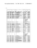 |  |
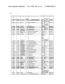 |  |
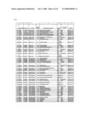 |  |
 |  |
 |  |
 | 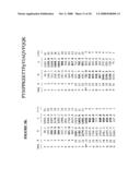 |
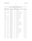 |  |
 | 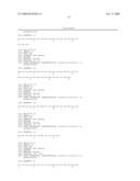 |
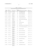 |  |
 |  |
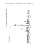 |  |
 | 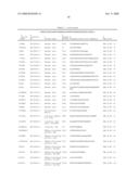 |
 | 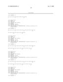 |
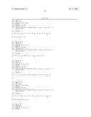 | 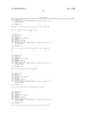 |
 | 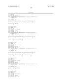 |
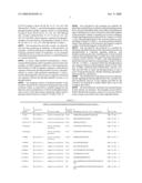 |  |
 | 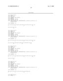 |
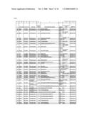 | 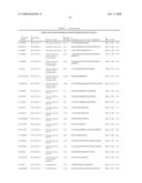 |
 | 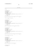 |
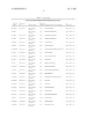 |  |
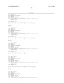 | 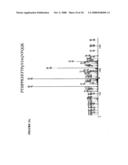 |
 | 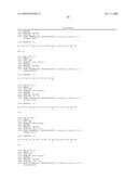 |
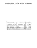 | 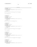 |
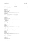 |  |
 | 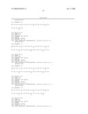 |
 | 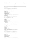 |
 | 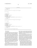 |
 |  |
 |  |
 | 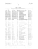 |
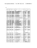 |  |
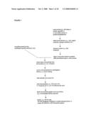 | 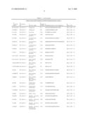 |
 |  |
 | 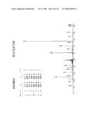 |
 | 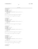 |
 |  |
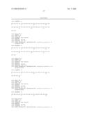 | 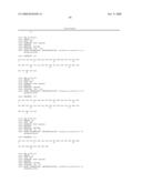 |
 |  |
 |  |
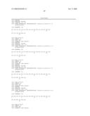 |  |
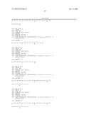 | 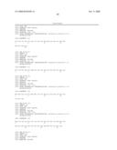 |
 | 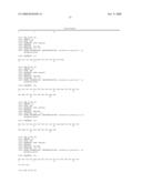 |
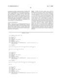 | 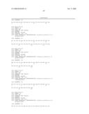 |
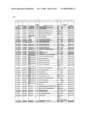 | 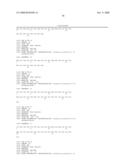 |
 |  |
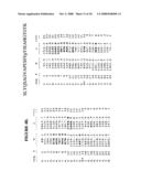 |  |
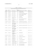 |  |
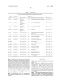 |  |
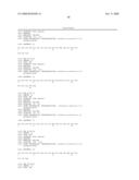 |  |
| Similar patent applications: | |
| Date | Title |
|---|---|
| 2013-06-27 | Enzyme regulating ether lipid signaling pathways |
| 2013-08-01 | In vitro process for the preparation of antibodies of the igg type |
| 2013-07-18 | Reference sample for quality control in molecular diagnosis |
| 2013-07-25 | Apparatus for polynucleotide detection and quantitation |
| 2013-02-14 | Arrangement for detection of hemolysis |
| New patent applications from these inventors: | |
| Date | Title |
|---|---|
| 2021-12-02 | Compositions for detecting mutant anaplastic lymphoma kinase in lung cancer |
| 2021-11-11 | Ros kinase in lung cancer |
| 2019-01-03 | Methods and reagents for creating monoclonal antibodies |
| 2018-06-07 | Methods of treating lung cancer by administration of an alk inhibitor |
| 2016-11-17 | Gene defects and mutant alk kinase in human solid tumors |
| Top Inventors for class "Chemistry: molecular biology and microbiology" | |
| Rank | Inventor's name |
|---|---|
| 1 | Marshall Medoff |
| 2 | Anthony P. Burgard |
| 3 | Mark J. Burk |
| 4 | Robin E. Osterhout |
| 5 | Rangarajan Sampath |